

Tested Applications
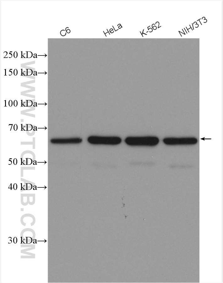
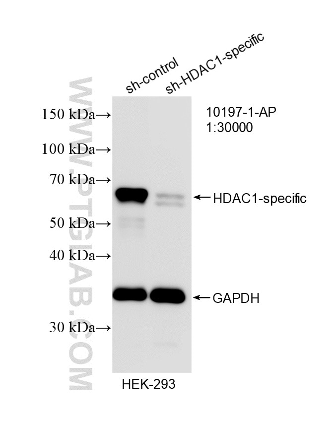
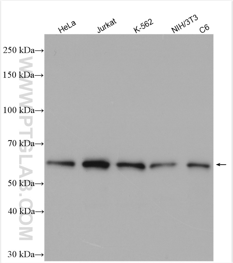
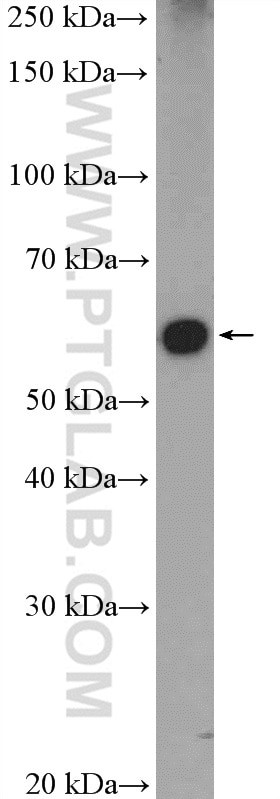
| Positive WB detected in | C6 cells, HEK-293 cells, HeLa cells, mouse testis tissue, K-562 cells, NIH/3T3 cells, Jurkat cells |
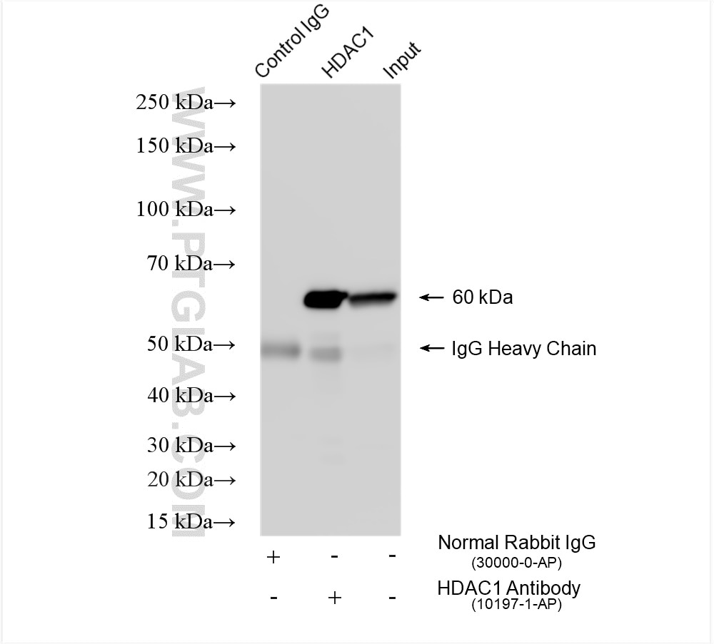
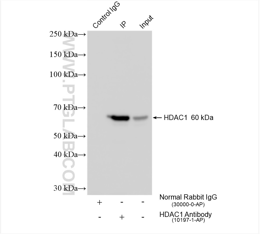
| Positive IP detected in | HeLa cells |
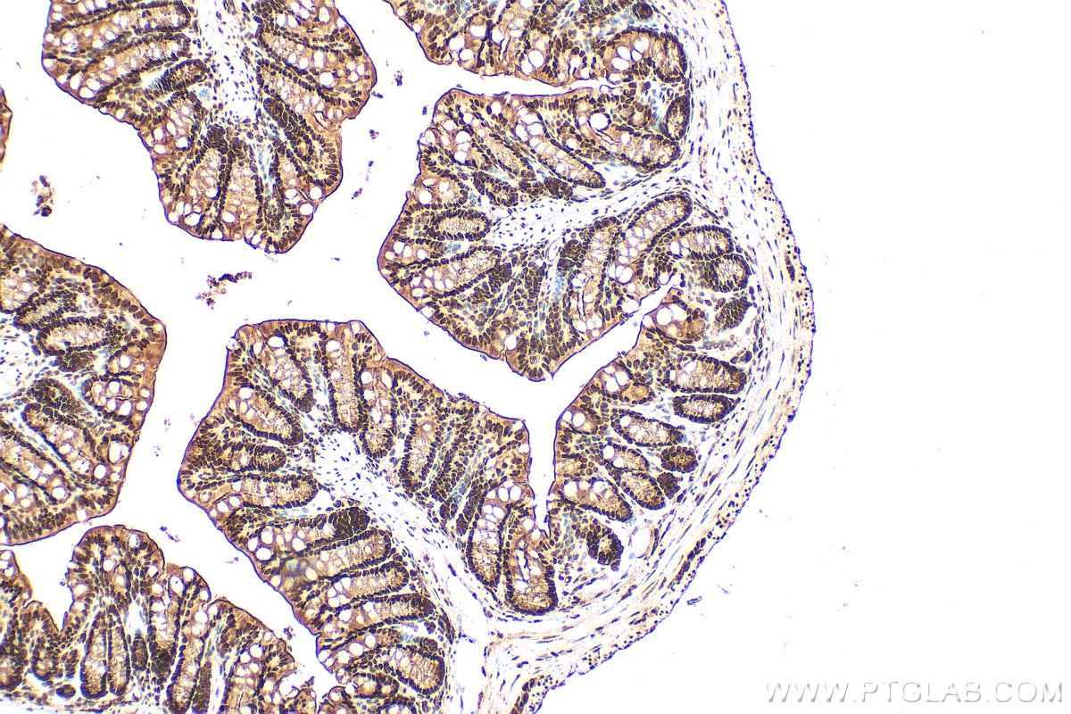
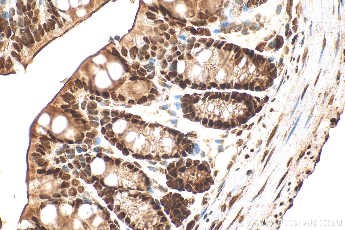
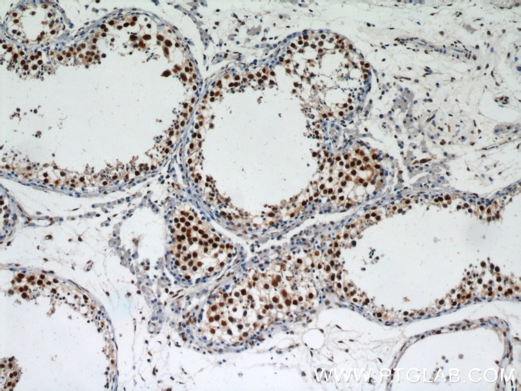
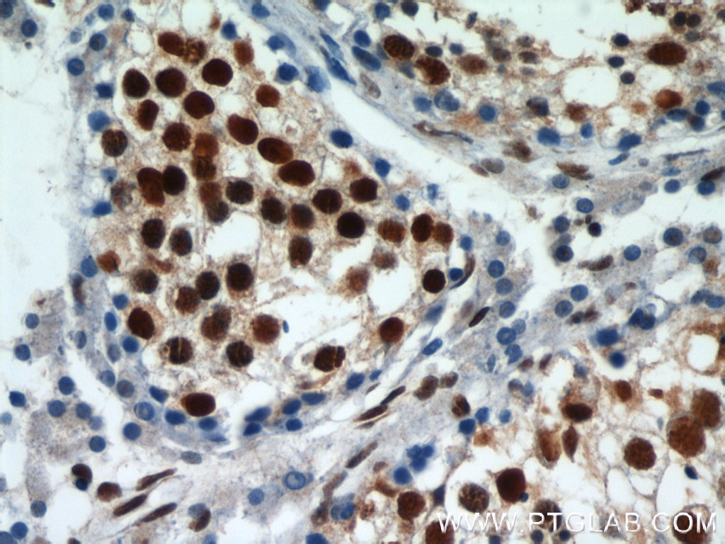
| Positive IHC detected in | mouse colon tissue, human testis tissue Note: suggested antigen retrieval with TE buffer pH 9.0; (*) Alternatively, antigen retrieval may be performed with citrate buffer pH 6.0 |
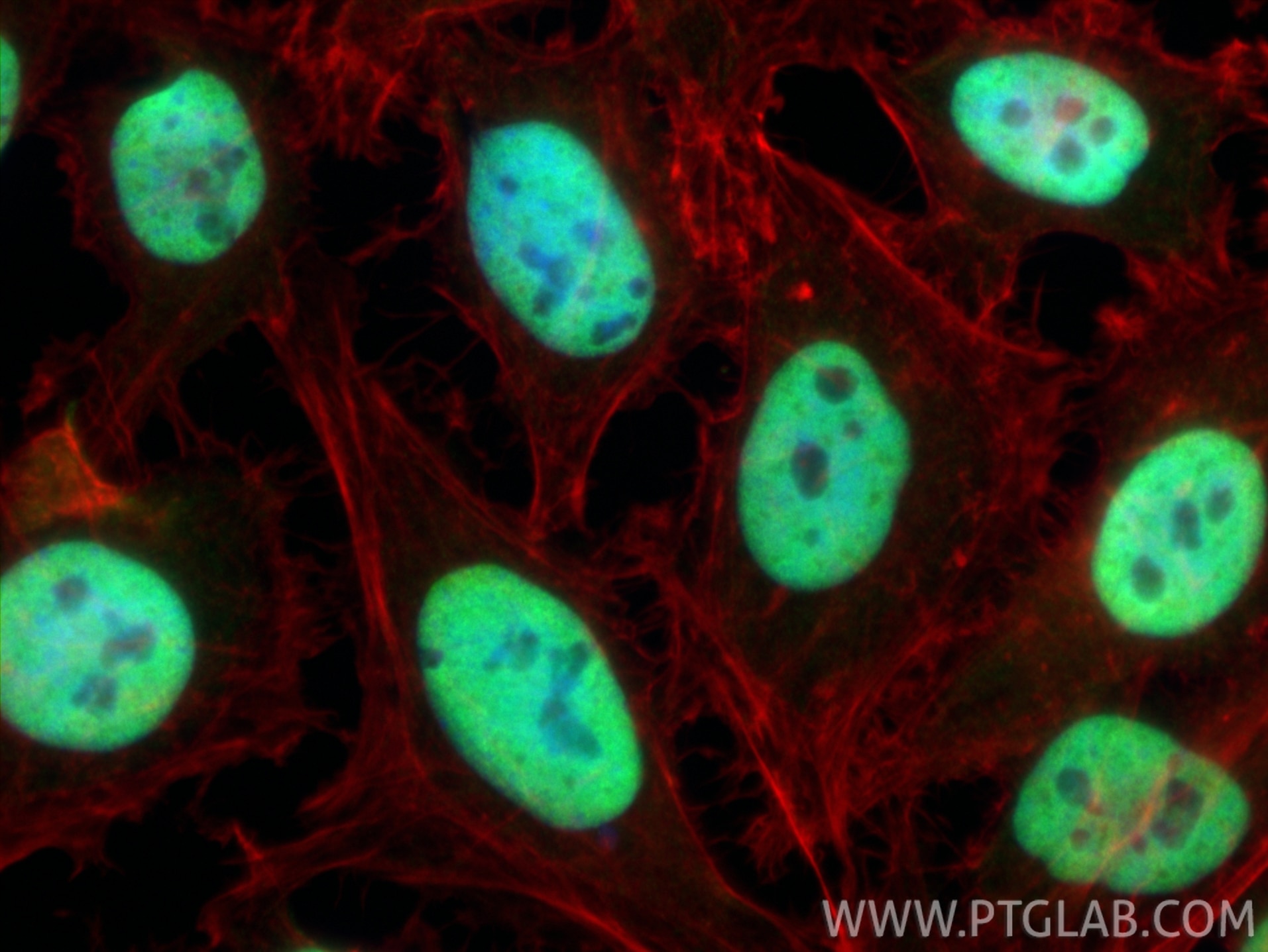
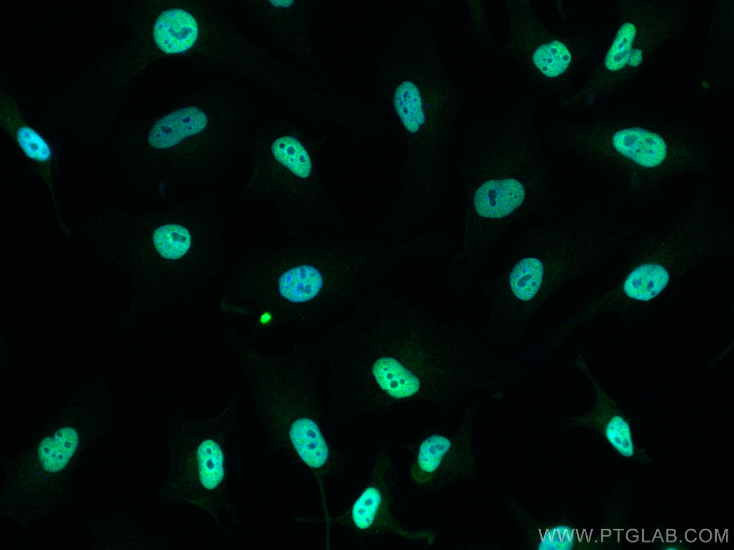
| Positive IF/ICC detected in | HeLa cells |
Recommended dilution
| Application | Dilution |
|---|---|
| Western Blot (WB) | WB : 1:8000-1:20000 |
| Immunoprecipitation (IP) | IP : 0.5-4.0 ug for 1.0-3.0 mg of total protein lysate |
| Immunohistochemistry (IHC) | IHC : 1:500-1:2000 |
| Immunofluorescence (IF)/ICC | IF/ICC : 1:200-1:800 |
| It is recommended that this reagent should be titrated in each testing system to obtain optimal results. | |
| Sample-dependent, Check data in validation data gallery. | |
Product Information
10197-1-AP targets HDAC1 in WB, IHC, IF/ICC, IP, CoIP, ChIP, RIP, ELISA, CUT&Tag applications and shows reactivity with human, mouse, rat samples.
| Tested Reactivity | human, mouse, rat |
| Cited Reactivity | human, mouse, rat, pig, rabbit |
| Host / Isotype | Rabbit / IgG |
| Class | Polyclonal |
| Type | Antibody |
| Immunogen |
CatNo: Ag0256 Product name: Recombinant human HDAC1 protein Source: e coli.-derived, PGEX-4T Tag: GST Domain: 184-482 aa of BC000301 Sequence: EEAFYTTDRVMTVSFHKYGEYFPGTGDLRDIGAGKGKYYAVNYPLRDGIDDESYEAIFKPVMSKVMEMFQPSAVVLQCGSDSLSGDRLGCFNLTIKGHAKCVEFVKSFNLPMLMLGGGGYTIRNVARCWTYETAVALDTEIPNELPYNDYFEYFGPDFKLHISPSNMTNQNTNEYLEKIKQRLFENLRMLPHAPGVQMQAIPEDAIPEESGDEDEDDPDKRISICSSDKRIACEEEFSDSEEEGEGGRKNSSNFKKAKRVKTEDEKEKDPEEKKEVTEEEKTKEEKPEAKGVKEEVKLA Predict reactive species |
| Full Name | histone deacetylase 1 |
| Calculated Molecular Weight | 55 kDa |
| Observed Molecular Weight | 60 kDa |
| GenBank Accession Number | BC000301 |
| Gene Symbol | HDAC1 |
| Gene ID (NCBI) | 3065 |
| RRID | AB_2118062 |
| Conjugate | Unconjugated |
| Form | Liquid |
| Purification Method | Antigen affinity purification |
| UNIPROT ID | Q13547 |
| Storage Buffer | PBS with 0.02% sodium azide and 50% glycerol, pH 7.3. |
| Storage Conditions | Store at -20°C. Stable for one year after shipment. Aliquoting is unnecessary for -20oC storage. 20ul sizes contain 0.1% BSA. |
Background Information
Background
Histone Deacetylase 1 (HDAC1) is a nuclear localized class I histone deacetylase that plays a role in the regulation of gene expression, mainly by repression of gene activity. Additionally, HDAC can mediate deacetylation of a subset of non-histone proteins, leading to their degradation. HDAC1 activity has an impact on cell growth, proliferation, and death.
1. What is the molecular weight of HDAC1?
The molecular size of HDAC1 is 60 kDa.
2. What is the subcellular localization of HDAC1?
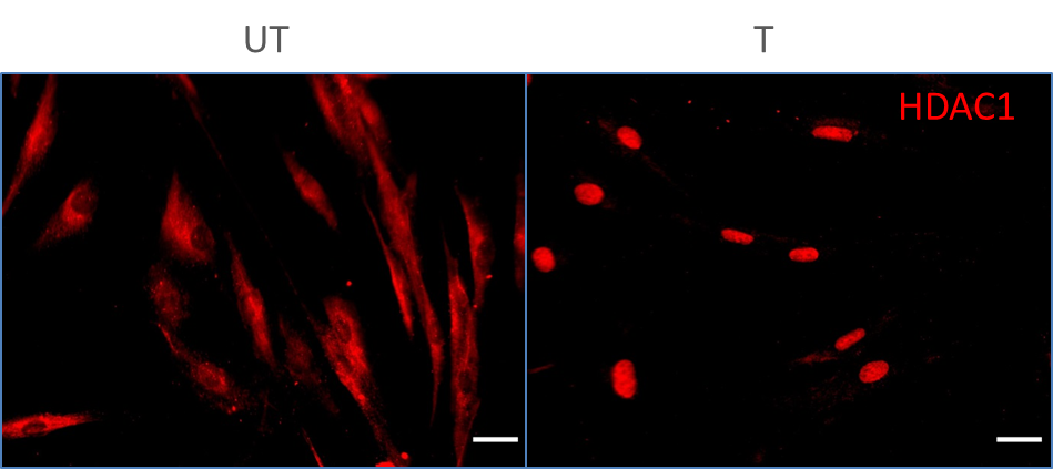
Unlike some HDACs, HDAC1 is present entirely in the nucleus. Our HDAC1 antibody has been broadly tested for IF/ICC.
3. I cannot detect an HDAC1 specific signal in my sample during western blotting.
Make sure that you efficiently extract nuclear proteins during your sample preparation. Some lysis buffers (e.g., based on Triton X-100) may not extract nuclear proteins. We recommend using RIPA buffer (https://www.ptglab.com/support/protocols/). We highly recommend using nuclear loading control antibodies, such as lamin B1 or PCNA (https://www.ptglab.com/news/blog/loading-control-antibodies-for-western-blotting/). Alternatively, you may consider performing cell fractionation.
4. How do I perform chromatin immunoprecipitation (ChiP) with the HDAC1 antibody?
We recommend using our standard chromatin immunoprecipitation protocol (https://www.ptglab.com/media/2708/web_chip-protocol.pdf).
5. Is HDAC1 post-translationally modified?
HDAC1 is a protein deacetylase but itself can also be a subject of post-translational modifications, including phosphorylation, acetylation, ubiquitination, SUMOylation, nitrosylation, and carbonylation (PMID: 21197454).
6. What is the role of HDAC1 in cancer?
The acetylation and deacetylation of histones play an important part in the epigenetic regulation of gene expression. Alterations in the balance between these two opposing processes changes chromosome remodeling and increased deacetylation leads to gene silencing. HDAC1 levels are increased in many cancer cell types and the role of HDAC1 in cancer is connected not only to histone deacetylation but also to the deacetylation of other proteins, such as Rb family proteins, estrogen receptors, and p53 protein (PMID: 19383284).
Protocols
| Product Specific Protocols | |
|---|---|
| IF protocol for HDAC1 antibody 10197-1-AP | Download protocol |
| IHC protocol for HDAC1 antibody 10197-1-AP | Download protocol |
| IP protocol for HDAC1 antibody 10197-1-AP | Download protocol |
| WB protocol for HDAC1 antibody 10197-1-AP | Download protocol |
| Standard Protocols | |
|---|---|
| Click here to view our Standard Protocols |
Publications
What published studies show
Anti-HDAC1 (Cat# 10197-1-AP) has accumulated 175 citations across journals including Nature, Nature Communications, Cell Metabolism, Molecular Cell, EMBO Journal, Cell Reports, Hepatology, Journal of Clinical Investigation, Advanced Science, and Oncogene. Research fields span epigenetics/histone modification, oncology (pancreatic, breast, liver, lung, colorectal cancers), cardiology, neuroscience, immunology, metabolism, and virology. Primary applications include Western blotting, immunoprecipitation, co-IP, IHC, and ChIP.
| Species | Application | Title |
|---|---|---|
Signal Transduct Target Ther B-lymphoid tyrosine kinase-mediated FAM83A phosphorylation elevates pancreatic tumorigenesis through interacting with β-catenin | ||
Nat Commun The methyltransferase METTL3 negatively regulates nonalcoholic steatohepatitis (NASH) progression. | ||
Nat Commun Exercise-induced histone lactylation in monocyte-derived macrophages restores cardiac immune homeostasis and function in sepsis-induced cardiomyopathy. |
Reviews
What customers say
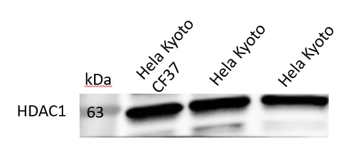
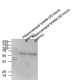
All five reviewers gave this antibody a perfect 5-star rating. Users report excellent performance in Western blotting across diverse cell types including mouse cells, human adipocytes, HeLa cells, hippocampal neurons, and primary fibroblasts. It also performs well in flow cytometry and immunofluorescence. Several reviewers shared detailed protocols, with common WB dilutions of 1:1000–1:2000 and overnight incubation at 4°C. For IF, one user achieved clear results at 1:50 dilution in human fibroblasts. Overall, customers consistently describe it as a reliable, high-quality reagent with strong specificity and sensitivity.
The reviews below have been submitted by verified Proteintech customers who received an incentive for providing their feedback.
Sara (Verified Customer) (10-06-2024) | I tried WB and FC with mouse cells, works well. I still need to give it a try for immunofluorescence, but it seems to work as well based on other reviews.
|
Mi (Verified Customer) (06-25-2023) | Works pretty well in human adipocyte cells.
|
Christos (Verified Customer) (04-03-2023) | Separating Gel: 10% poly-acrylamide Transfer at 400mA for 2hr onto nitrocellulose membrane at 4oC Blocking and antibodies dilutions: 5% non-fat milk in PBS-Tween20 for 1hr Incubation with primary Abs: overnight at 4oC Incubation with secondary Abs: 1hr at 4oC
 |
Dipen (Verified Customer) (07-20-2020) | Great antibody for Western blotting.
 |
Uxoa (Verified Customer) (02-19-2020) | HDAC1 in human primary fibroblasts untreated vs treated. Cells fixed with 4% PFA 15 min RT. HDAC1 incubation 1:50 in PBST (0.1% triton) + 10% DS O/N 4ºC. Donkey anti-rabbit Alexa fluor 555 1:500 1h RT.
 |


