"DYKDDDDK tag Antibodies" Comparison
View side-by-side comparison of DYKDDDDK tag antibodies from other vendors to find the one that best suits your research needs.
Tested Applications
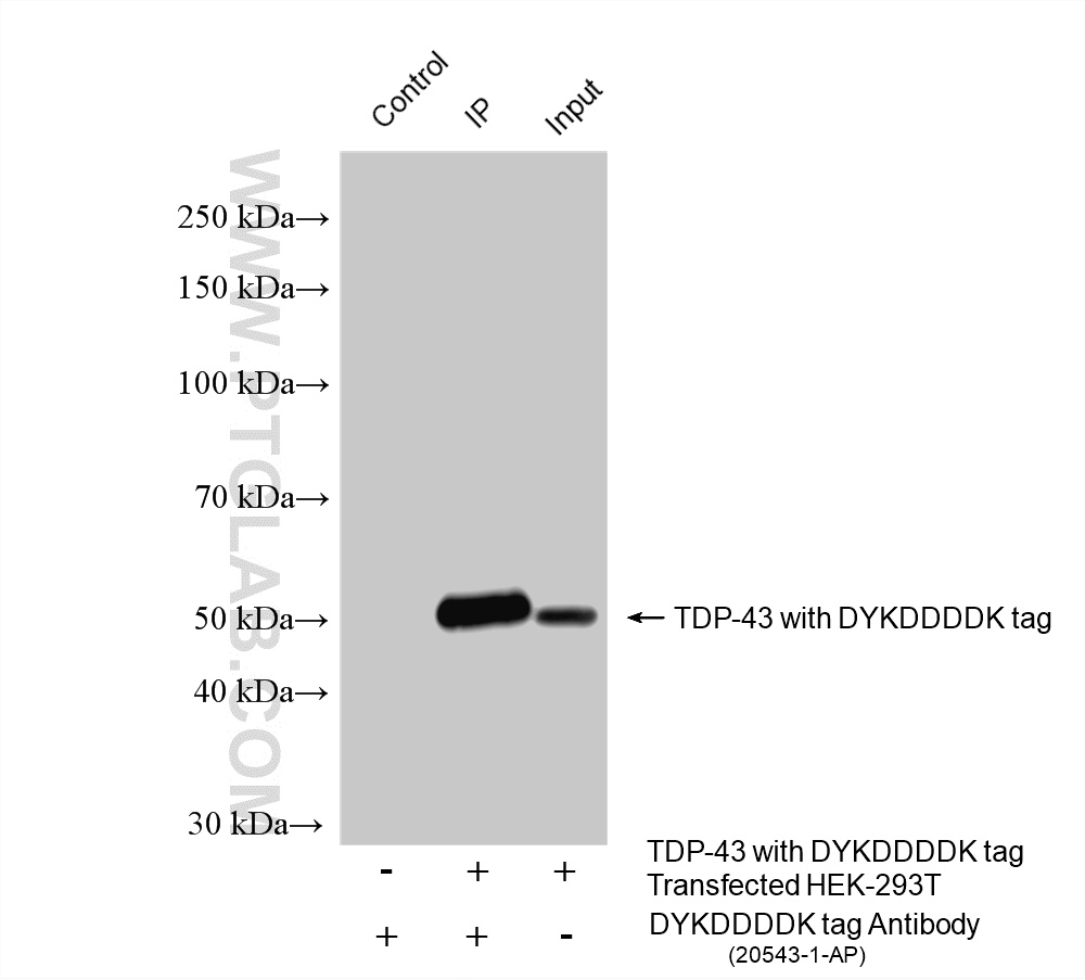
| Positive WB detected in | Transfected HEK-293T cells |
| Positive IP detected in | Transfected HEK-293T cells |
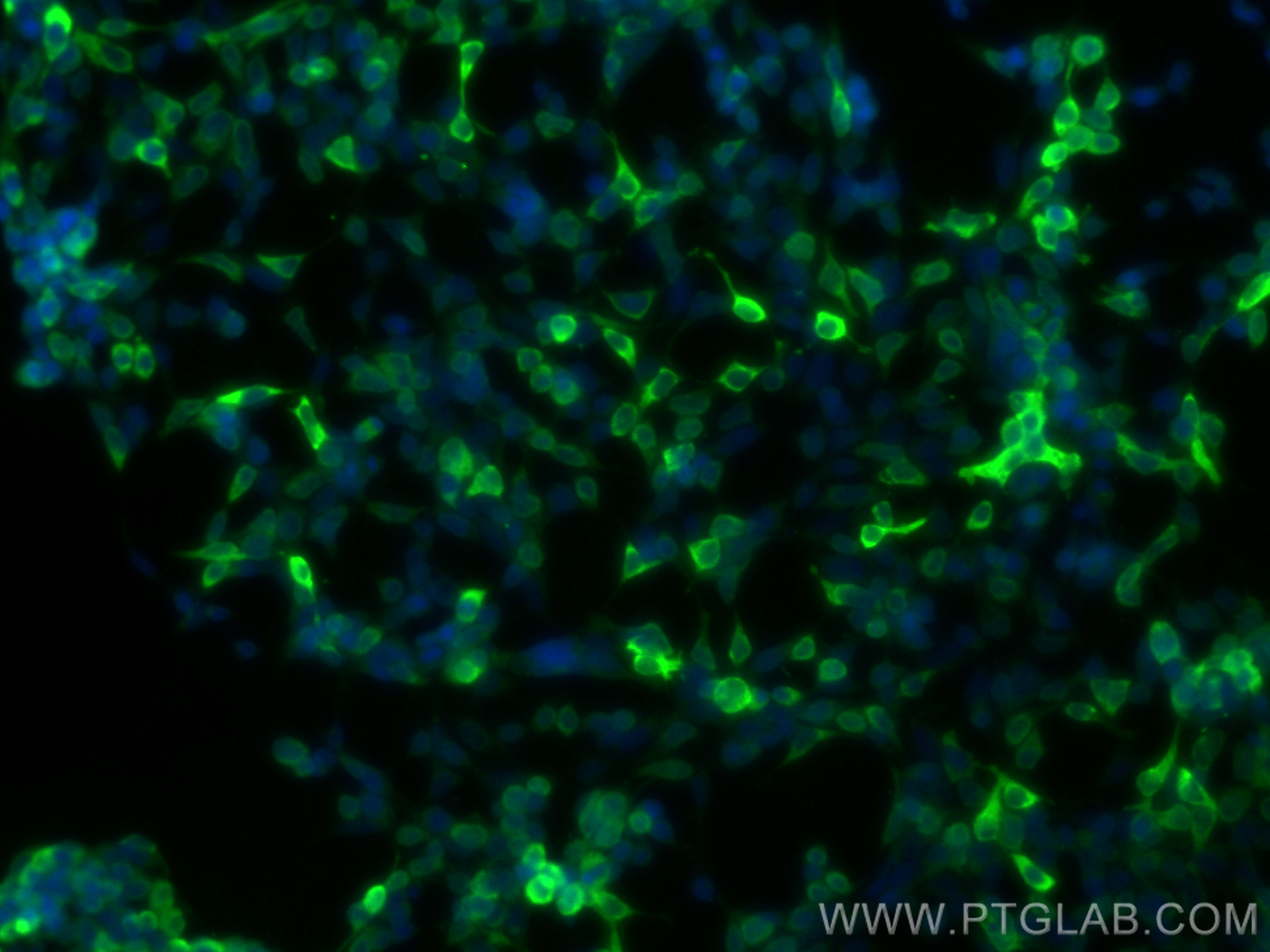
| Positive IF/ICC detected in | Transfected HEK-293 cells |
Recommended dilution
| Application | Dilution |
|---|---|
| Western Blot (WB) | WB : 1:20000-1:100000 |
| Immunoprecipitation (IP) | IP : 0.5-4.0 ug for 1.0-3.0 mg of total protein lysate |
| Immunofluorescence (IF)/ICC | IF/ICC : 1:200-1:800 |
| It is recommended that this reagent should be titrated in each testing system to obtain optimal results. | |
| Sample-dependent, Check data in validation data gallery. | |
Product Information
20543-1-AP targets DYKDDDDK tag in WB, IHC, IF/ICC, IP, CoIP, ChIP, RIP, ELISA applications and shows reactivity with recombinant protein samples.
| Tested Reactivity | recombinant protein |
| Cited Reactivity | human, mouse, pig, silkworm, recombinant protein |
| Host / Isotype | Rabbit / IgG |
| Class | Polyclonal |
| Type | Antibody |
| Immunogen |
CatNo: Ag2329 Product name: Recombinant DYKDDDDK tag protein Source: e coli.-derived, PGEX-4T Tag: GST Sequence: DYKDDDDK Predict reactive species |
| Full Name | DYKDDDDK tag |
| Gene Symbol | |
| Gene ID (NCBI) | |
| RRID | AB_11232216 |
| Conjugate | Unconjugated |
| Form | Liquid |
| Purification Method | Antigen affinity purification |
| UNIPROT ID | FLAGTAG |
| Storage Buffer | PBS with 0.02% sodium azide and 50% glycerol, pH 7.3. |
| Storage Conditions | Store at -20°C. Stable for one year after shipment. Aliquoting is unnecessary for -20oC storage. 20ul sizes contain 0.1% BSA. |
Background Information
Protein tags are protein or peptide sequences located either on the C- or N- terminal of the target protein, which facilitates one or several of the following characteristics: solubility, detection, purification, localization and expression. The DYKDDDDK(FLAG) peptide has been used extensively as a general tag in expression vectors. This peptide can be expressed and detected with the protein of interest as an amino-terminal or carboxy-terminal fusion. N-terminal DDDDK vectors provide an Ek cleavage site for removal of the fusion tag. The DDDDK peptide is likely to be located on the surface of a fusion protein because of its hydrophilic nature. As a result, the DDDDK peptide is more likely to be accessible to antibodies. A DDDDK-tag can be used in many different assays that require recognition by an antibody, such as western blotting, immunocytochemistry, immunoprecipitation, flow cytometry, protein purification, and in the study of protein-protein interactions, cell ultrastructure, and protein localization and so on. This antibody is a rabbit polyclonal antibody raised against 3xFlag (3xDYKDDDDKT) sequence and recognizes the (1x) and (3x)DYKDDDDK peptide and detects DDDDK-tagged proteins. Anti-FLAG is a registered trademark of Sigma-Aldrich Biotechnology.
Protocols
| Product Specific Protocols | |
|---|---|
| IF protocol for DYKDDDDK tag antibody 20543-1-AP | Download protocol |
| IP protocol for DYKDDDDK tag antibody 20543-1-AP | Download protocol |
| WB protocol for DYKDDDDK tag antibody 20543-1-AP | Download protocol |
| Standard Protocols | |
|---|---|
| Click here to view our Standard Protocols |
Publications
| Species | Application | Title |
|---|---|---|
Science Ubiquitination of G3BP1 mediates stress granule disassembly in a context-specific manner. | ||
Signal Transduct Target Ther Trogocytosis of CAR molecule regulates CAR-T cell dysfunction and tumor antigen escape | ||
Cell Ca2+ sensor-mediated ROS scavenging suppresses rice immunity and is exploited by a fungal effector. | ||
Nat Immunol Sparcl1 mitigates abdominal aortic aneurysm through inhibiting lymphangiogenesis-mediated TLS formation | ||
Protein Cell Senktide blocks aberrant RTN3 interactome to retard memory decline and tau pathology in social isolated Alzheimer's disease mice |
Reviews
The reviews below have been submitted by verified Proteintech customers who received an incentive for providing their feedback.
FH Dan (Verified Customer) (02-27-2026) | transfection for ORF1P-Flag (~40KD) done on 293T cells and 40ug loaded for western blot.
 |
FH Hong (Verified Customer) (01-26-2026) | good
 |
FH Mason (Verified Customer) (01-26-2026) | This antibody worked great, and I could even cut the dilution back to 1:10,000 since the signal was so strong and clear.
|
FH YINGJIAN (Verified Customer) (05-27-2025) | This primary antibody performs very well, even at a low concentration (1:10000).
 |
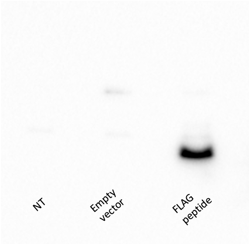
FH Jon (Verified Customer) (02-28-2023) | The antibody works fine in WB in the range fo 1:1000-1:5000 dilution in 5% milk TBST. In the image the samples are HEK293 lysates on a tricine SDS PAGE with primary antiFLAG antibody 1:5000 overnight at 4 ºC. It is true that the antibody could show considerable nonspecific background depending on the concentrations and cells used. In my experience, works perfectly fine and is quite sensitive in HEK293 cells, although in my EndoC-ßH1 cells signal is weaker and backgorund is very notable. However, I assume it is due to the EndoC cells, which are a pain for many applications. I also used this antibody for some IF experiments. In the EndoC line, no matter the antibody concentration, I always get non specific signal. I'm worried the antibody binds to something in particular in these cells. In HEK293 cells however, I managed to obtain good results for overexpression of a FLAG tagged peptide, same as the one on Western Blot
 |
FH Brogan (Verified Customer) (11-09-2022) | Also used in a pul down assay for FLAG tag plasmids
|
FH Chun (Verified Customer) (06-19-2022) | This antibody worked very well in both ECL and Li-Cor procedures.
|
FH WEI (Verified Customer) (05-11-2022) | Good for western blot of knock-in mice but not good for immunostaining.
|
FH Barbara (Verified Customer) (03-28-2022) | reliable antibody for detection of FLAG tag by western blot
|
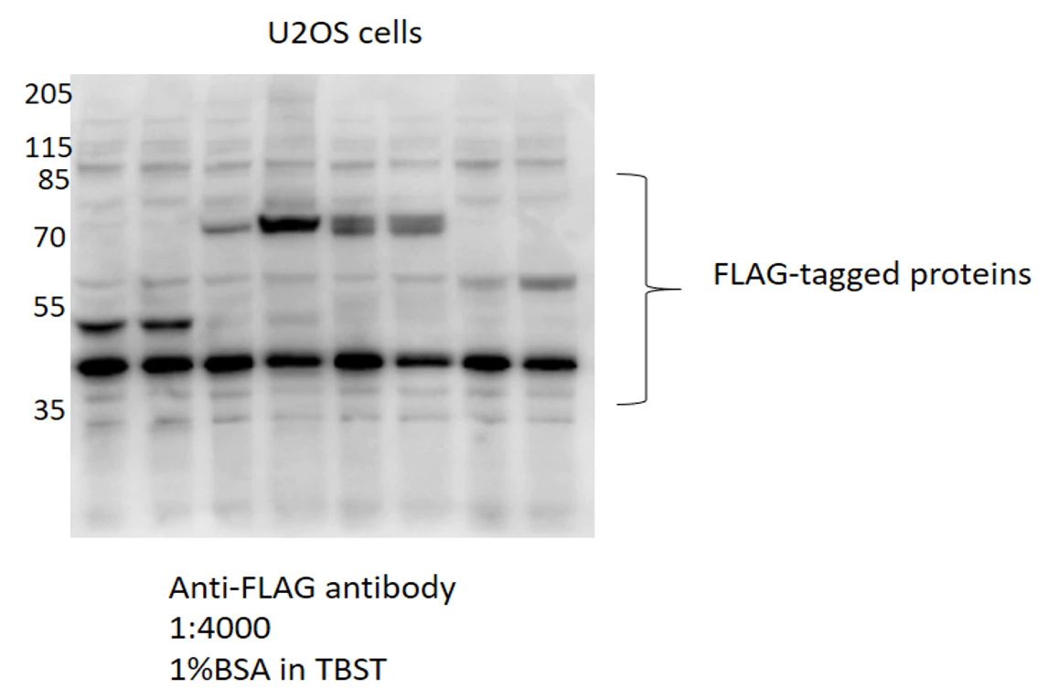
FH ISABELLE CRISTINE (Verified Customer) (01-14-2022) | This antibody is extremely concentrated and works very well for detecting FLAG-tagged proteins in U2OS and HEK293 cells.
 |
FH X (Verified Customer) (01-05-2022) | It is Ok to use it in WB but with some non-specific bands.
 |
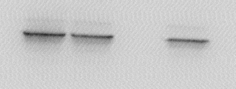
FH Christine (Verified Customer) (10-13-2021) | Antibody used on HEK293T transfected with a FLAG tagged protein and western blots compared with 3 other FLAG antibodies. This one gives the strongest signal for the tagged protein (which is a about 34 kDa) so it might be very useful for poorly expressed proteins. However it also has the most background bands, with 2 main additional background bands (but with a lower intensity than my tagged protein of interest): one at around 50 kDa (just above 47 kDa marker) and another one between 118 and 85 kDa markers. So this antibody is worth a try depending on the size of your protein of interest. It gives the strongest signal of the 4 antibodies tested.
|
FH Boyan (Verified Customer) (02-05-2021) | Very strong signal, but with some weak non specific bands.
|
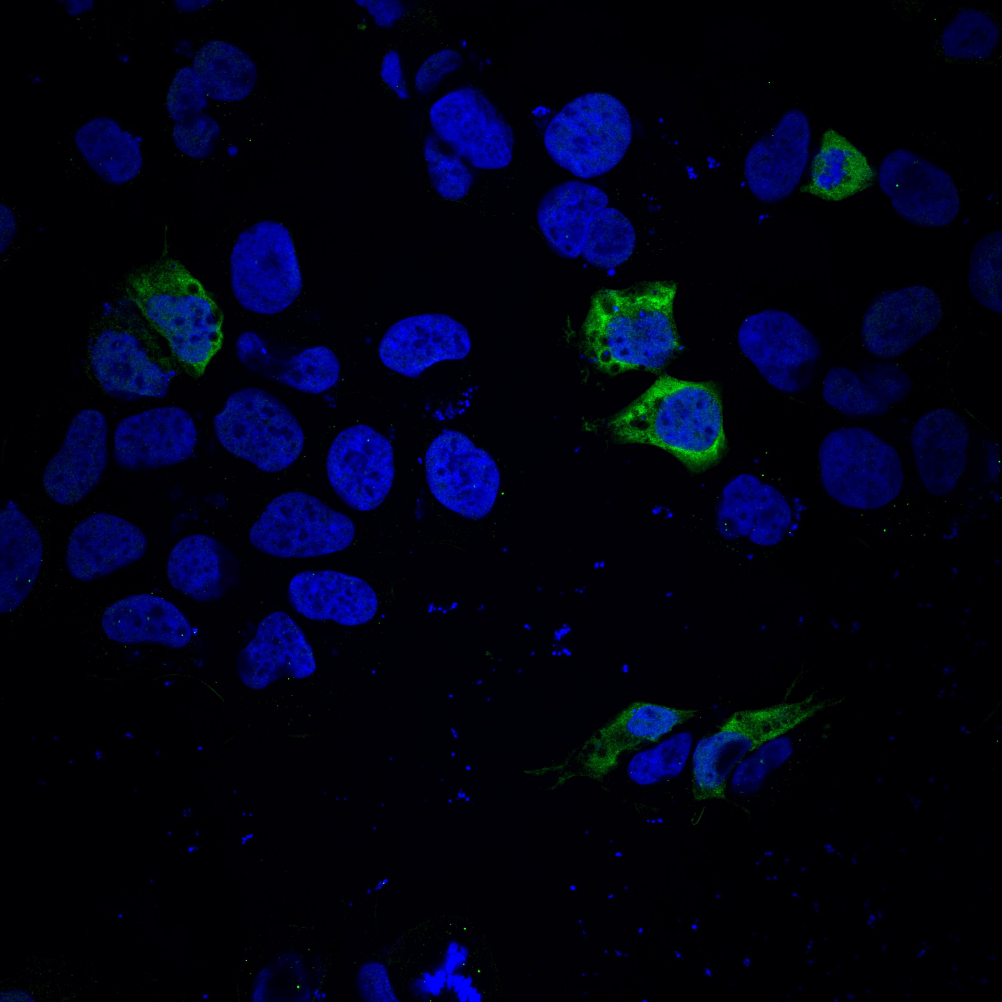
FH Lahiru Chamara (Verified Customer) (11-06-2020) | HEK293 cells were transfected with gene of interested. Primary antibody incubation was performed at 4C for an overnight and Alexa Fluor 488 secondary antibody incubation was performed at room temperature for an hour. Cells were stained with Hoechst for DNA/ nuclei . Images were taken from Zeiss LSM780 microscope.
 |
FH Tongbin (Verified Customer) (08-25-2020) | For western blots, this FLAG antibody is comparable to Sigma's M2 FLAG antibody, which has been the gold standard for FLAG.
|
FH Dipen (Verified Customer) (11-11-2019) | Great antibody for Western blotting. I used it at 1:1000 and got a super strong signal. Will use at a lower concentration next time.
 |
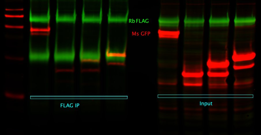
FH Sara (Verified Customer) (10-24-2019) | Works really well for both IP of overexpressed FLAG-tagged proteins and WB!
 |
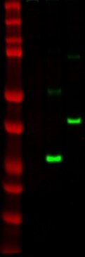
FH Nikhil (Verified Customer) (10-16-2019) | Flag tagged p53 expressed in H1299 cells.
 |
FH Shivani (Verified Customer) (09-26-2019) | Works better than another brand I had tested.
|
FH Corey (Verified Customer) (08-23-2019) | This antibody has worked well for western blotting as well as immunoprecipitation. The lots have been consistent so far, we will definitely be ordering more.
|
FH Kishor (Verified Customer) (06-23-2019) | Works very good for WB at 1:1000 and IP at 1:200.
|
FH David (Verified Customer) (06-19-2019) | This is working very well for me. I ran out of another brand and got this one because I saw you had it and it was a better value. After testing it in the lab, it works just as well, if not better than the previous one. A very strong signal in my experiments.
|
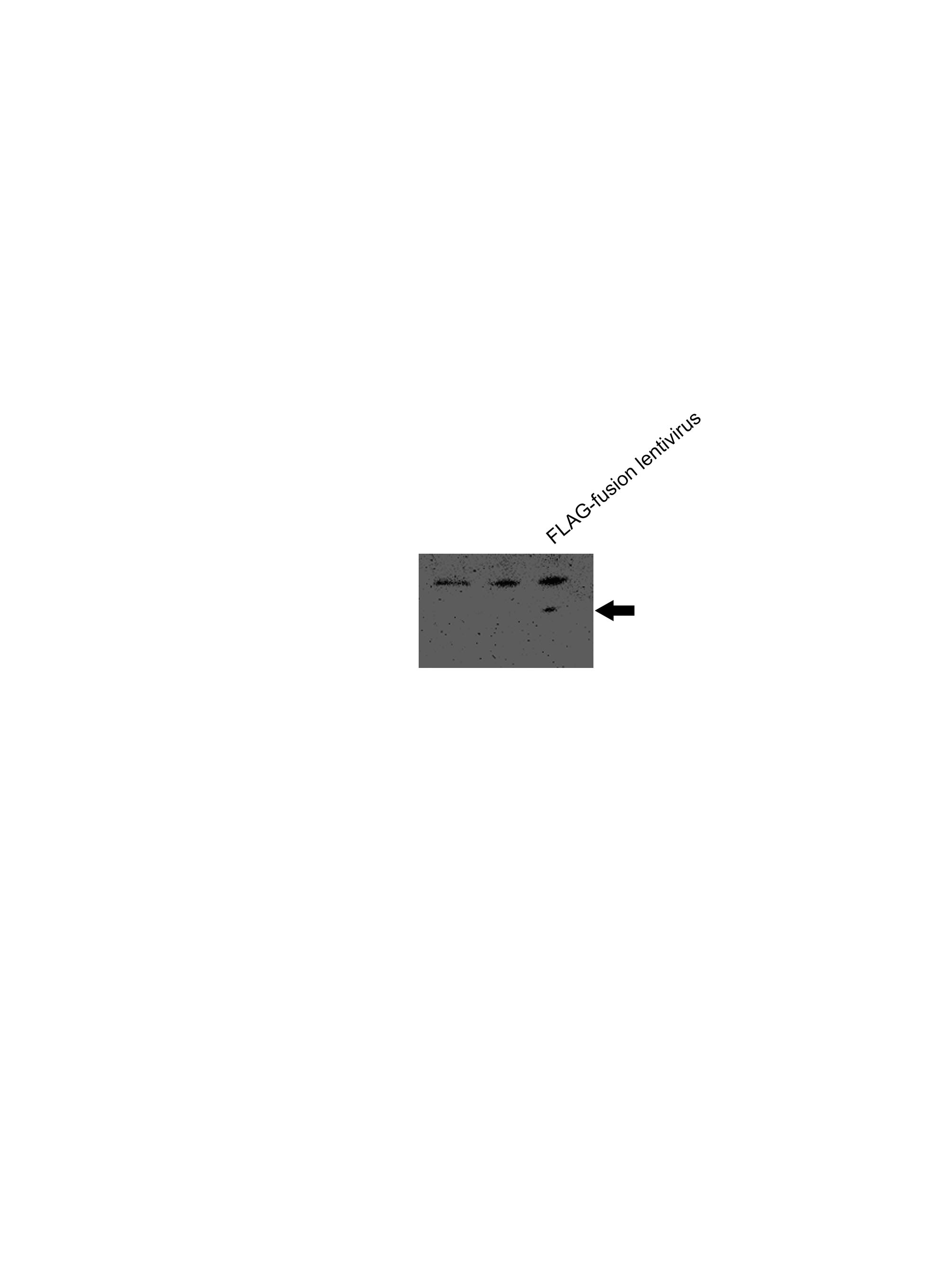
FH Lana (Verified Customer) (04-20-2019) | SDS-PAGE:10 ug/ul RIPA lysate of whole cells4-12% Bis-tris gradient gelTransfer:Immobilon-FL transfer membranes (Millipore) O/N at 30V, 4CBlocking:SEA Block Blocking Buffer 1hPrimary Ab:DDDDK pRb Cat# 20543-1-APO/N incubation at 4CSecondary Ab:IRDye 800CW Goat anti-Rabbit1 h incubation at room temperatureLines on WB:1. BioRad Precision Plus Protein standards2. Whole cell lysate of HEK293t cells (negative transfection)3. Whole cell lysate of HEK293t cells transfected with FLAG-tag construct 27 kDa3. Whole cell lysate of HEK293t cells transfected with FLAG-tag construct 40 kDa
 |
FH Luce (Verified Customer) (11-12-2018) |
|