
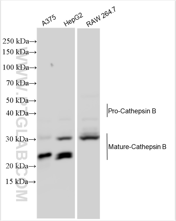
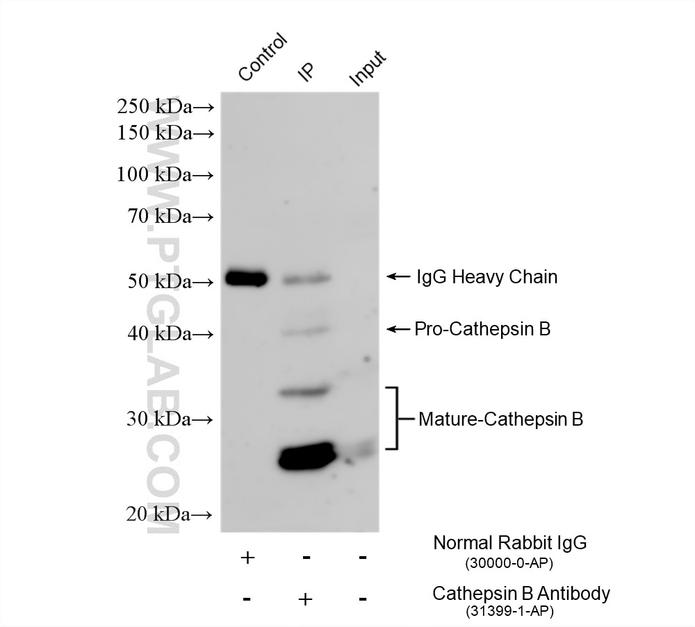
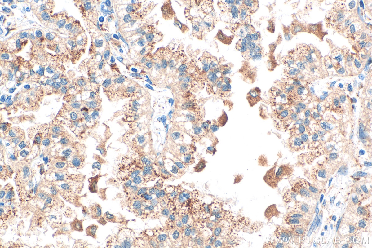

Tested Applications
| Positive WB detected in | A375 cells, HepG2 cells, RAW 264.7 cells |
| Positive IP detected in | HepG2 cells |
| Positive IHC detected in | human lung cancer tissue Note: suggested antigen retrieval with TE buffer pH 9.0; (*) Alternatively, antigen retrieval may be performed with citrate buffer pH 6.0 |
Recommended dilution
| Application | Dilution |
|---|---|
| Western Blot (WB) | WB : 1:500-1:2000 |
| Immunoprecipitation (IP) | IP : 0.5-4.0 ug for 1.0-3.0 mg of total protein lysate |
| Immunohistochemistry (IHC) | IHC : 1:50-1:500 |
| It is recommended that this reagent should be titrated in each testing system to obtain optimal results. | |
| Sample-dependent, Check data in validation data gallery. | |
Product Information
31399-1-AP targets Cathepsin B in WB, IHC, IP, ELISA applications and shows reactivity with human, mouse samples.
| Tested Reactivity | human, mouse |
| Cited Reactivity | mouse |
| Host / Isotype | Rabbit / IgG |
| Class | Polyclonal |
| Type | Antibody |
| Immunogen |
CatNo: Eg1020 Product name: Recombinant human Cathepsin B protein Source: mammalian cells-derived, pHZ-KIsec-C-6*HIS Tag: C-10*His Domain: 18-339 aa of NM_001908.5 Sequence: RSRPSFHPLSDELVNYVNKRNTTWQAGHNFYNVDMSYLKRLCGTFLGGPKPPQRVMFTEDLKLPASFDAREQWPQCPTIKEIRDQGSCGSCWAFGAVEAISDRICIHTNAHVSVEVSAEDLLTCCGSMCGDGCNGGYPAEAWNFWTRKGLVSGGLYESHVGCRPYSIPPCEHHVNGSRPPCTGEGDTPKCSKICEPGYSPTYKQDKHYGYNSYSVSNSEKDIMAEIYKNGPVEGAFSVYSDFLLYKSGVYQHVTGEMMGGHAIRILGWGVENGTPYWLVANSWNTDWGDNGFFKILRGQDHCGIESEVVAGIPRTDQYWEKI Predict reactive species |
| Full Name | cathepsin B |
| Calculated Molecular Weight | 38kDa |
| Observed Molecular Weight | 25 kDa and 31 kDa |
| GenBank Accession Number | NM_001908.5 |
| Gene Symbol | Cathepsin B |
| Gene ID (NCBI) | 1508 |
| RRID | AB_3669964 |
| Conjugate | Unconjugated |
| Form | Liquid |
| Purification Method | Antigen affinity Purification |
| UNIPROT ID | P07858 |
| Storage Buffer | PBS with 0.02% sodium azide and 50% glycerol, pH 7.3. |
| Storage Conditions | Store at -20°C. Stable for one year after shipment. Aliquoting is unnecessary for -20oC storage. 20ul sizes contain 0.1% BSA. |
Background Information
CTSB(Cathepsin B) is also named as CPSB and belongs to the peptidase C1 family. It participates in intracellular degradation and turnover of proteins. Cathepsin B precursors found in human malignant ascites fluid do not possess mannose-rich carbohydrates suggesting that a defect in the post translational processing of carbohydrate moieties on tumor. Cathepsin B exists as both glycosylated and unglycosylated forms(PMID:1637335). In rat macrophages and hepatocytes pro- cathepsin B is 39 kDa (unglycosylated =35 kDa), whereas in human fibroblasts procathepsin B is 44.5-46kDa (unglycosylated=39kDa)(PMID:2097084). It can be detected the 43 kDa form of pro-CTSB, 31 kDa and 25 kDa mature forms in mouse brain by western blot(PMID:20616152).
Protocols
| Product Specific Protocols | |
|---|---|
| IHC protocol for Cathepsin B antibody 31399-1-AP | Download protocol |
| IP protocol for Cathepsin B antibody 31399-1-AP | Download protocol |
| WB protocol for Cathepsin B antibody 31399-1-AP | Download protocol |
| Standard Protocols | |
|---|---|
| Click here to view our Standard Protocols |
Publications
What published studies show
The Cathepsin B Polyclonal antibody (Cat# 31399-1-AP) has 2 citations. These publications appear in Metab Brain Dis and Cell Signal. The research fields encompass neurodegenerative disease (aggrephagy in ALS) and neuroinflammation (microglial pyroptosis and lysosomal membrane permeabilization following spinal cord injury). Both studies utilized the antibody for Western blot in mouse models.
| Species | Application | Title |
|---|---|---|
Metab Brain Dis Unraveling the regulatory nexus of aggrephagy in ALS: identification of novel candidate biomarkers and molecular triggers. | ||
Cell Signal DRAM1 activates microglial pyroptosis and neuroinflammation through TMEM9-mediated lysosomal membrane permeabilization after spinal cord injury. |

