Various lysates were subjected to SDS PAGE followed by western blot with 20882-1-AP (CUL4B antibody) at dilution of 1:1500 incubated at room temperature for 1.5 hours.
WB analysis of HeLa cells using 20882-1-AP
WB result of CUL4B antibody (20882-1-AP, 1:500) with si-Control and si-CUL4B transfected HeLa cells.
WB analysis using 20882-1-AP
Various lysates were subjected to SDS PAGE followed by western blot with 20882-1-AP (CUL4B antibody) at dilution of 1:2500 incubated at room temperature for 1.5 hours.
WB analysis of Y79 using 20882-1-AP
Y79 cells were subjected to SDS PAGE followed by western blot with 20882-1-AP (CUL4B antibody) at dilution of 1:500 incubated at room temperature for 1.5 hours.
WB analysis of SH-SY5Y using 20882-1-AP
SH-SY5Y cells were subjected to SDS PAGE followed by western blot with 20882-1-AP (CUL4B antibody) at dilution of 1:800 incubated at room temperature for 1.5 hours.
IP experiment of mouse brain using 20882-1-AP
IP result of anti-CUL4B (IP:20882-1-AP, 4ug; Detection:20882-1-AP 1:300) with mouse brain tissue lysate 4000ug.
IHC staining of human tonsillitis using 20882-1-AP
Immunohistochemical analysis of paraffin-embedded human tonsillitis tissue slide using 20882-1-AP (CUL4B Antibody) at dilution of 1:200 (under 10x lens). Heat mediated antigen retrieval with Tris-EDTA buffer (pH 9.0).
IHC staining of human tonsillitis using 20882-1-AP
Immunohistochemical analysis of paraffin-embedded human tonsillitis tissue slide using 20882-1-AP (CUL4B Antibody) at dilution of 1:200 (under 40x lens). Heat mediated antigen retrieval with Tris-EDTA buffer (pH 9.0).
IHC staining of human gliomas using 20882-1-AP
Immunohistochemical analysis of paraffin-embedded human gliomas tissue slide using 20882-1-AP (CUL4B Antibody) at dilution of 1:200 (under 10x lens). Heat mediated antigen retrieval with Tris-EDTA buffer (pH 9.0).
IHC staining of human gliomas using 20882-1-AP
Immunohistochemical analysis of paraffin-embedded human gliomas tissue slide using 20882-1-AP (CUL4B Antibody) at dilution of 1:200 (under 40x lens). Heat mediated antigen retrieval with Tris-EDTA buffer (pH 9.0).
IHC staining of human gliomas using 20882-1-AP
Immunohistochemical analysis of paraffin-embedded human gliomas tissue slide using 20882-1-AP (CUL4B antibody) at dilution of 1:200 (under 10x lens). Heat mediated antigen retrieval with Tris-EDTA buffer (pH 9.0).
IHC staining of human gliomas using 20882-1-AP
Immunohistochemical analysis of paraffin-embedded human gliomas tissue slide using 20882-1-AP (CUL4B antibody) at dilution of 1:200 (under 40x lens). Heat mediated antigen retrieval with Tris-EDTA buffer (pH 9.0).
IF Staining of HeLa using 20882-1-AP
Immunofluorescent analysis of (4% PFA) fixed HeLa cells using CUL4B antibody (20882-1-AP) at dilution of 1:1000 and CoraLite®488-Conjugated Goat Anti-Rabbit IgG(H+L) (SA00013-2), CL594-Phalloidin (red).
IF Staining of HepG2 using 20882-1-AP
Immunofluorescent analysis of (4% PFA) fixed HepG2 cells using CUL4B antibody (20882-1-AP) at dilution of 1:1000 and CoraLite®488-Conjugated Goat Anti-Rabbit IgG(H+L) (SA00013-2), CL594-Phalloidin (red).
IF Staining of HeLa using 20882-1-AP
Immunofluorescent analysis of Hela cells, using CUL4B antibody 20882-1-AP at 1:25 dilution and Rhodamine-labeled goat anti-rabbit IgG (red).
The Proteintech guarantee covers Proteintech antibodies in any species and any application, including those not listed on the datasheet. If the antibody doesn’t perform, you can receive a hassle-free refund or credit note.
mouse thymus tissue, Jurkat cells, Y79 cells, SH-SY5Y cells, HeLa cells, rat thymus tissue
Positive IP detected in
mouse brain tissue
Positive IHC detected in
human tonsillitis tissue, human gliomas tissue Note: suggested antigen retrieval with TE buffer pH 9.0; (*) Alternatively, antigen retrieval may be performed with citrate buffer pH 6.0
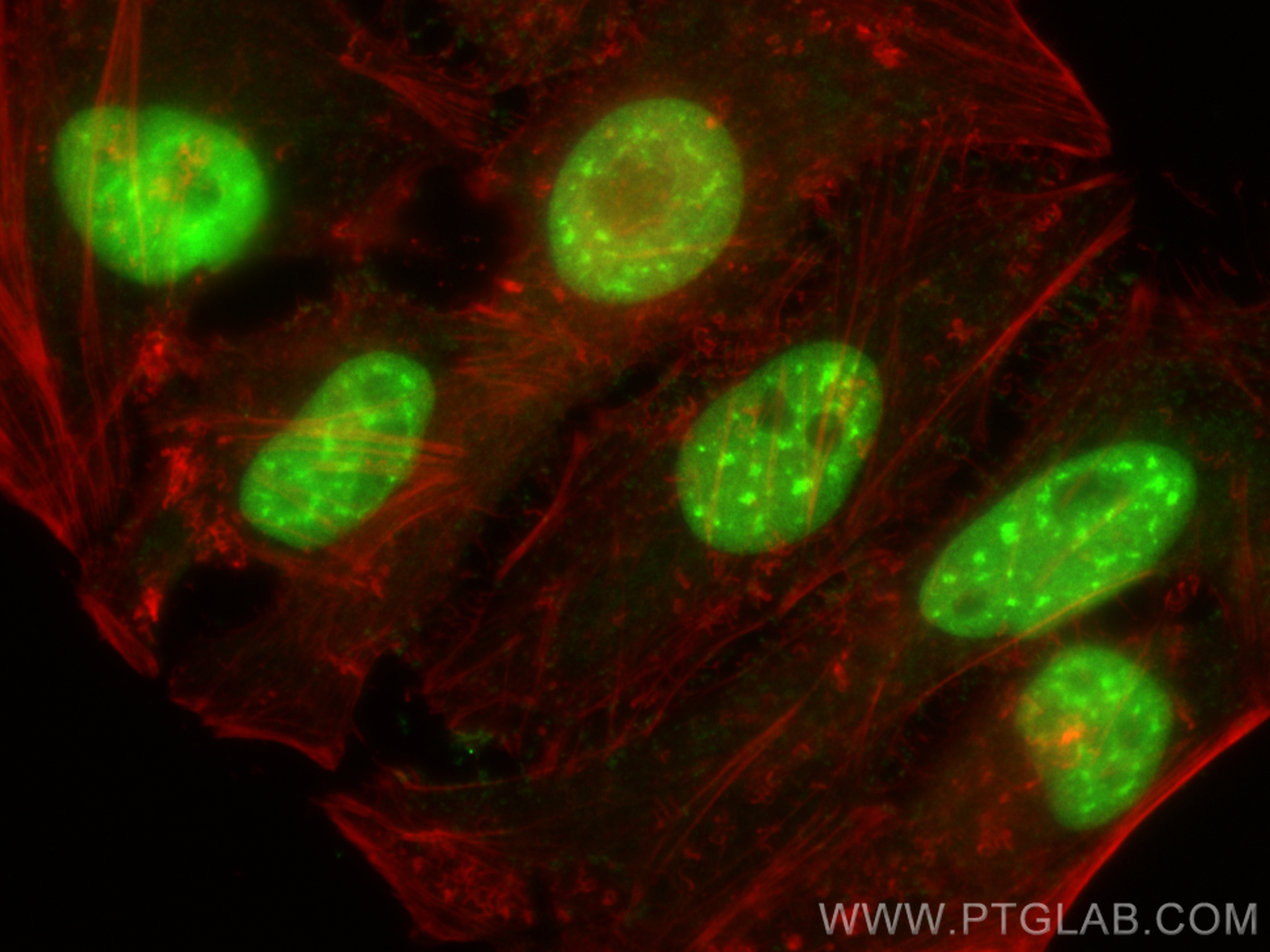
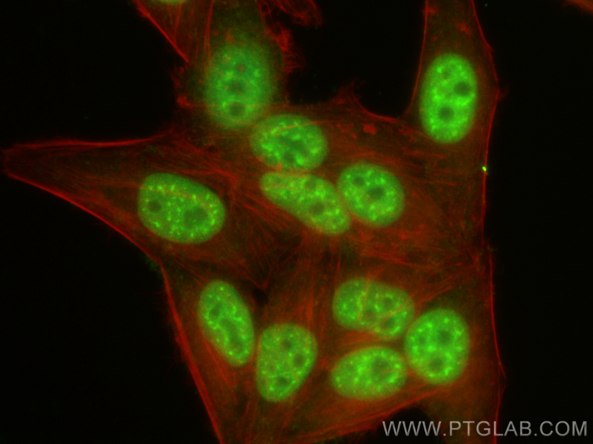
Positive IF/ICC detected in
HepG2 cells, HeLa cells
Recommended dilution
Application
Dilution
Western Blot (WB)
WB : 1:500-1:3000
Immunoprecipitation (IP)
IP : 0.5-4.0 ug for 1.0-3.0 mg of total protein lysate
Immunohistochemistry (IHC)
IHC : 1:50-1:500
Immunofluorescence (IF)/ICC
IF/ICC : 1:500-1:2000
It is recommended that this reagent should be titrated in each testing system to obtain optimal results.
Sample-dependent, Check data in validation data gallery.
PBS with 0.02% sodium azide and 50% glycerol pH 7.3.
Storage Conditions
Store at -20°C. Stable for one year after shipment. Aliquoting is unnecessary for -20oC storage. 20ul sizes contain 0.1% BSA.
Background Information
Cullin-RING ligases (CRLs) complexes participate in the regulation of diverse cellular processes, including cell cycle progression, transcription, signal transduction and development (PMID: 21816341)(PMID: 21554755). Serving as the scaffold protein, cullins are crucial for the assembly of ligase complexes, which recognize and target various substrates for proteosomal degradation. Two cullin 4 (CUL4) proteins, CUL4A (87 kDa) and CUL4B(104 kDa), are two members in cullin family with 83% of identity. Mutations in human CUL4B are one of the major causes of X-linked mental retardation. Cul4b knockout mice demonstrated that CUL4B is indispensable for embryonic development in the mouse (PMID: 22606329). Proteintech's CUL4B antibody 20882-1-AP can specifically recognize CUL4B.
Various lysates were subjected to SDS PAGE followed by western blot with 20882-1-AP (CUL4B antibody) at dilution of 1:1500 incubated at room temperature for 1.5 hours.
WB analysis of HeLa cells using 20882-1-AP
WB result of CUL4B antibody (20882-1-AP, 1:500) with si-Control and si-CUL4B transfected HeLa cells.
WB analysis using 20882-1-AP
Various lysates were subjected to SDS PAGE followed by western blot with 20882-1-AP (CUL4B antibody) at dilution of 1:2500 incubated at room temperature for 1.5 hours.
WB analysis of Y79 using 20882-1-AP
Y79 cells were subjected to SDS PAGE followed by western blot with 20882-1-AP (CUL4B antibody) at dilution of 1:500 incubated at room temperature for 1.5 hours.
WB analysis of SH-SY5Y using 20882-1-AP
SH-SY5Y cells were subjected to SDS PAGE followed by western blot with 20882-1-AP (CUL4B antibody) at dilution of 1:800 incubated at room temperature for 1.5 hours.
IHC Figures
IHC staining of human tonsillitis using 20882-1-AP
Immunohistochemical analysis of paraffin-embedded human tonsillitis tissue slide using 20882-1-AP (CUL4B Antibody) at dilution of 1:200 (under 10x lens). Heat mediated antigen retrieval with Tris-EDTA buffer (pH 9.0).
IHC staining of human tonsillitis using 20882-1-AP
Immunohistochemical analysis of paraffin-embedded human tonsillitis tissue slide using 20882-1-AP (CUL4B Antibody) at dilution of 1:200 (under 40x lens). Heat mediated antigen retrieval with Tris-EDTA buffer (pH 9.0).
IHC staining of human gliomas using 20882-1-AP
Immunohistochemical analysis of paraffin-embedded human gliomas tissue slide using 20882-1-AP (CUL4B Antibody) at dilution of 1:200 (under 10x lens). Heat mediated antigen retrieval with Tris-EDTA buffer (pH 9.0).
IHC staining of human gliomas using 20882-1-AP
Immunohistochemical analysis of paraffin-embedded human gliomas tissue slide using 20882-1-AP (CUL4B Antibody) at dilution of 1:200 (under 40x lens). Heat mediated antigen retrieval with Tris-EDTA buffer (pH 9.0).
IHC staining of human gliomas using 20882-1-AP
Immunohistochemical analysis of paraffin-embedded human gliomas tissue slide using 20882-1-AP (CUL4B antibody) at dilution of 1:200 (under 10x lens). Heat mediated antigen retrieval with Tris-EDTA buffer (pH 9.0).
IHC staining of human gliomas using 20882-1-AP
Immunohistochemical analysis of paraffin-embedded human gliomas tissue slide using 20882-1-AP (CUL4B antibody) at dilution of 1:200 (under 40x lens). Heat mediated antigen retrieval with Tris-EDTA buffer (pH 9.0).
IP Figures
IP experiment of mouse brain using 20882-1-AP
IP result of anti-CUL4B (IP:20882-1-AP, 4ug; Detection:20882-1-AP 1:300) with mouse brain tissue lysate 4000ug.
IF/ICC Figures
IF Staining of HeLa using 20882-1-AP
Immunofluorescent analysis of (4% PFA) fixed HeLa cells using CUL4B antibody (20882-1-AP) at dilution of 1:1000 and CoraLite®488-Conjugated Goat Anti-Rabbit IgG(H+L) (SA00013-2), CL594-Phalloidin (red).
IF Staining of HepG2 using 20882-1-AP
Immunofluorescent analysis of (4% PFA) fixed HepG2 cells using CUL4B antibody (20882-1-AP) at dilution of 1:1000 and CoraLite®488-Conjugated Goat Anti-Rabbit IgG(H+L) (SA00013-2), CL594-Phalloidin (red).
IF Staining of HeLa using 20882-1-AP
Immunofluorescent analysis of Hela cells, using CUL4B antibody 20882-1-AP at 1:25 dilution and Rhodamine-labeled goat anti-rabbit IgG (red).
IF Staining of HepG2 using 20882-1-AP
Immunofluorescent analysis of HepG2 cells, using CUL4B antibody 20882-1-AP at 1:25 dilution and Rhodamine-labeled goat anti-rabbit IgG (red).
The species listed in Tested Reactivity are in-house verified and applicable species. For unlisted species, please refer to the homology analysis of the immunogen sequence and related species. For rabbit polyclonal antibodies, homology >70% is recommended. For mouse monoclonal antibodies and rabbit recombinant antibodies, homology >90% is recommended. Generally, the higher the homology, the greater the applicability. However, there will be certain differences in protein expression in different species, tissues or cells. Therefore, the homology analysis results are for reference only and do not serve as a guarantee.
At Proteintech, we pride ourselves on our antibody quality, customer service and transparency. As such, we are comparing our antibodies with other vendors, enabling easy identification and comparisons of key data to help you choose the suitable antibody for your needs.
We have selected the top cited antibodies from these vendors for you to compare.
Proteintech
KD/KO VALIDATED
CUL4B Polyclonal antibody
Catalog Number
20882-1-AP
Citations
3
Dilutions
WB : 1:500-1:3000 IP : 0.5-4.0 ug for IP and 0.5-4.0 ug for 1.0-3.0 mg of total protein lysate for WB IHC : 1:50-1:500 IF/ICC : 1:500-1:2000
Applications
WB, IHC, IF/ICC, IP, ELISA
Reactivity
human, mouse, rat
Product Guarantee
Covers any species including not listed on datasheet
Covers any applications including not listed on datasheet