
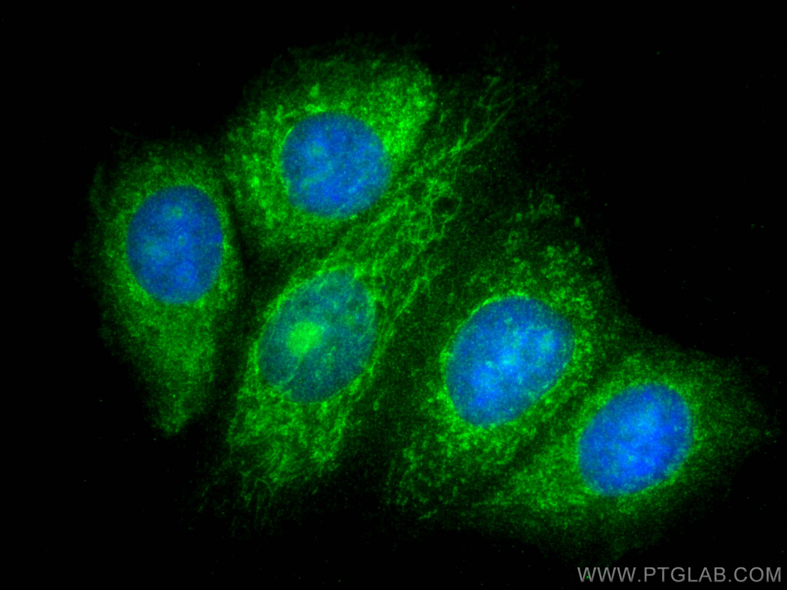

Tested Applications
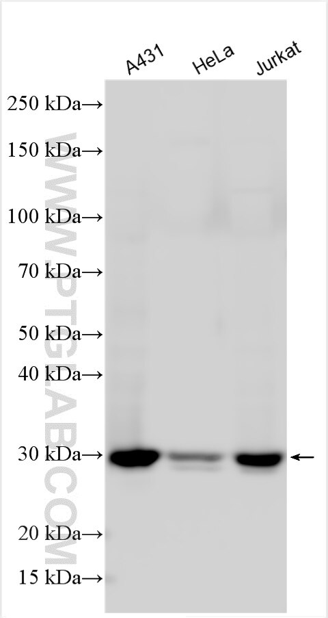
| Positive WB detected in | A431 cells, HeLa cells, Jurkat cells |
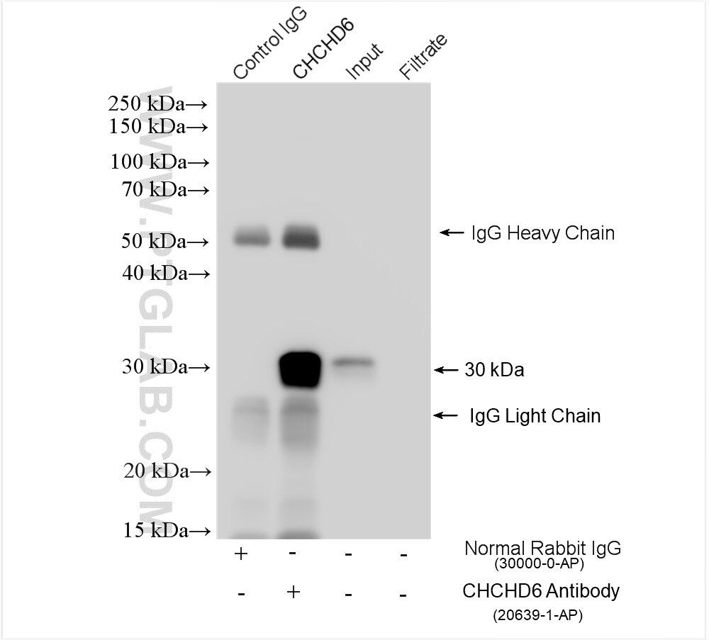
| Positive IP detected in | Jurkat cells |
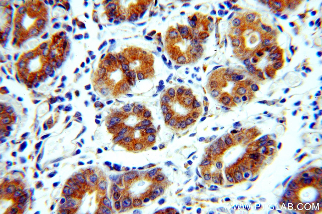
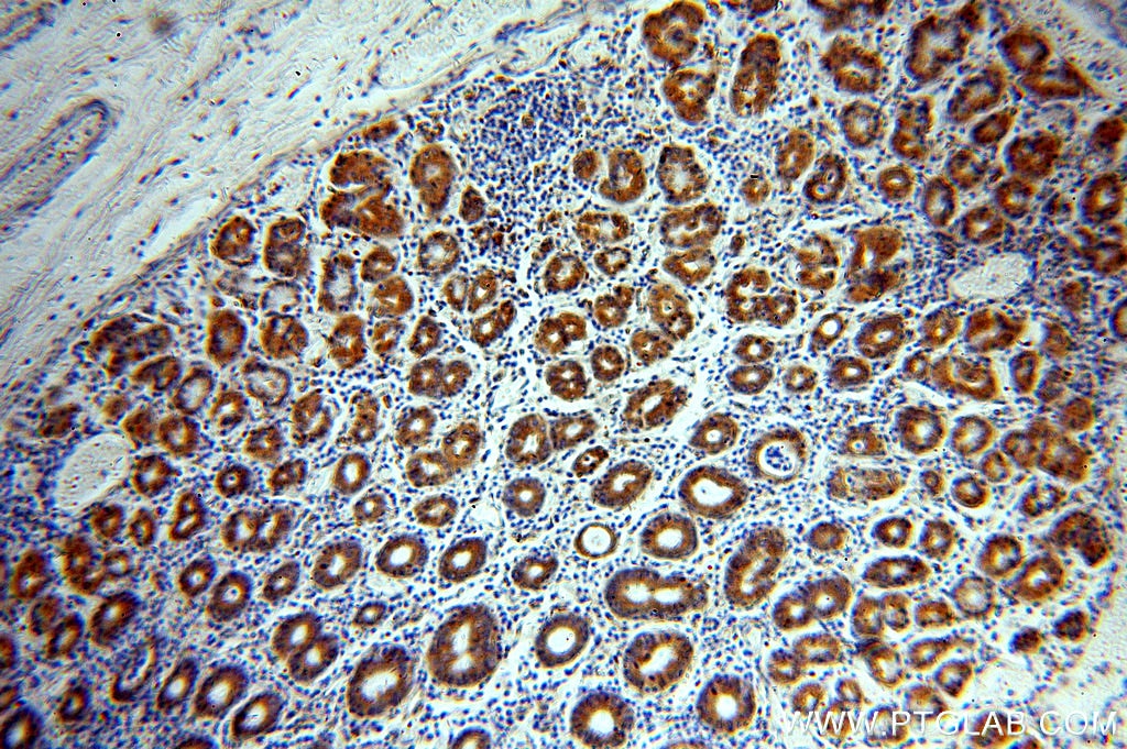
| Positive IHC detected in | human stomach tissue Note: suggested antigen retrieval with TE buffer pH 9.0; (*) Alternatively, antigen retrieval may be performed with citrate buffer pH 6.0 |
| Positive IF/ICC detected in | MCF-7 cells |
Recommended dilution
| Application | Dilution |
|---|---|
| Western Blot (WB) | WB : 1:1000-1:4000 |
| Immunoprecipitation (IP) | IP : 0.5-4.0 ug for 1.0-3.0 mg of total protein lysate |
| Immunohistochemistry (IHC) | IHC : 1:20-1:200 |
| Immunofluorescence (IF)/ICC | IF/ICC : 1:200-1:800 |
| It is recommended that this reagent should be titrated in each testing system to obtain optimal results. | |
| Sample-dependent, Check data in validation data gallery. | |
Product Information
20639-1-AP targets CHCHD6 in WB, IHC, IF/ICC, IP, CoIP, ELISA applications and shows reactivity with human samples.
| Tested Reactivity | human |
| Cited Reactivity | human, mouse |
| Host / Isotype | Rabbit / IgG |
| Class | Polyclonal |
| Type | Antibody |
| Immunogen |
CatNo: Ag14700 Product name: Recombinant human CHCHD6 protein Source: e coli.-derived, PGEX-4T Tag: GST Domain: 1-235 aa of BC006123 Sequence: MGSTESSEGRRVSFGVDEEERVRVLQGVRLSENVVNRMKEPSSPPPAPTSSTFGLQDGNLRAPHKESTLPRSGSSGGQQPSGMKEGVKRYEQEHAAIQDKLFQVAKREREAATKHSKASLPTGEGSISHEEQKSVRLARELESREAELRRRDTFYKEQLERIERKNAEMYKLSSEQFHEAASKMESTIKPRRVEPVCSGLQAQILHCYRDRPHEVLLCSDLVKAYQRCVSAAHKG Predict reactive species |
| Full Name | coiled-coil-helix-coiled-coil-helix domain containing 6 |
| Calculated Molecular Weight | 235 aa, 26 kDa |
| Observed Molecular Weight | 26-29 kDa |
| GenBank Accession Number | BC006123 |
| Gene Symbol | CHCHD6 |
| Gene ID (NCBI) | 84303 |
| RRID | AB_10697667 |
| Conjugate | Unconjugated |
| Form | Liquid |
| Purification Method | Antigen affinity purification |
| UNIPROT ID | Q9BRQ6 |
| Storage Buffer | PBS with 0.02% sodium azide and 50% glycerol, pH 7.3. |
| Storage Conditions | Store at -20°C. Stable for one year after shipment. Aliquoting is unnecessary for -20oC storage. 20ul sizes contain 0.1% BSA. |
Protocols
| Product Specific Protocols | |
|---|---|
| IF protocol for CHCHD6 antibody 20639-1-AP | Download protocol |
| IHC protocol for CHCHD6 antibody 20639-1-AP | Download protocol |
| IP protocol for CHCHD6 antibody 20639-1-AP | Download protocol |
| WB protocol for CHCHD6 antibody 20639-1-AP | Download protocol |
| Standard Protocols | |
|---|---|
| Click here to view our Standard Protocols |
Publications
What published studies show
Anti-MIC60 (Mitofilin) Antibody, Cat# 20639-1-AP has 19 total citations. Citations appear in journals including Molecular Cell, Cell Death & Differentiation, Science Bulletin, Advanced Science, Acta Neuropathologica, Journal of Cell Biology, EMBO Reports, Scientific Reports, iScience, Journal of Biological Chemistry, Life Science Alliance, BBA-Biomembranes, eLife, and bioRxiv. Research fields span mitochondrial cristae structure, MICOS complex assembly, mitochondrial dynamics, mitophagy, ER–mitochondria contact sites, Alzheimer's disease, glioblastoma, and respiratory supercomplex organization.
| Species | Application | Title |
|---|---|---|
Sci Bull (Beijing) Mitochondria serve as a source of mineral precursors initiating early cartilage calcification in osteoarthritis. | ||
Mol Cell BRD4-mediated ER membrane contact creates functionally distinct mitochondrial subtypes. | ||
Adv Sci (Weinh) LRRC4 Orchestrates AP2A1-Containing Clathrin-Coated Vesicles to Disrupt Mitochondrial Cristae and Restrict Glioblastoma Progression | ||
Cell Death Differ Mic60/Mitofilin determines MICOS assembly essential for mitochondrial dynamics and mtDNA nucleoid organization.
| ||
Acta Neuropathol A CHCHD6-APP axis connects amyloid and mitochondrial pathology in Alzheimer's disease
| ||
J Cell Biol The outer mitochondrial membrane protein TMEM11 demarcates spatially restricted BNIP3/BNIP3L-mediated mitophagy |
Reviews
What customers say
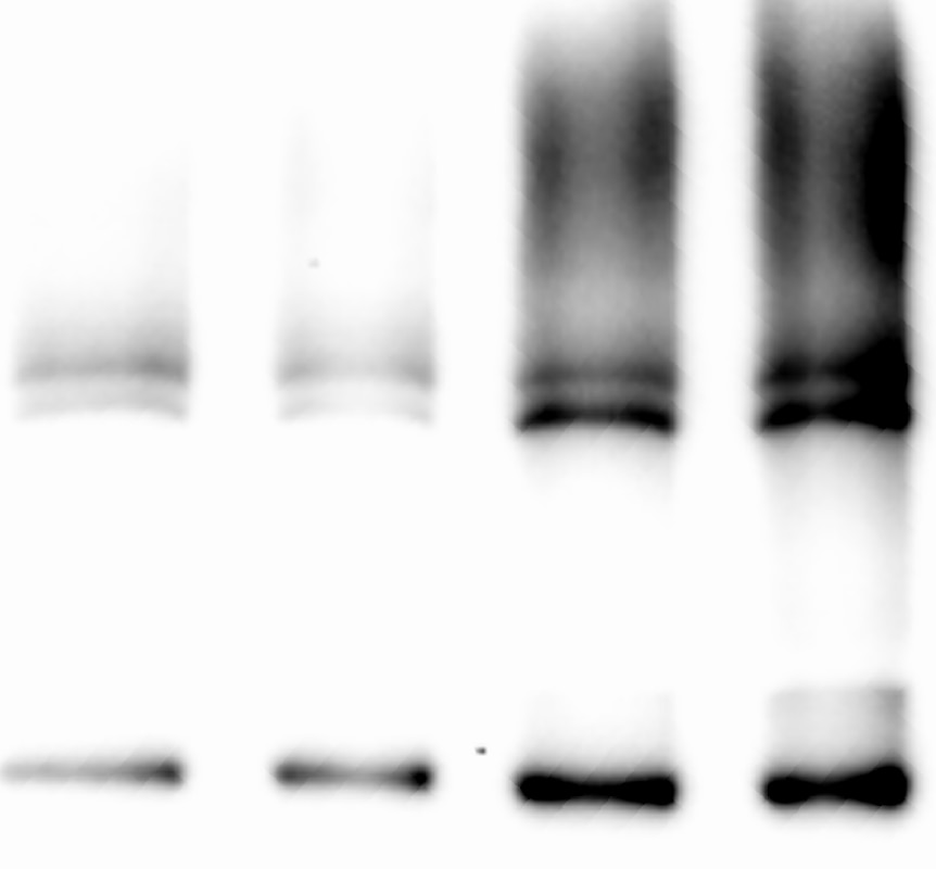
All four user reviews are positive, with ratings of 4–5 stars. Every reviewer used the antibody for Western Blot at a 1:1000 dilution. Users reported clear bands and strong performance across different sample types, including heart tissue (human and mouse) and cancer tissue. Common feedback highlights include "very clear band," "very good antibody," and "worked very well." Overall, users express high satisfaction with the antibody's specificity and reliability in WB applications.
The reviews below have been submitted by verified Proteintech customers who received an incentive for providing their feedback.
Angela (Verified Customer) (12-27-2024) | good
 |
angela (Verified Customer) (12-27-2024) | very good antibody
 |
SITING (Verified Customer) (07-13-2020) | very clear band in our wester blot expose result
|
Jingwen (Verified Customer) (01-28-2020) | This antibody worked very well when I performed western blot.
|


