

Tested Applications
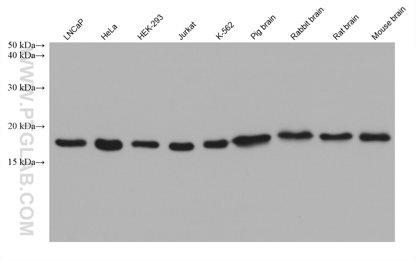
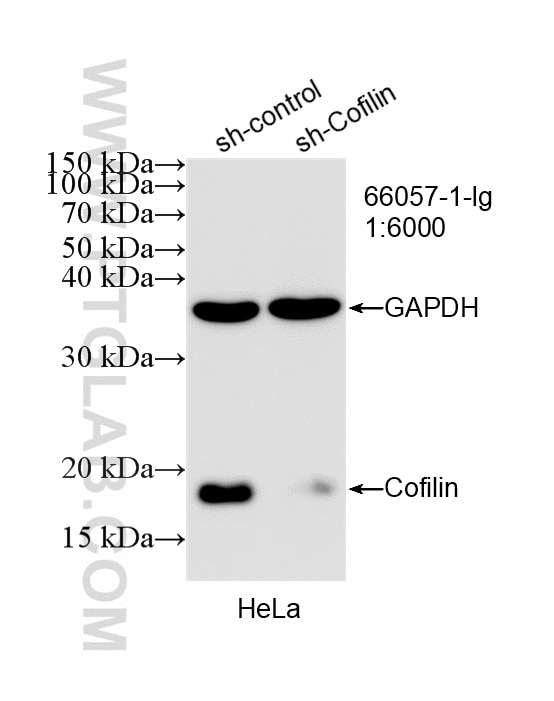
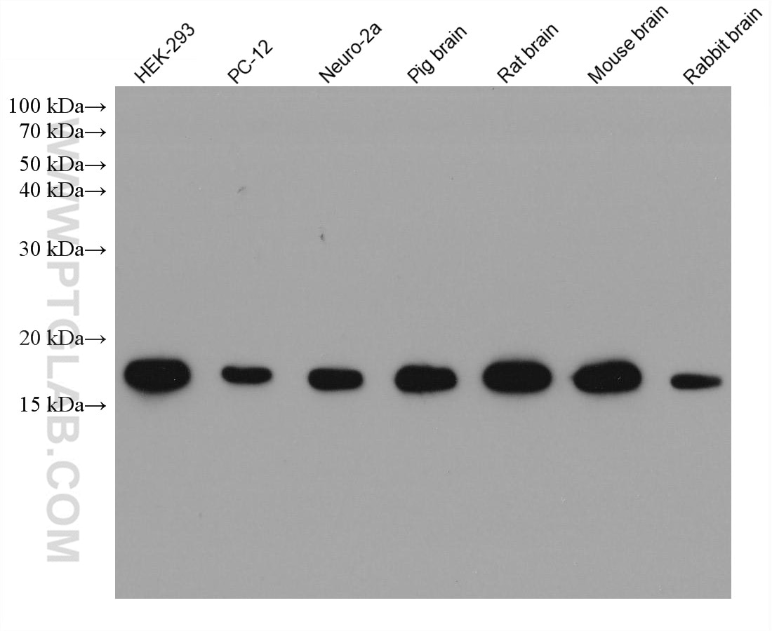
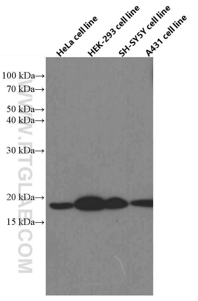
| Positive WB detected in | LNCaP cells, HEK-293 cells, HeLa cells, PC-12 cells, Neuro-2a cells, pig brain, rat brain, mouse brain, rabbit brain, SH-SY5Y cells, A431 cells, Jurkat cells, K-562 cells, pig brain tissue, rabbit brain tissue, rat brain tissue, mouse brain tissue |
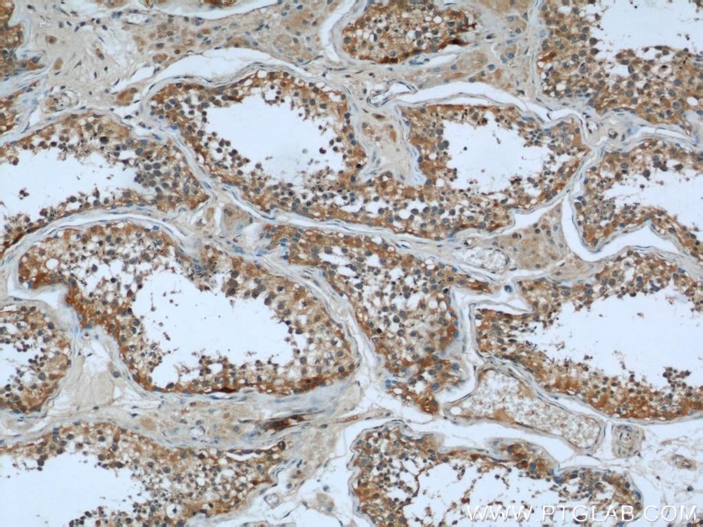
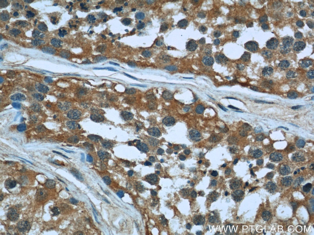
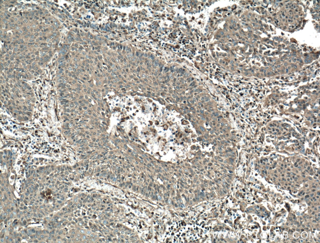
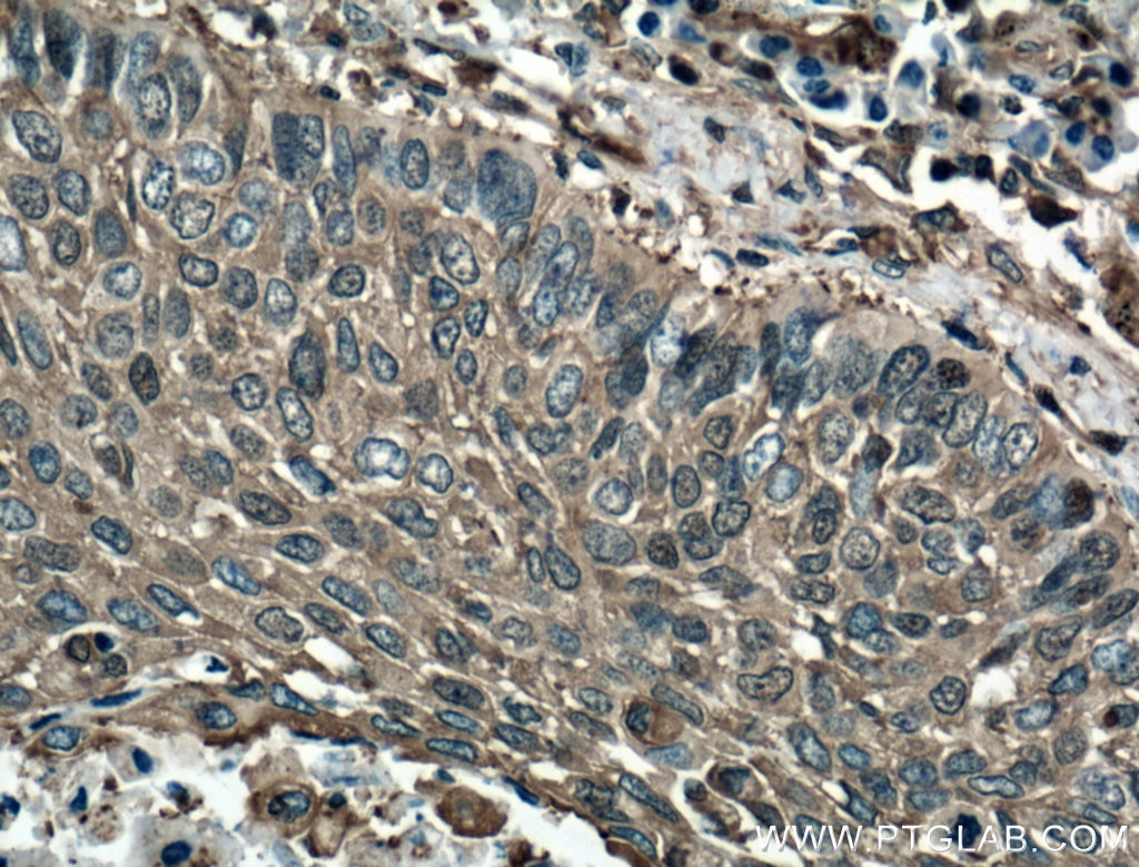
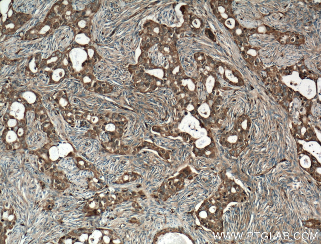
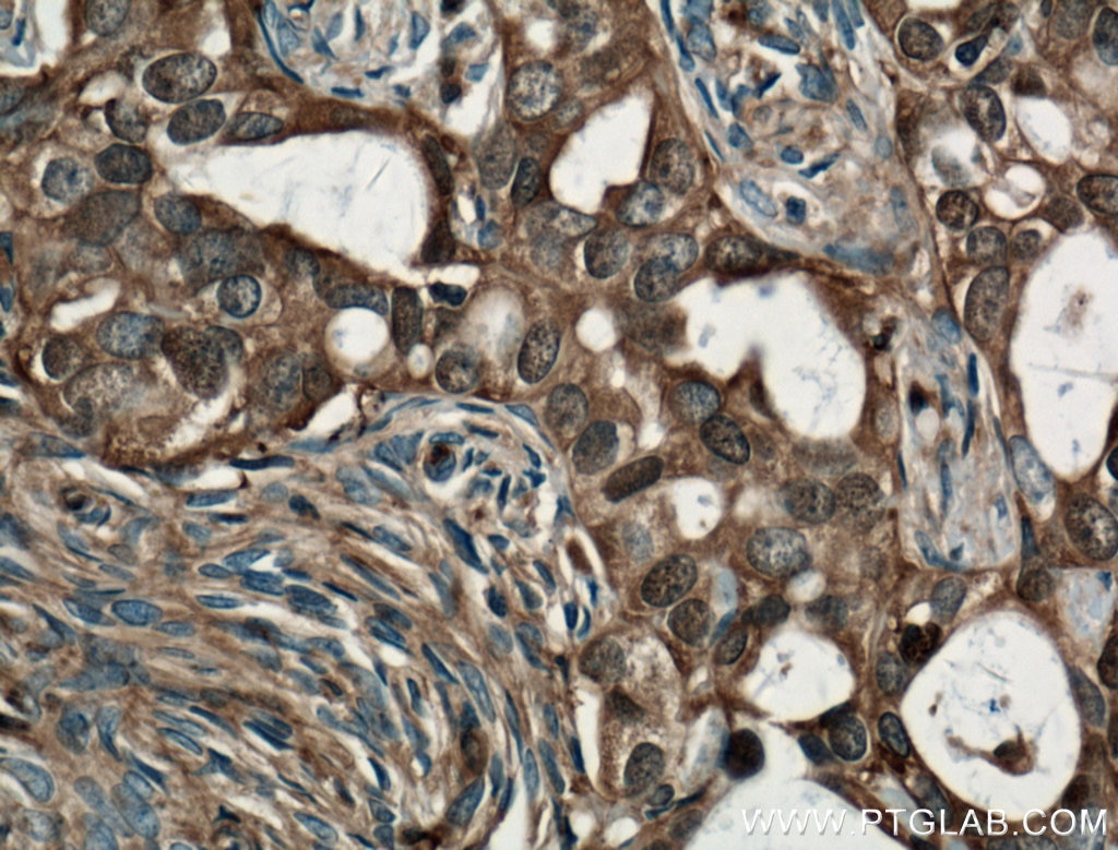
| Positive IHC detected in | human testis tissue, human lung cancer tissue, human ovary tumor tissue Note: suggested antigen retrieval with TE buffer pH 9.0; (*) Alternatively, antigen retrieval may be performed with citrate buffer pH 6.0 |
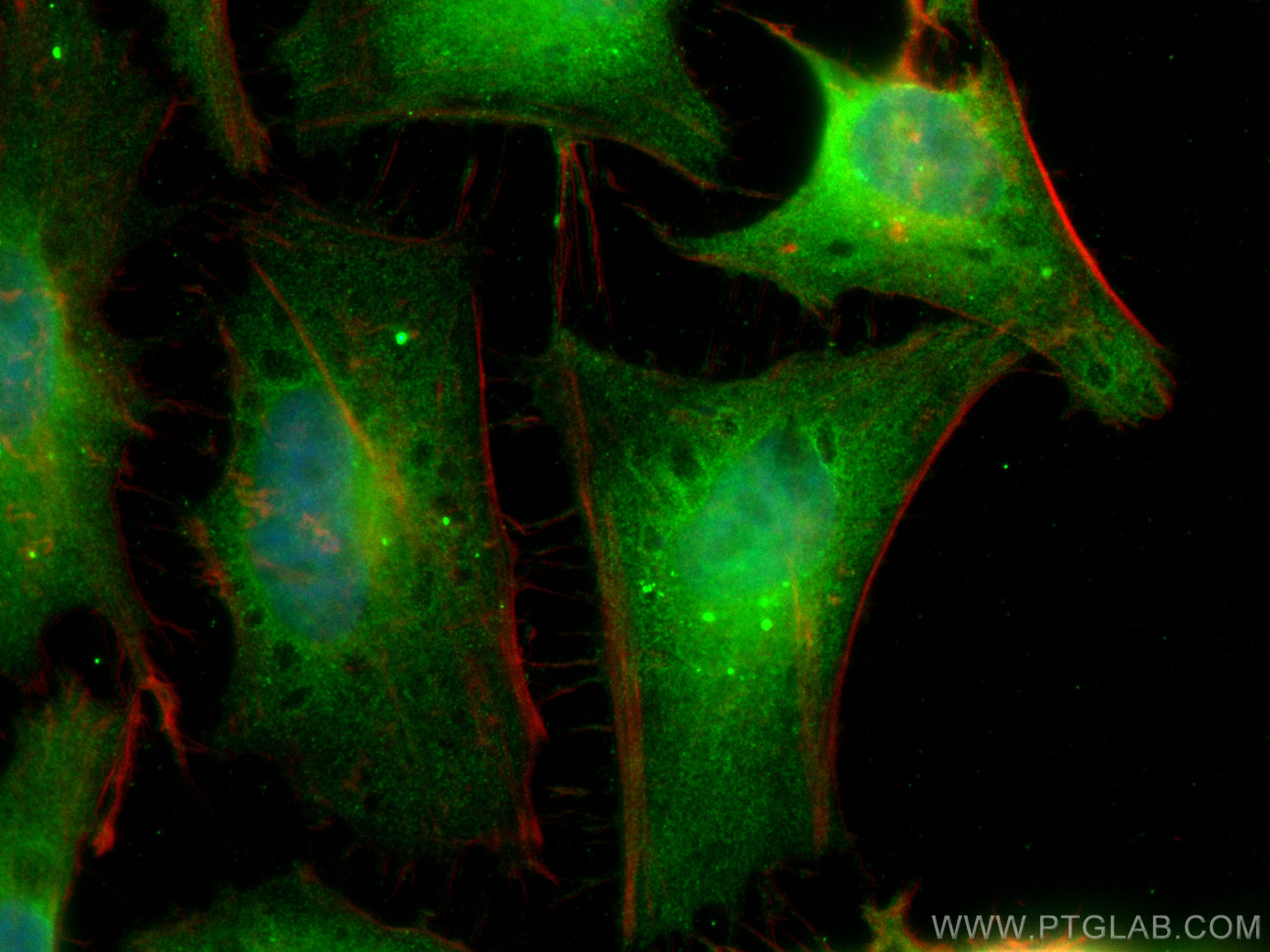
| Positive IF/ICC detected in | HeLa cells |
Recommended dilution
| Application | Dilution |
|---|---|
| Western Blot (WB) | WB : 1:5000-1:50000 |
| Immunohistochemistry (IHC) | IHC : 1:20-1:200 |
| Immunofluorescence (IF)/ICC | IF/ICC : 1:400-1:1600 |
| It is recommended that this reagent should be titrated in each testing system to obtain optimal results. | |
| Sample-dependent, Check data in validation data gallery. | |
Product Information
66057-1-Ig targets Cofilin in WB, IHC, IF/ICC, IP, ELISA applications and shows reactivity with human, mouse, rat, pig, rabbit samples.
| Tested Reactivity | human, mouse, rat, pig, rabbit |
| Cited Reactivity | human, mouse, rat |
| Host / Isotype | Mouse / IgG2a |
| Class | Monoclonal |
| Type | Antibody |
| Immunogen |
CatNo: Ag18044 Product name: Recombinant human Cofilin protein Source: e coli.-derived, PET28a Tag: 6*His Domain: 1-166 aa of BC012318 Sequence: MASGVAVSDGVIKVFNDMKVRKSSTPEEVKKRKKAVLFCLSEDKKNIILEEGKEILVGDVGQTVDDPYATFVKMLPDKDCRYALYDATYETKESKKEDLVFIFWAPESAPLKSKMIYASSKDAIKKKLTGIKHELQANCYEEVKDRCTLAEKLGGSAVISLEGKPL Predict reactive species |
| Full Name | cofilin 1 (non-muscle) |
| Calculated Molecular Weight | 18 kDa |
| Observed Molecular Weight | 18 kDa |
| GenBank Accession Number | BC012318 |
| Gene Symbol | Cofilin |
| Gene ID (NCBI) | 1072 |
| RRID | AB_11043339 |
| Conjugate | Unconjugated |
| Form | Liquid |
| Purification Method | Protein A purification |
| UNIPROT ID | P23528 |
| Storage Buffer | PBS with 0.02% sodium azide and 50% glycerol, pH 7.3. |
| Storage Conditions | Store at -20°C. Stable for one year after shipment. Aliquoting is unnecessary for -20oC storage. 20ul sizes contain 0.1% BSA. |
Background Information
Cofilin is a ubiquitous actin-binding protein required for the reorganization of actin filaments. It is a member of ADF (actin-depolymerizing factor)/cofilin family that is a key regulator of actin dynamics and essential for cellular motility, cytokinesis, and endocytosis. Cofilin activity is tightly regulated by phosphorylation and dephosphorylation. Phosphorylation at Ser3 can inhibit its activity, also causing translocation from the nucleus to the cytoplasm.
Protocols
| Product Specific Protocols | |
|---|---|
| WB protocol for Cofilin antibody 66057-1-Ig | Download protocol |
| IF protocol for Cofilin antibody 66057-1-Ig | Download protocol |
| IHC protocol for Cofilin antibody 66057-1-Ig | Download protocol |
| Standard Protocols | |
|---|---|
| Click here to view our Standard Protocols |
Publications
What published studies show
The Cofilin antibody (Cat# 66057-1-Ig) has 32 total citations published in journals including Nature Neuroscience, Nature Communications, Advanced Science, Science Advances, Cell Death & Differentiation, Molecular Psychiatry, eLife, Human Molecular Genetics, iScience, and others. Research fields span neuroscience (actin dynamics, synapses, neurodegeneration), cancer biology (migration, invasion, EMT), cellular actin/cytoskeleton regulation, developmental biology (oocyte spindle checkpoint), virology, and cardiovascular disease.
| Species | Application | Title |
|---|---|---|
Nat Commun N-terminal α-amino SUMOylation promotes phosphorylation-independent cofilin-1 translocation to the mitochondrial matrix and induces apoptosis.
| ||
Nat Commun Dendritic autophagy degrades postsynaptic proteins and is required for long-term synaptic depression in mice. | ||
Nat Commun N-terminal α-amino SUMOylation of cofilin-1 is critical for its regulation of actin depolymerization | ||
Nat Commun Nuclear-capture of endosomes depletes nuclear G-actin to promote SRF/MRTF activation and cancer cell invasion. | ||
Adv Sci (Weinh) A Microtubule-Associated Protein Functions in Preventing Oocytes from Evading the Spindle Assembly Checkpoint |
Reviews
What customers say
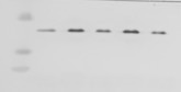
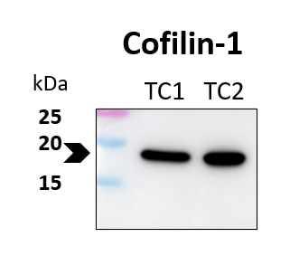
Both reviewers gave this antibody a perfect 5-star rating for Western Blot. One user reported excellent performance at a 1:3000 dilution in HEK293T cells. Another used it at 1:5000 on human glioblastoma tumor cell lines (TC1 and TC2) with a 4–15% gradient gel, overnight primary incubation at 4°C, and 1-hour secondary incubation at room temperature, noting that bands appeared at the expected molecular weight. Overall, customers find this antibody reliable and effective for WB applications across different cell types and dilutions.
The reviews below have been submitted by verified Proteintech customers who received an incentive for providing their feedback.
Udesh (Verified Customer) (08-19-2024) | Worked well at 1:3000
 |
Marina (Verified Customer) (03-12-2024) | Samples are two tumour (glioblastoma) cell lines (TC1, TC2). Gradient gel 4-15 %. Dilution 1:5000, primary antibody incubation 4ºC overnight, secondary antibody incubation 1 h room temperature. The bands appear at the expected size of the target protein.
 |


