Tested Applications
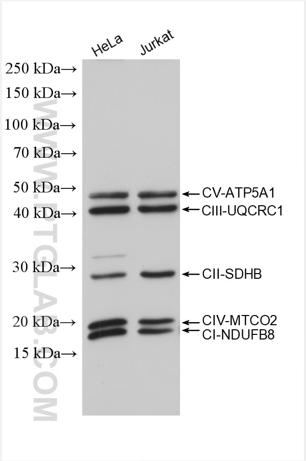
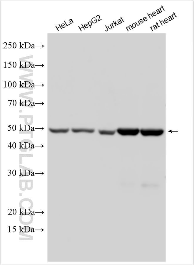
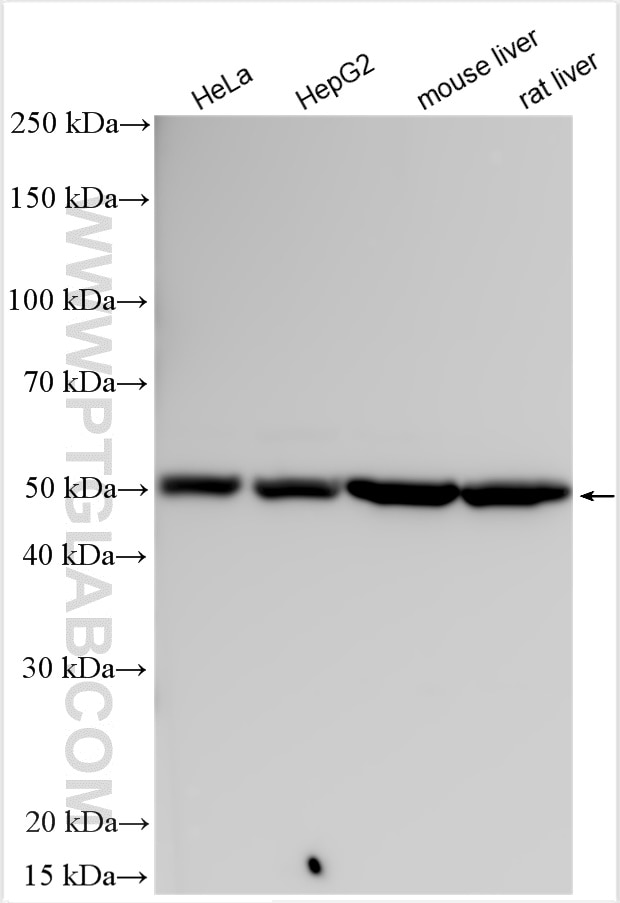
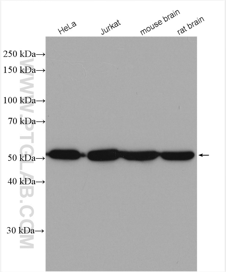
| Positive WB detected in | HeLa cells, various samples, HepG2 cells, Jurkat cells, mouse heart tissue, rat heart tissue, mouse liver tissue, rat liver tissue, mouse brain tissue, rat brain tissue |
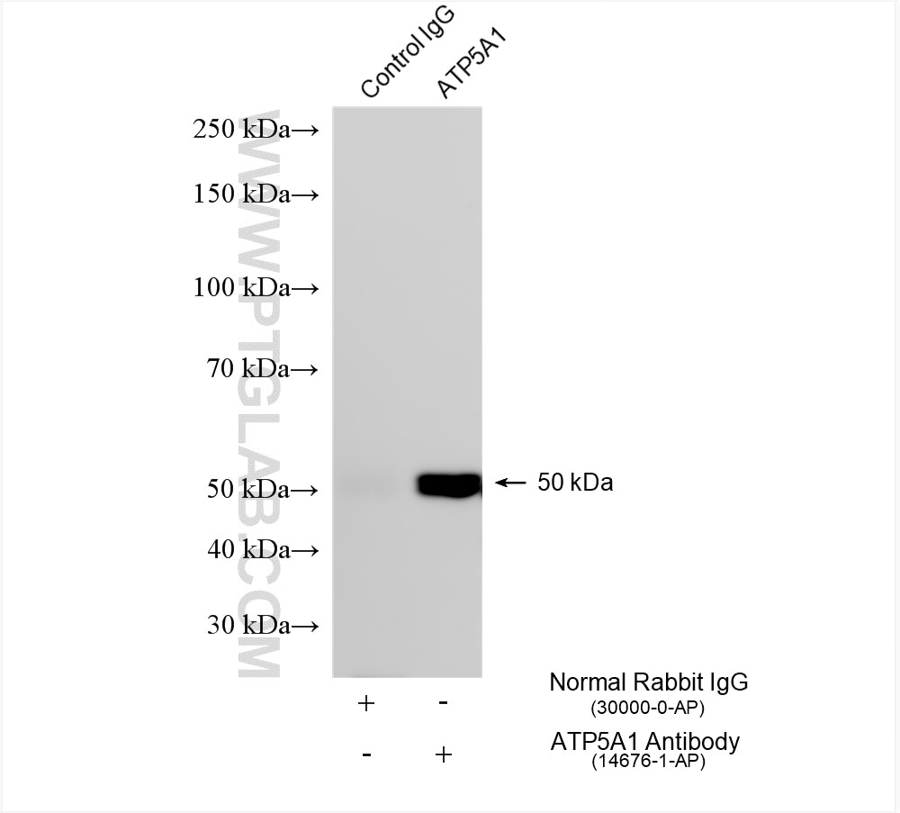
| Positive IP detected in | HeLa cells |
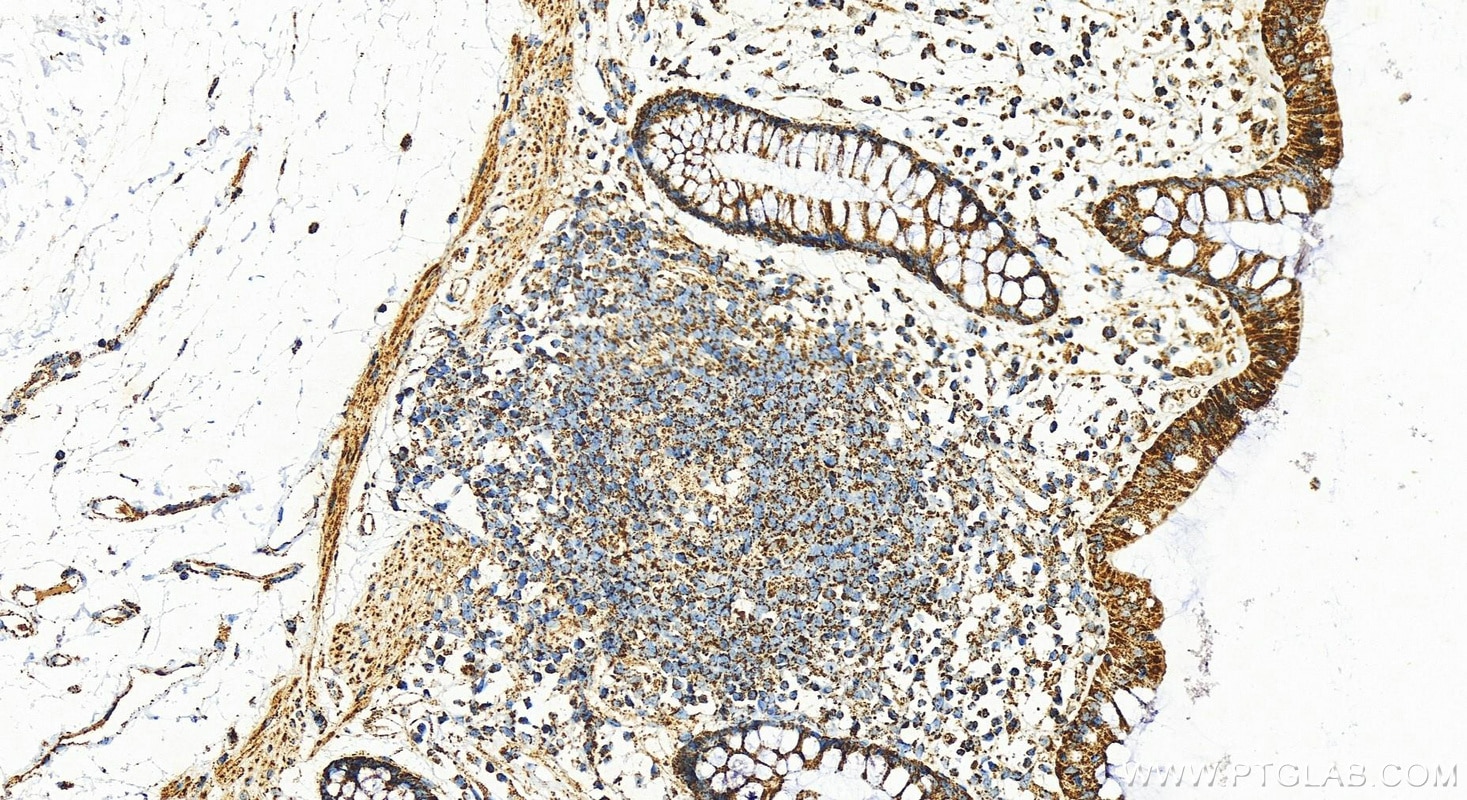
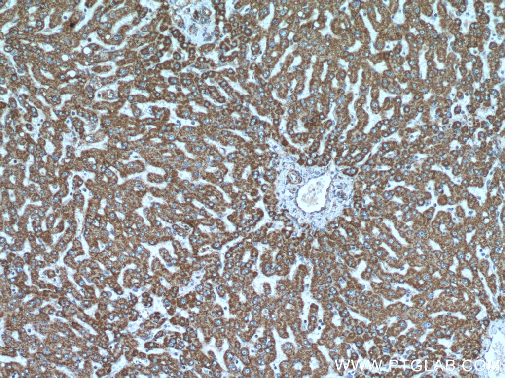
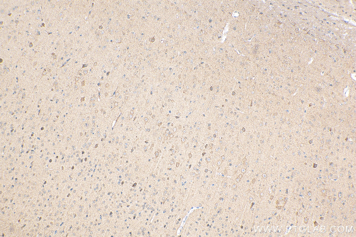
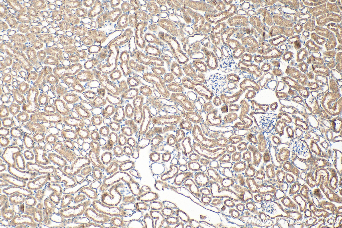
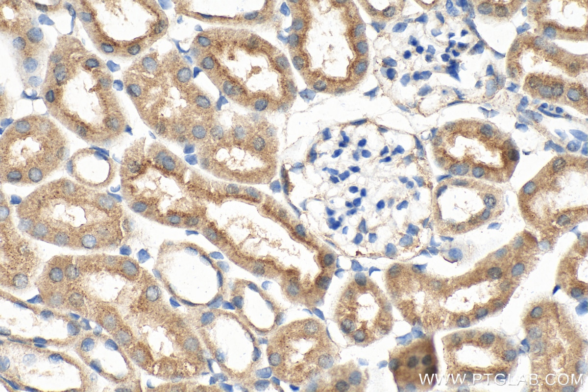
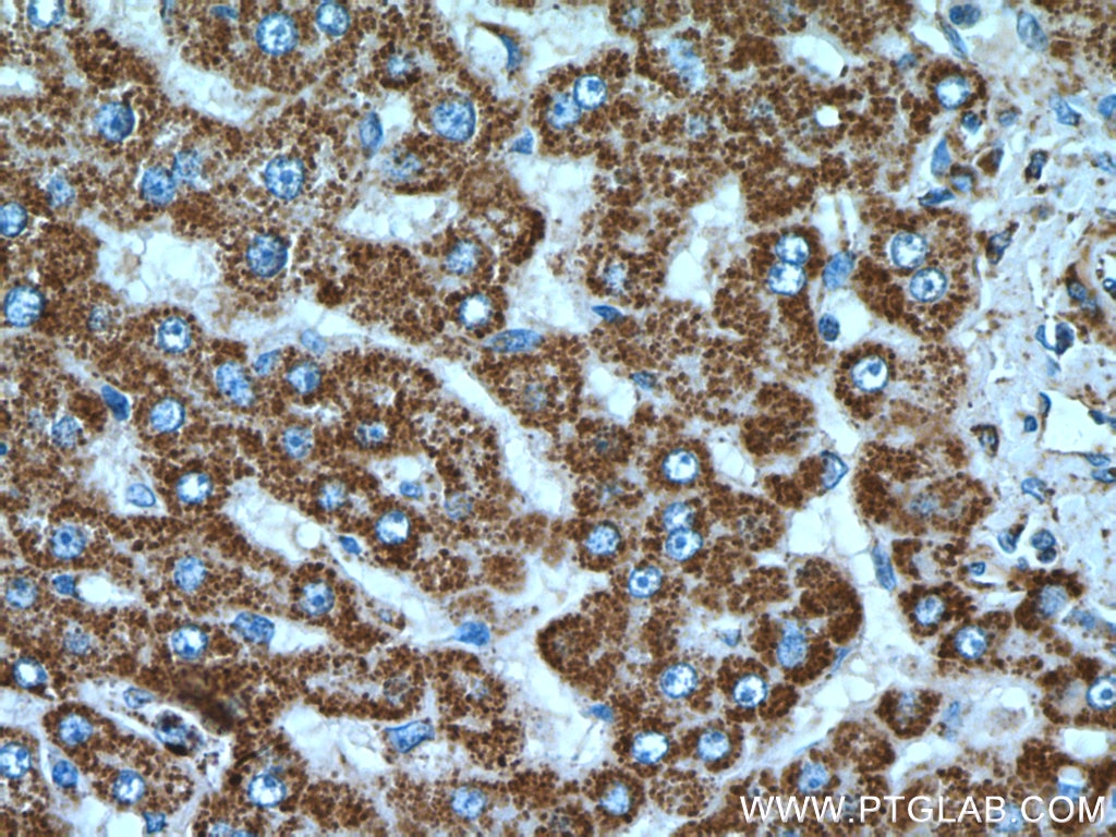
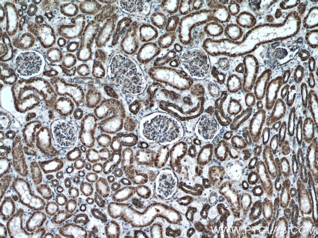
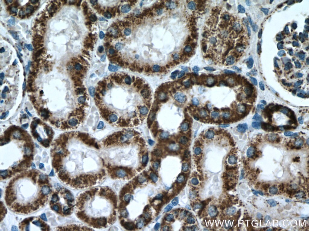
| Positive IHC detected in | mouse kidney tissue, human colon tissue, human kidney tissue, human liver tissue, mouse brain tissue Note: suggested antigen retrieval with TE buffer pH 9.0; (*) Alternatively, antigen retrieval may be performed with citrate buffer pH 6.0 |
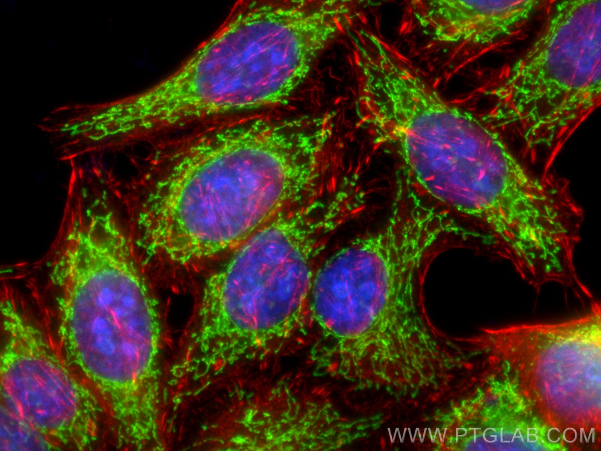
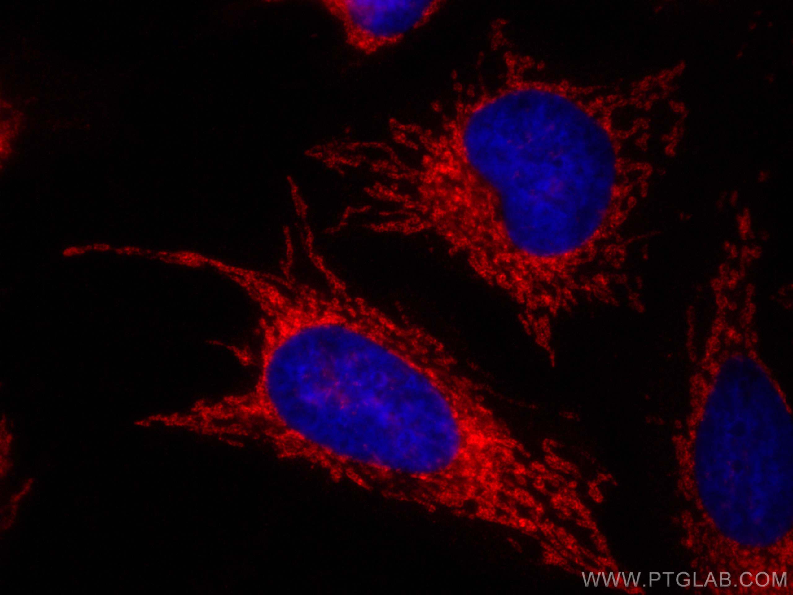
| Positive IF/ICC detected in | HeLa cells, HepG2 cells |
Recommended dilution
| Application | Dilution |
|---|---|
| Western Blot (WB) | WB : 1:5000-1:50000 |
| Immunoprecipitation (IP) | IP : 0.5-4.0 ug for 1.0-3.0 mg of total protein lysate |
| Immunohistochemistry (IHC) | IHC : 1:500-1:2000 |
| Immunofluorescence (IF)/ICC | IF/ICC : 1:400-1:1600 |
| It is recommended that this reagent should be titrated in each testing system to obtain optimal results. | |
| Sample-dependent, Check data in validation data gallery. | |
Published Applications
| WB | See 150 publications below |
| IHC | See 5 publications below |
| IF | See 21 publications below |
| IP | See 3 publications below |
| CoIP | See 2 publications below |
Product Information
14676-1-AP targets ATP5A1 in WB, IHC, IF/ICC, IP, CoIP, ELISA applications and shows reactivity with human, mouse, rat samples.
| Tested Reactivity | human, mouse, rat |
| Cited Reactivity | human, mouse, rat, monkey, zebrafish, hamster |
| Host / Isotype | Rabbit / IgG |
| Class | Polyclonal |
| Type | Antibody |
| Immunogen |
CatNo: Ag6385 Product name: Recombinant human ATP5A1 protein Source: e coli.-derived, PGEX-4T Tag: GST Domain: 203-553 aa of BC064562 Sequence: GRGQRELIIGDRQTGKTSIAIDTIINQKRFNDGSDEKKKLYCIYVAIGQKRSTVAQLVKRLTDADAMKYTIVVSATASDAAPLQYLAPYSGCSMGEYFRDNGKHALIIYDDLSKQAVAYRQMSLLLRRPPGREAYPGDVFYLHSRLLERAAKMNDAFGGGSLTALPVIETQAGDVSAYIPTNVISITDGQIFLETELFYKGIRPAINVGLSVSRVGSAAQTRAMKQVAGTMKLELAQYREVAAFAQFGSDLDAATQQLLSRGVRLTELLKQGQYSPMAIEEQVAVIYAGVRGYLDKLEPSKITKFENAFLSHVVSQHQALLGTIRADGKISEQSDAKLKEIVTNFLAGFEA Predict reactive species |
| Full Name | ATP synthase, H+ transporting, mitochondrial F1 complex, alpha subunit 1, cardiac muscle |
| Calculated Molecular Weight | 60 kDa |
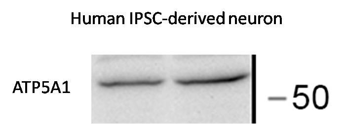
| Observed Molecular Weight | 50-55 kDa |
| GenBank Accession Number | BC064562 |
| Gene Symbol | ATP5A1 |
| Gene ID (NCBI) | 498 |
| RRID | AB_2061761 |
| Conjugate | Unconjugated |
| Form | Liquid |
| Purification Method | Antigen affinity purification |
| UNIPROT ID | P25705 |
| Storage Buffer | PBS with 0.02% sodium azide and 50% glycerol, pH 7.3. |
| Storage Conditions | Store at -20°C. Stable for one year after shipment. Aliquoting is unnecessary for -20oC storage. 20ul sizes contain 0.1% BSA. |
Background Information
The ATP5A1 gene encodes the α subunit of mitochondrial ATP synthase which produces ATP from ADP in the presence of a proton gradient across the membrane. The mitochondrial ATP synthase, also known as Complex V or F1F0 ATP synthase, is a multi-subunit enzyme complex consisting of two functional domains, the F1-containing the catalytic core and the Fo-containing the membrane proton channel. F0 domain has 10 subunits: a,b, c, d, e, f, g, OSCP, A6L, and F6. F1 is composed of subunits α, β, γ, δ, ε, and a loosely attached inhibitor protein IF1. Recently defect in ATP5A1 has been linked to the fatal neonatal mitochondrial encephalopathy. ATP5A1 is localized in the mitochondria and anti-ATP5A1 can be used as the loading control for mitochondrial or Complex V proteins. This antibody recognizes the endogenous ATP5A1 protein in lysates from various cell lines and tissues. The predicted MW of ATP5A1 is 60 kDa, while it undergoes the transit peptide cleavage to become a mature form around 50-55 kDa. Several isoforms of ATP5A1 exist due to the alternative splicing.
Protocols
| Product Specific Protocols | |
|---|---|
| IF protocol for ATP5A1 antibody 14676-1-AP | Download protocol |
| IHC protocol for ATP5A1 antibody 14676-1-AP | Download protocol |
| IP protocol for ATP5A1 antibody 14676-1-AP | Download protocol |
| WB protocol for ATP5A1 antibody 14676-1-AP | Download protocol |
| Standard Protocols | |
|---|---|
| Click here to view our Standard Protocols |
Publications
| Species | Application | Title |
|---|---|---|
Cell Res Disruption of ER ion homeostasis maintained by an ER anion channel CLCC1 contributes to ALS-like pathologies | ||
Mol Cell RAPIDASH: Tag-free enrichment of ribosome-associated proteins reveals composition dynamics in embryonic tissue, cancer cells, and macrophages | ||
Mol Cell Global mitochondrial protein import proteomics reveal distinct regulation by translation and translocation machinery | ||
Nat Chem Biol Small molecule agonist of mitochondrial fusion repairs mitochondrial dysfunction | ||
Cell Rep Med Targeting mitochondrial complex I of CD177+ neutrophils alleviates lung ischemia-reperfusion injury | ||
Sci Adv Mitochondrial proteostasis stress in muscle drives a long-range protective response to alleviate dietary obesity independently of ATF4. |
Reviews
The reviews below have been submitted by verified Proteintech customers who received an incentive for providing their feedback.
FH Edward (Verified Customer) (11-15-2022) | Used with 12 ug protein lysate ATP5A1 works well, both with ECL detection and fluorescent secondary antibodies it gives a clear band for K562 and KCL22, though no band was apparent in THP-1 (this could be cell line specific).
|
FH XB (Verified Customer) (10-29-2020) | It probes ATP5A1 clearly with a right MW 55 kD.
 |
FH Siting (Verified Customer) (09-04-2020) | Good antibody
 |

