
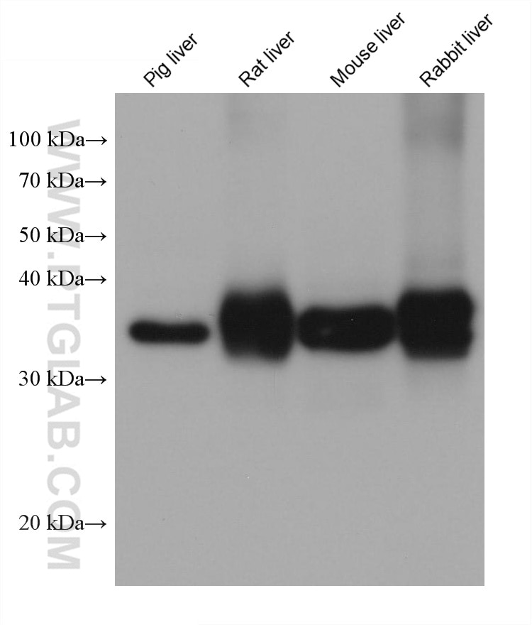
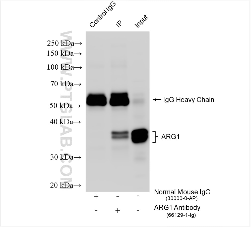
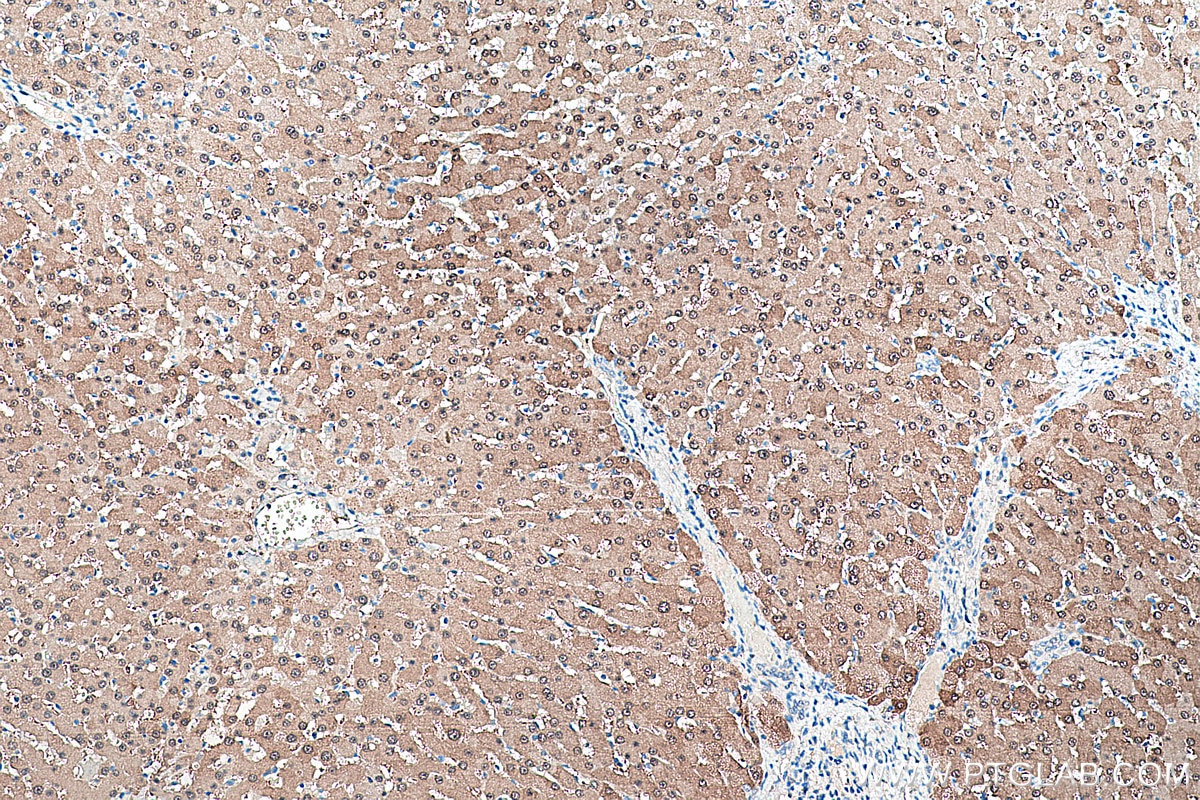
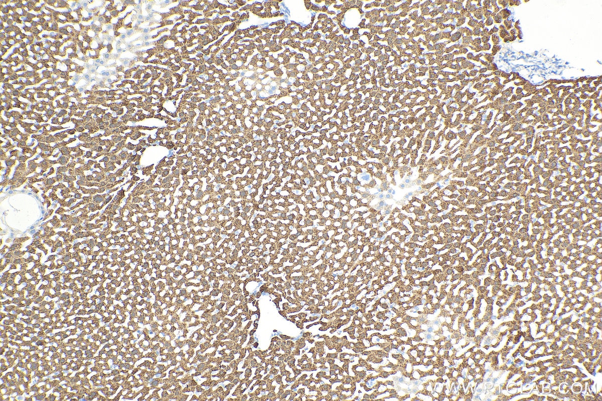

Tested Applications
| Positive WB detected in | pig liver tissue, rat liver tissue, mouse liver tissue, rabbit liver tissue |
| Positive IP detected in | mouse liver tissue |
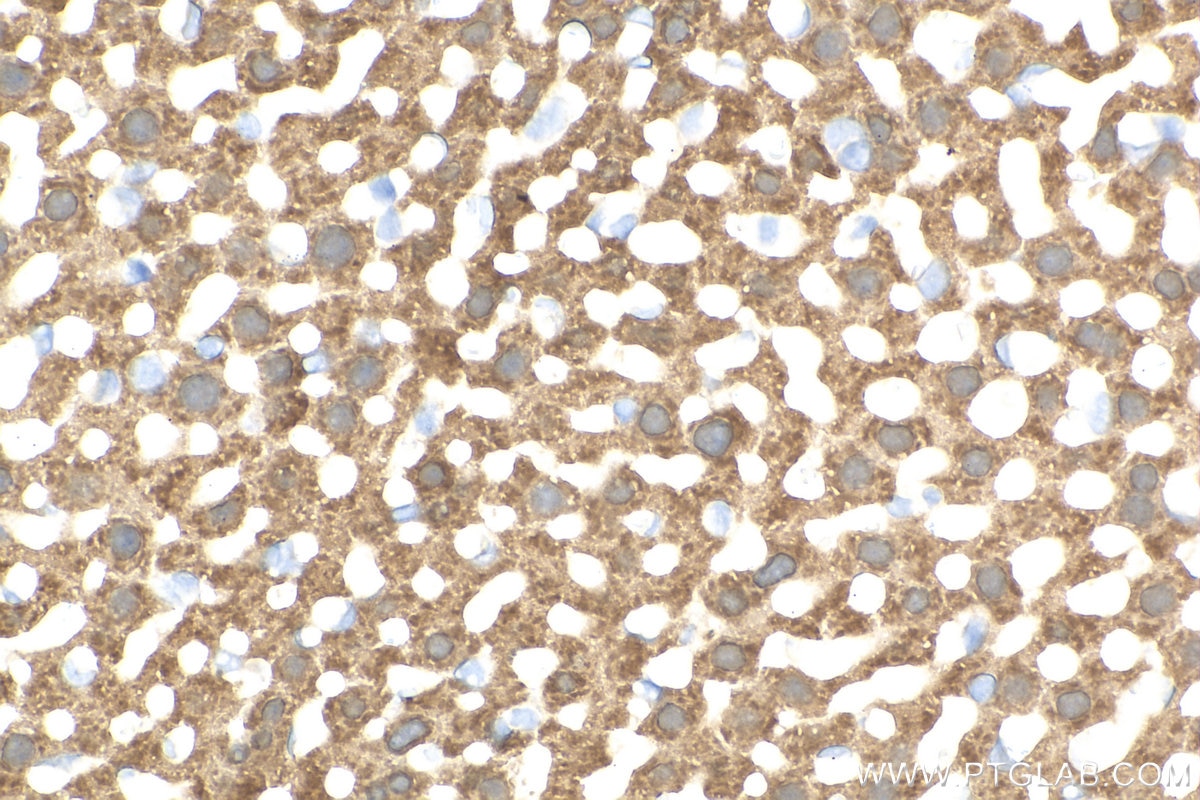
| Positive IHC detected in | mouse liver tissue, human liver tissue Note: suggested antigen retrieval with TE buffer pH 9.0; (*) Alternatively, antigen retrieval may be performed with citrate buffer pH 6.0 |
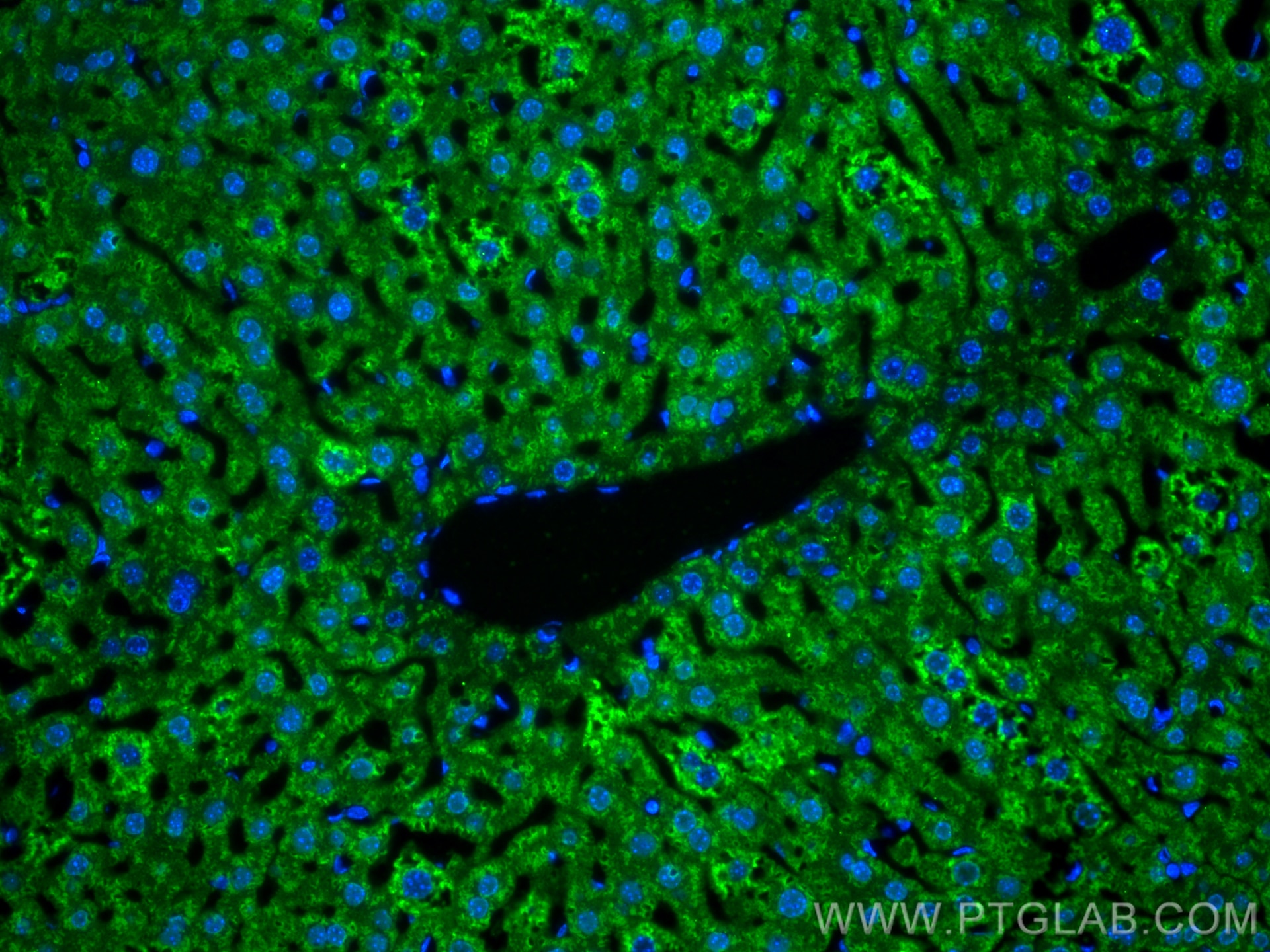
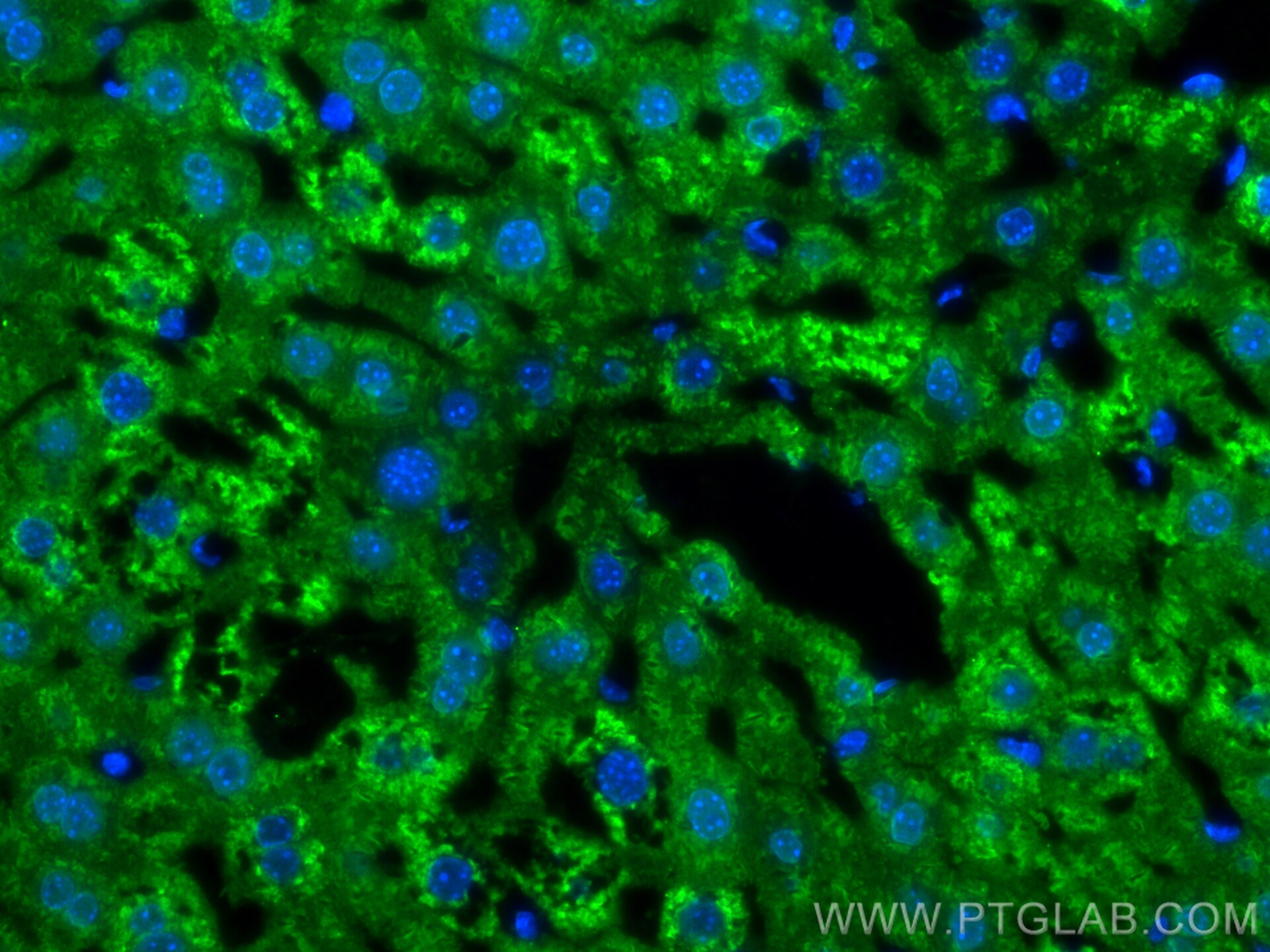
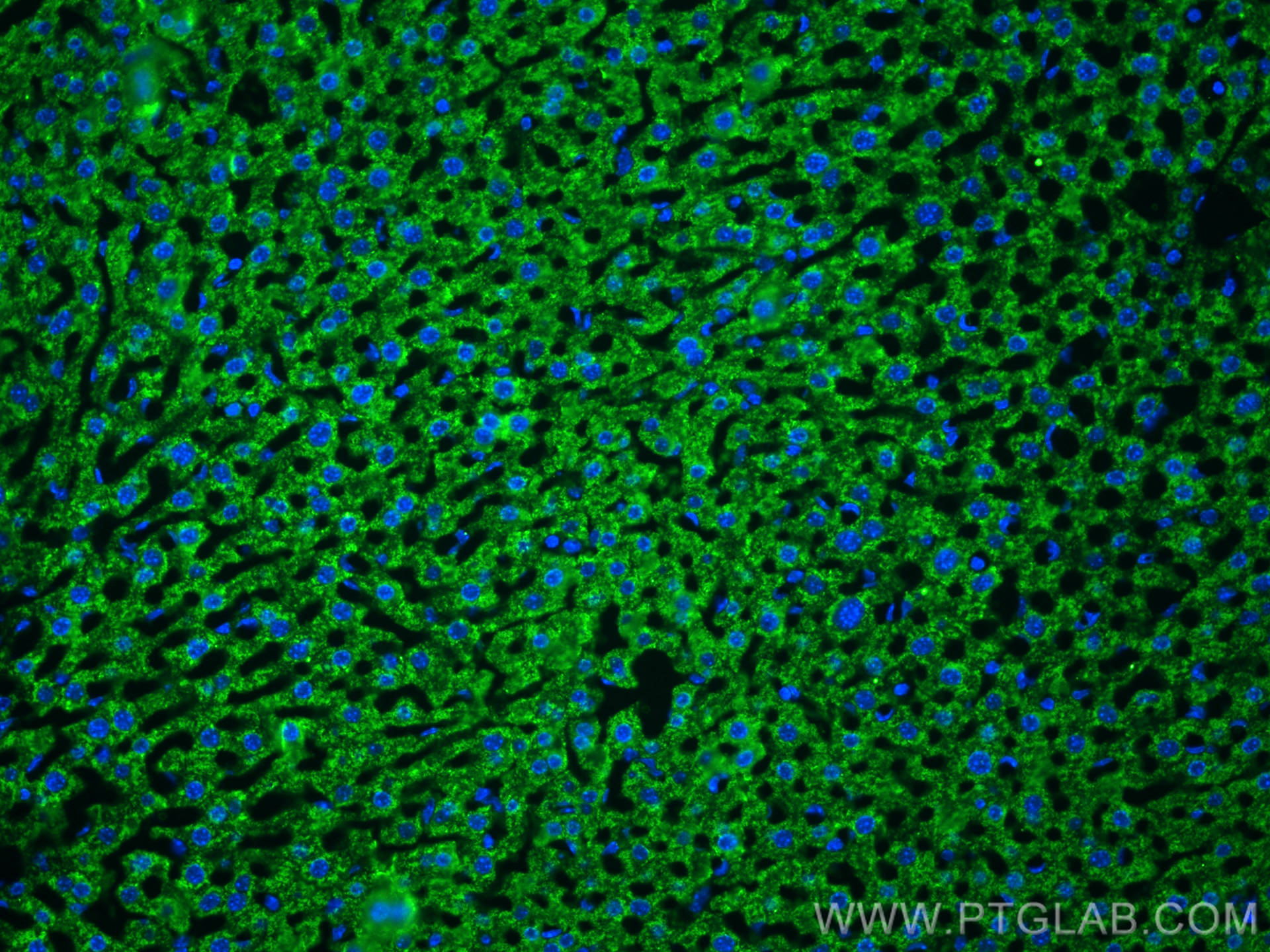
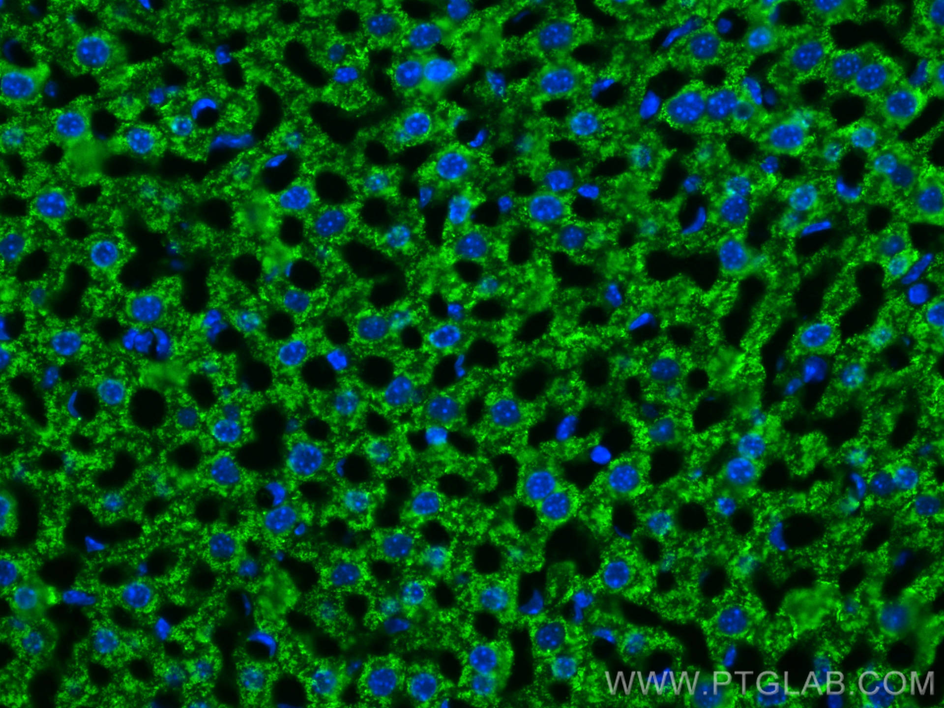
| Positive IF-P detected in | mouse liver tissue |
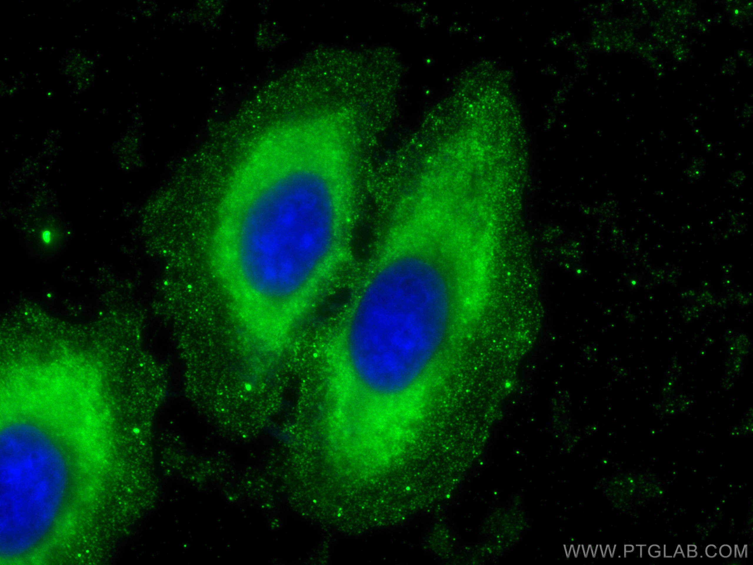
| Positive IF/ICC detected in | HepG2 cells, mouse liver tissue |
Recommended dilution
| Application | Dilution |
|---|---|
| Western Blot (WB) | WB : 1:5000-1:50000 |
| Immunoprecipitation (IP) | IP : 0.5-4.0 ug for 1.0-3.0 mg of total protein lysate |
| Immunohistochemistry (IHC) | IHC : 1:2000-1:5000 |
| Immunofluorescence (IF)-P | IF-P : 1:200-1:800 |
| Immunofluorescence (IF)/ICC | IF/ICC : 1:400-1:1600 |
| It is recommended that this reagent should be titrated in each testing system to obtain optimal results. | |
| Sample-dependent, Check data in validation data gallery. | |
Product Information
66129-1-Ig targets Arginase-1 in WB, IHC, IF/ICC, IF-P, IP, ELISA applications and shows reactivity with human, mouse, rat, pig, rabbit samples.
| Tested Reactivity | human, mouse, rat, pig, rabbit |
| Cited Reactivity | human, mouse, rat, pig, rabbit |
| Host / Isotype | Mouse / IgG1 |
| Class | Monoclonal |
| Type | Antibody |
| Immunogen |
CatNo: Ag8810 Product name: Recombinant human ARG1 protein Source: e coli.-derived, PET28a Tag: 6*His Domain: 1-236 aa of BC005321 Sequence: MSAKSRTIGIIGAPFSKGQPRGGVEEGPTVLRKAGLLEKLKEQECDVKDYGDLPFADIPNDSPFQIVKNPRSVGKASEQLAGKVAEVKKNGRISLVLGGDHSLAIGSISGHARVHPDLGVIWVDAHTDINTPLTTTSGNLHGQPVSFLLKELKGKIPDVPGFSWVTPCISAKDIVYIGLRDVDPGEHYILKTLGIKYFSMTEVTRTVNTAVAITLACFGLAREGNHKPIDYLNPPK Predict reactive species |
| Full Name | arginase, liver |
| Calculated Molecular Weight | 236aa,25 kDa; 322aa,35 kDa |
| Observed Molecular Weight | 36 kDa |
| GenBank Accession Number | BC005321 |
| Gene Symbol | Arginase-1 |
| Gene ID (NCBI) | 383 |
| RRID | AB_2881528 |
| Conjugate | Unconjugated |
| Form | Liquid |
| Purification Method | Protein G purification |
| UNIPROT ID | P05089 |
| Storage Buffer | PBS with 0.02% sodium azide and 50% glycerol, pH 7.3. |
| Storage Conditions | Store at -20°C. Stable for one year after shipment. Aliquoting is unnecessary for -20oC storage. 20ul sizes contain 0.1% BSA. |
Background Information
Arginase-1 (Liver arginase) belongs to the arginase family. ARG1 is a novel immunohistochemical marker of hepatocellular differentiation in fine needle aspiration cytology and a marker of hepatocytes and hepatocellular neoplasms. ARG1 is closely associated with alternative macrophage activation and ARG1 has been shown to protectmotor neurons from trophic factor deprivation and allow sensory neurons to overcome neurite outgrowth inhibition by myelin proteins (PMID: 20071539, PMID:12098359). It can exsit as a homotrimer and it has 3 isoforms produced by alternative splicing (PMID:16141327). Defects in ARG1 are the cause of argininemia (ARGIN). Deletion or TNF-mediated restriction of ARG1 unleashes the production of NO by NOS2, which is critical for pathogen control (PMID:27117406). Before stroke, ARG1 mainly expressed in neurons in a normal brain (PMID: 23311438). The expression of ARG1 increases in microglia/macrophages and astrocytes early after CNS injuries. ARG1 has been regarded as a marker for beneficial microglia/macrophages and possesses antiinflammatory and tissue repair properties under various pathological conditions (PMID: 26538310, PMID: 31619589).
Protocols
| Product Specific Protocols | |
|---|---|
| IF protocol for Arginase-1 antibody 66129-1-Ig | Download protocol |
| IHC protocol for Arginase-1 antibody 66129-1-Ig | Download protocol |
| IP protocol for Arginase-1 antibody 66129-1-Ig | Download protocol |
| WB protocol for Arginase-1 antibody 66129-1-Ig | Download protocol |
| Standard Protocols | |
|---|---|
| Click here to view our Standard Protocols |
Publications
What published studies show
The Arginase-1 Monoclonal antibody (5D6D12), catalog number 66129-1-Ig, has accumulated 210 citations across journals including Immunity, Nature Communications, Advanced Materials, Advanced Science, Biomaterials, JCI, Theranostics, and Journal of Neuroinflammation. Research fields span macrophage/microglial M2 polarization, neuroinflammation and neurodegeneration (Parkinson's, Alzheimer's, spinal cord injury, stroke), wound healing, cancer immunotherapy, bone regeneration, cardiovascular disease, liver fibrosis, ferroptosis, and biomaterials-based nanomedicine.
| Species | Application | Title |
|---|---|---|
Immunity Excessive Polyamine Generation in Keratinocytes Promotes Self-RNA Sensing by Dendritic Cells in Psoriasis. | ||
Adv Mater Ultrasound-Activated Sonophage Synergizes Sonodynamic Therapy and Saltoptosis for Solid Tumor Eradication. | ||
Adv Mater Photothermal and Photocatalytic Glycol Chitosan and Polydopamine-Grafted Oxygen Vacancy Bismuth Oxyiodide (BiO1-x I) Nanoparticles for the Diagnosis and Targeted Therapy of Diabetic Wounds | ||
Bioact Mater 3D printing of interferon γ-preconditioned NSC-derived exosomes/collagen/chitosan biological scaffolds for neurological recovery after TBI | ||
Bioact Mater Immune-modulated adhesive hydrogel for enhancing osteochondral graft adhesion and cartilage repair | ||
Reviews
What customers say
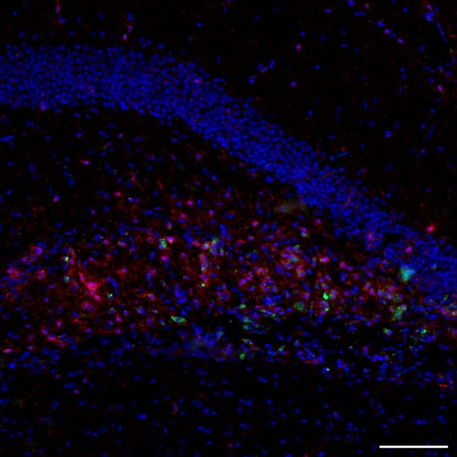
One reviewer (Marion B., November 2024) gave this antibody a perfect 5-star rating after using it at a 1:600 dilution for immunofluorescence on mouse brain tissue. In an Alzheimer's disease model, the antibody successfully stained Arg1, which colocalized with Iba1 to identify M2-type microglia. The reviewer expressed satisfaction with the results. Overall, the single review reflects a positive experience with strong performance in IF applications on neural tissue.
The reviews below have been submitted by verified Proteintech customers who received an incentive for providing their feedback.
Marion (Verified Customer) (11-24-2024) | DAPI (blue) + Iba1 (Red) + Arg1 (green) Arg1 colocalizes with Iba1 staining M2-type microglia in this model of AD. We are satisfied with the results.
 |


