Anticorps Polyclonal de lapin anti-VWF
VWF Polyclonal Antibody for WB, IHC, IF-P, ELISA
Hôte / Isotype
Lapin / IgG
Réactivité testée
Humain, rat, souris
Applications
WB, IHC, IF-P, ELISA
Conjugaison
Non conjugué
N° de cat : 27186-1-AP
Synonymes
Galerie de données de validation
Applications testées
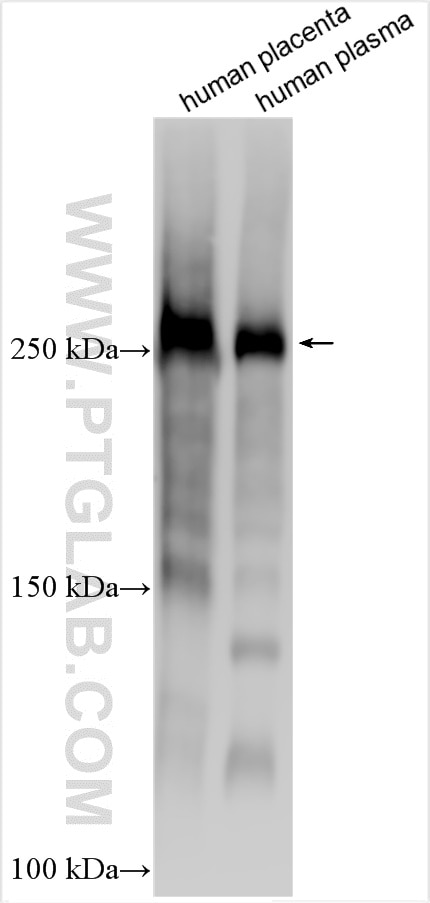
| Résultats positifs en WB | tissu placentaire humain, plasma humain |
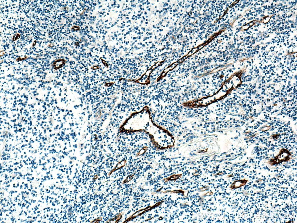
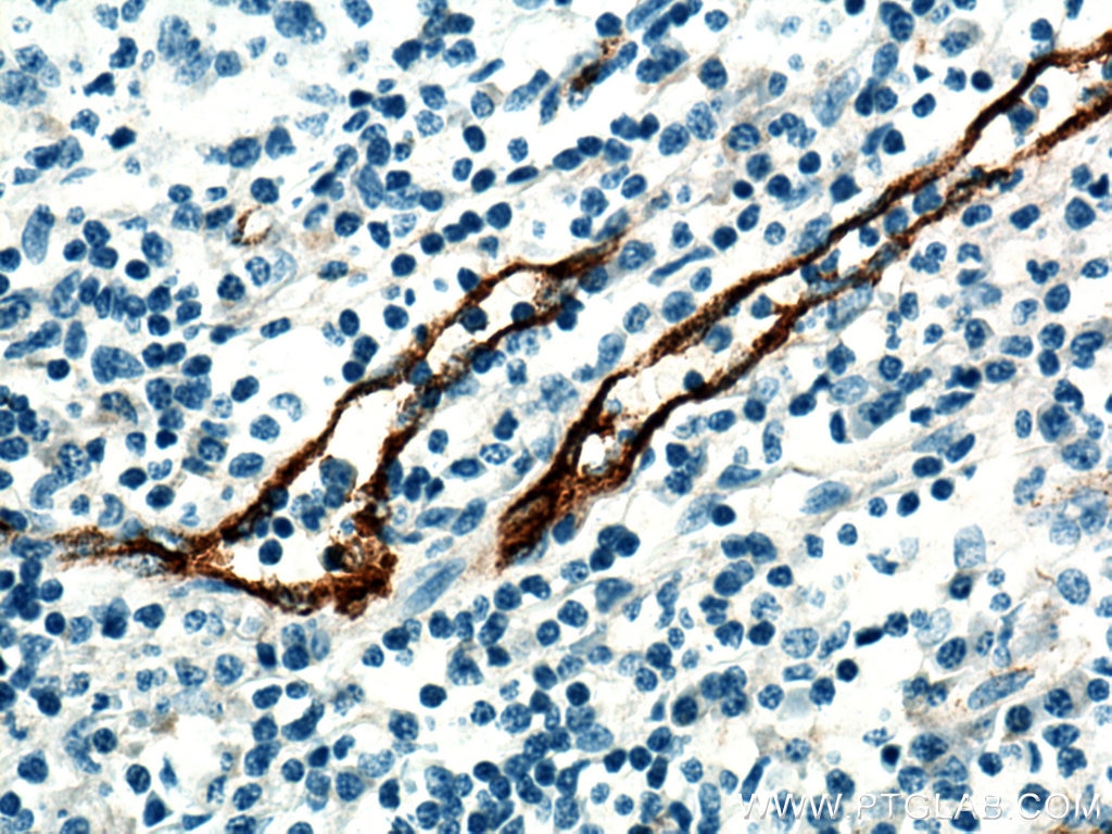
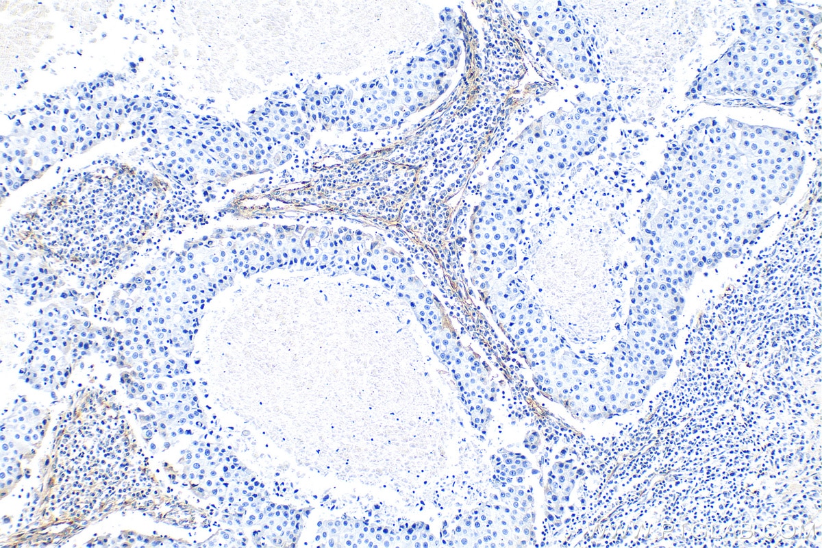
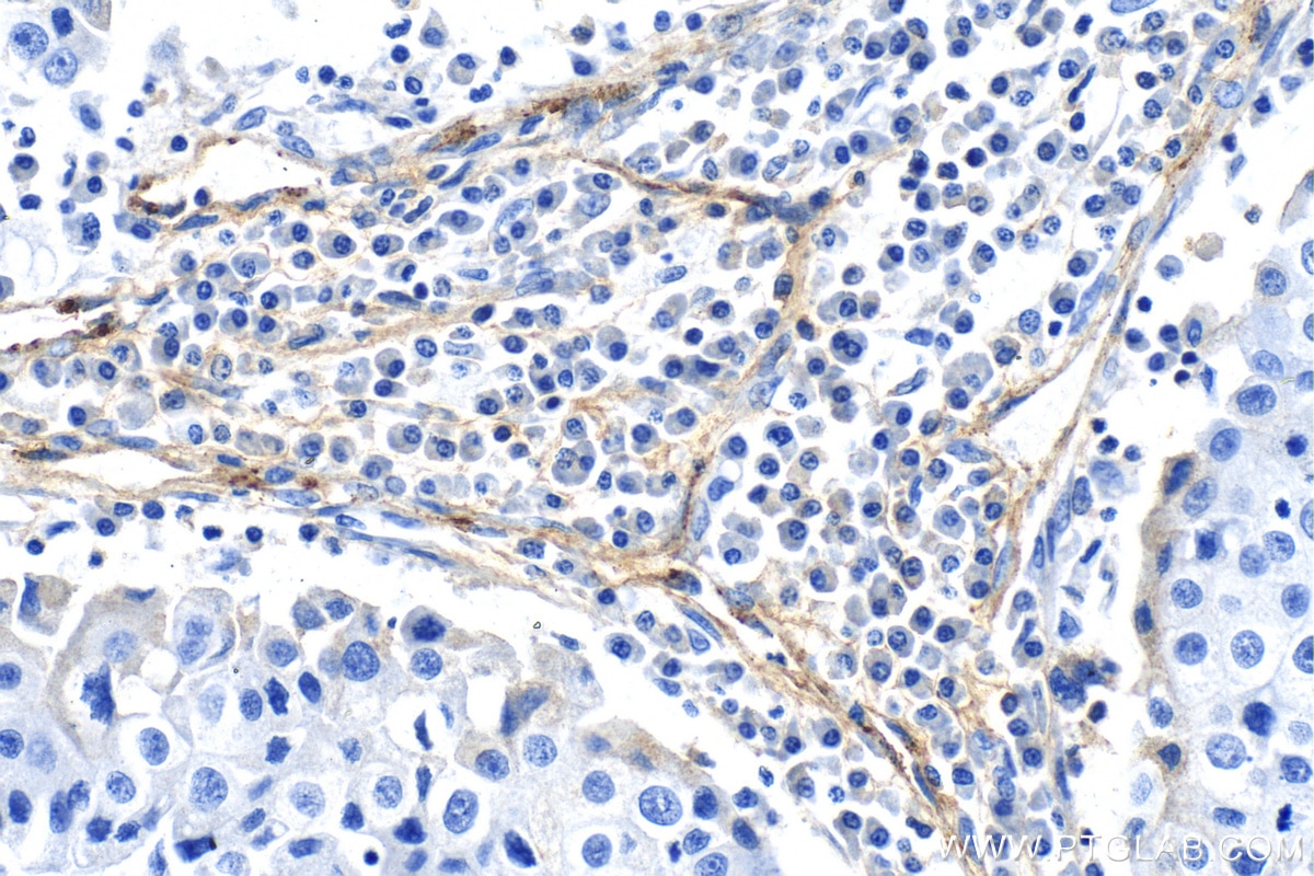
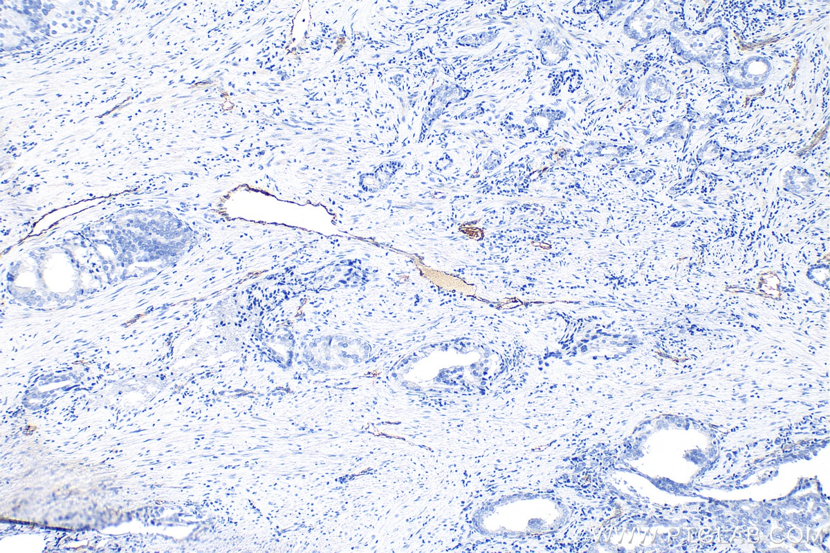
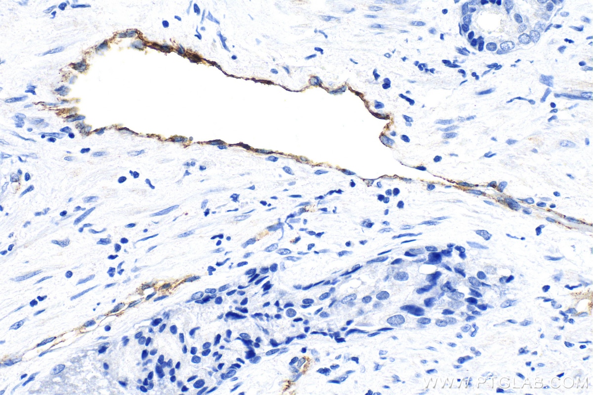
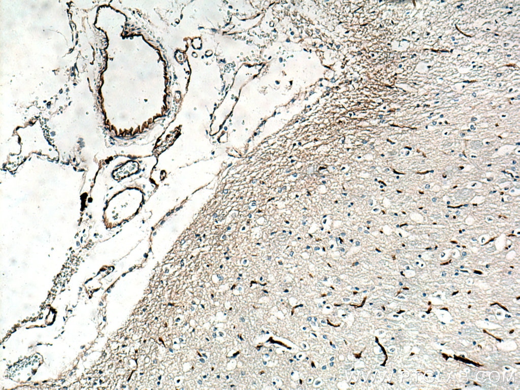
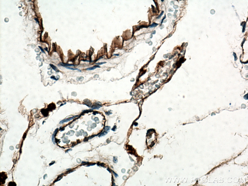
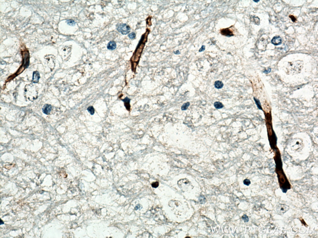
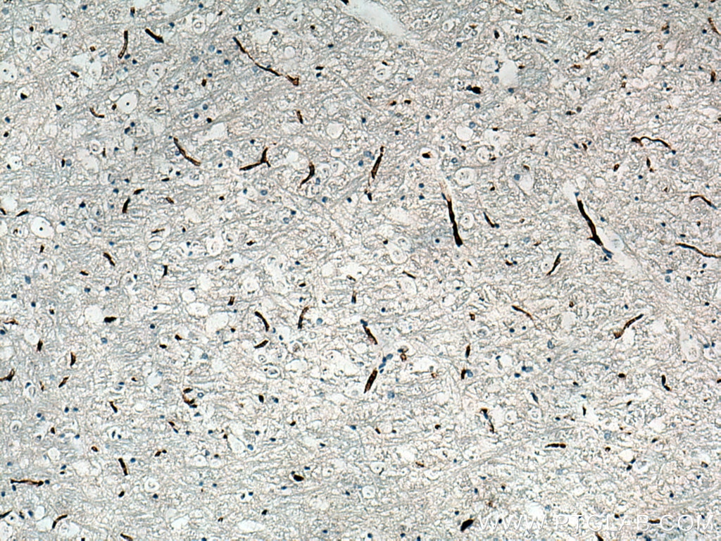
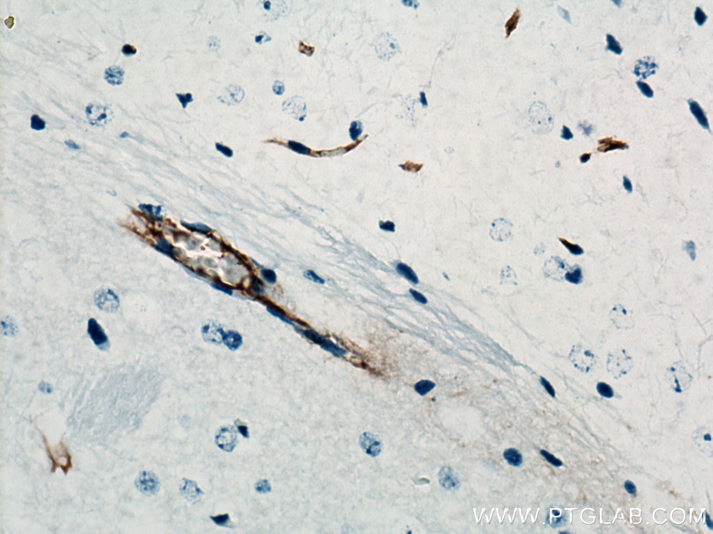
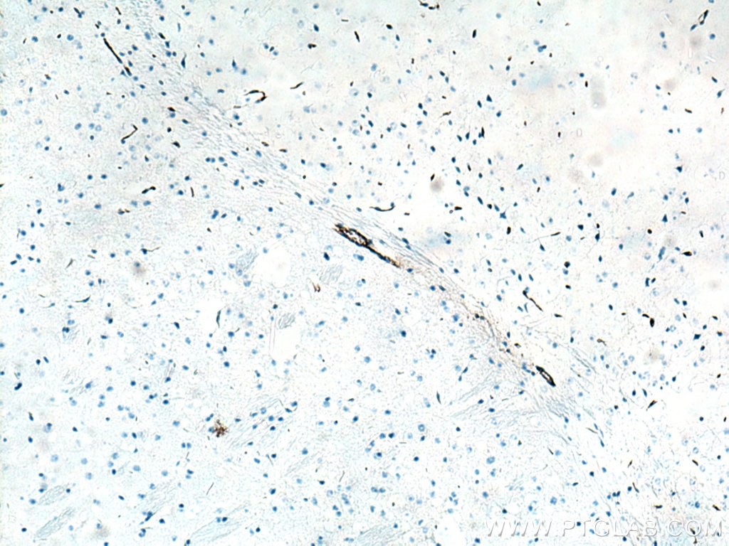
| Résultats positifs en IHC | tissu d'amygdalite humain, tissu cérébral de rat, tissu cérébral de souris, tissu de cancer de la prostate humain, tissu de cancer du sein humain il est suggéré de démasquer l'antigène avec un tampon de TE buffer pH 9.0; (*) À défaut, 'le démasquage de l'antigène peut être 'effectué avec un tampon citrate pH 6,0. |
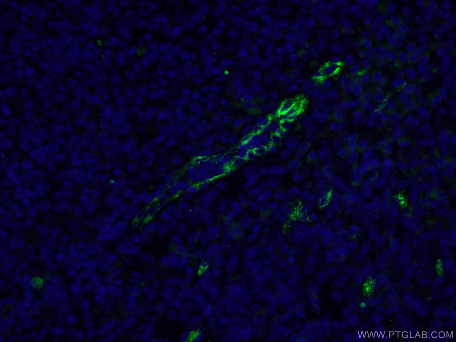
| Résultats positifs en IF-P | tissu d'amygdalite humain, |
Dilution recommandée
| Application | Dilution |
|---|---|
| Western Blot (WB) | WB : 1:500-1:1000 |
| Immunohistochimie (IHC) | IHC : 1:50-1:500 |
| Immunofluorescence (IF)-P | IF-P : 1:50-1:500 |
| It is recommended that this reagent should be titrated in each testing system to obtain optimal results. | |
| Sample-dependent, check data in validation data gallery | |
Applications publiées
| WB | See 3 publications below |
| IHC | See 7 publications below |
| IF | See 34 publications below |
Informations sur le produit
27186-1-AP cible VWF dans les applications de WB, IHC, IF-P, ELISA et montre une réactivité avec des échantillons Humain, rat, souris
| Réactivité | Humain, rat, souris |
| Réactivité citée | rat, Humain, souris |
| Hôte / Isotype | Lapin / IgG |
| Clonalité | Polyclonal |
| Type | Anticorps |
| Immunogène | VWF Protéine recombinante Ag25578 |
| Nom complet | von Willebrand factor |
| Poids moléculaire observé | 220-250 kDa |
| Symbole du gène | VWF |
| Identification du gène (NCBI) | 7450 |
| Conjugaison | Non conjugué |
| Forme | Liquide |
| Méthode de purification | Purification par affinité contre l'antigène |
| Tampon de stockage | PBS with 0.02% sodium azide and 50% glycerol |
| Conditions de stockage | Stocker à -20°C. Stable pendant un an après l'expédition. L'aliquotage n'est pas nécessaire pour le stockage à -20oC Les 20ul contiennent 0,1% de BSA. |
Informations générales
Von Willebrand factor (VWF) is a large multimeric glycoprotein found in blood plasma involved in hemostasis following vascular injury.
What is the molecular weight of VWF?
Due to the multimeric nature of VWF, it can range in size from 500 to 20,000 kDa due to the differences in the number of subunits comprising the protein. Each subunit is approximately 250 kDa (PMID: 9759493).
What is the tissue specificity of VWF?
The biosynthesis of VWF in vivo is limited to endothelial cells (PMID: 4209883) and megakaryocytes (PMID: 2413071). VWF synthesized in endothelial cells is either released directly into the plasma via a secretory pathway, or tubulized and stored in organelles unique to this cell type called Weibel-Palade bodies (PMID: 16459301). Whereas VWF synthesized in megakaryocytes is stored in the alpha granules of platelets (PMID: 2046403).
What is the function of VWF?
The primary function of VWF is as an adhesive plasma glycoprotein, particularly factor VIII; an essential blood-clotting protein (PMID: 6982084). VWF is also important in platelet adhesion to wound sites by binding specifically to type I and type III collagen (PMID: 11098050), with larger VWF multimers being most effective (PMID: 24448155).
What is the importance of post-translational modifications (PTMs) on VWF function?
During the synthesis of VWF, it must undergo extensive PTMs. The VWF monomers are subsequently N-glycosylated with the addition of a 12 N-linked high-mannose-containing oligosaccharide after the cleavage of the signal peptide in the endoplasmic reticulum. Dimerization of the pro-VWF follows glycosylation via carboxyl-termini disulphide bond formation. In the Golgi apparatus, the oligosaccharide chains are further modified into complex carbohydrates and multimerization of pro-VWF dimers occurs after another round of disulphide bond formation at the amino-termini end of the subunits (PMID: 6334089).
Protocole
| Product Specific Protocols | |
|---|---|
| WB protocol for VWF antibody 27186-1-AP | Download protocol |
| IHC protocol for VWF antibody 27186-1-AP | Download protocol |
| IF protocol for VWF antibody 27186-1-AP | Download protocol |
| Standard Protocols | |
|---|---|
| Click here to view our Standard Protocols |
Publications
| Species | Application | Title |
|---|---|---|
Biomaterials Oriented nanofibrous P(MMD-co-LA)/Deferoxamine nerve scaffold facilitates peripheral nerve regeneration by regulating macrophage phenotype and revascularization. | ||
Adv Healthc Mater Platelet Membrane-Fused Circulating Extracellular Vesicles Protect the Heart from Ischemia/Reperfusion Injury | ||
Acta Biomater Selective inhibition of glycolysis in hepatic stellate cells and suppression of liver fibrogenesis with vitamin A-derivative decorated camptothecin micelles | ||
Front Immunol Fatty acid synthase inhibition improves hypertension-induced erectile dysfunction by suppressing oxidative stress and NLRP3 inflammasome-dependent pyroptosis through activating the Nrf2/HO-1 pathway | ||
Regen Biomater Microvascular network based on the Hilbert curve for nutrient transport in thick tissue | ||
Aging (Albany NY) Extracorporeal shockwave relieves endothelial injury and dysfunction in steroid-induced osteonecrosis of the femoral head via miR-135b targeting FOXO1: in vitro and in vivo studies. |
