Anticorps Polyclonal de lapin anti-Villin
Villin Polyclonal Antibody for WB, IHC, IF/ICC, IF-P, FC (Intra), IP, ELISA
Hôte / Isotype
Lapin / IgG
Réactivité testée
Humain, rat, souris et plus (1)
Applications
WB, IHC, IF/ICC, IF-P, FC (Intra), IP, ELISA
Conjugaison
Non conjugué
N° de cat : 16488-1-AP
Synonymes
Galerie de données de validation
Applications testées
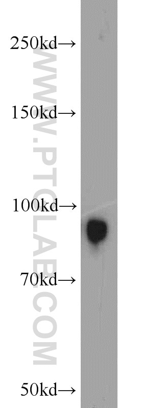
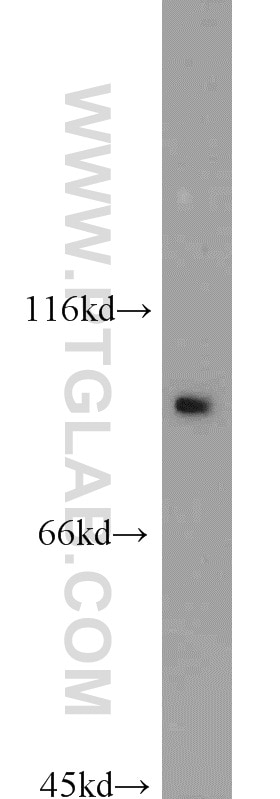
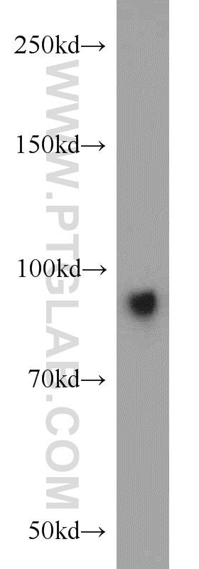
| Résultats positifs en WB | tissu rénal de souris, tissu de côlon de souris, tissu hépatique de souris |
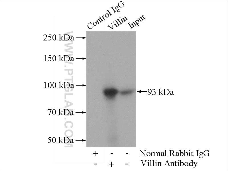
| Résultats positifs en IP | tissu rénal de souris |
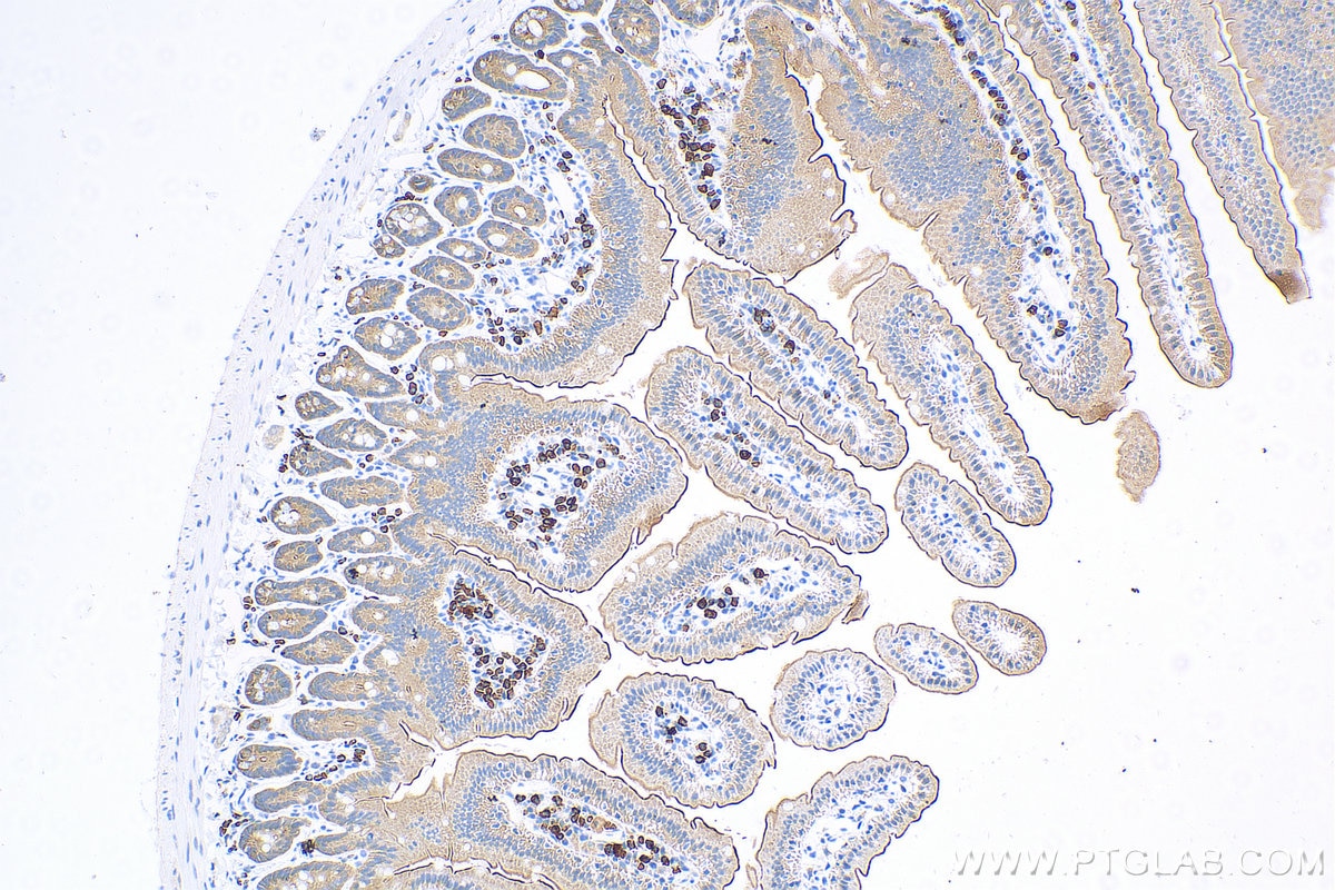
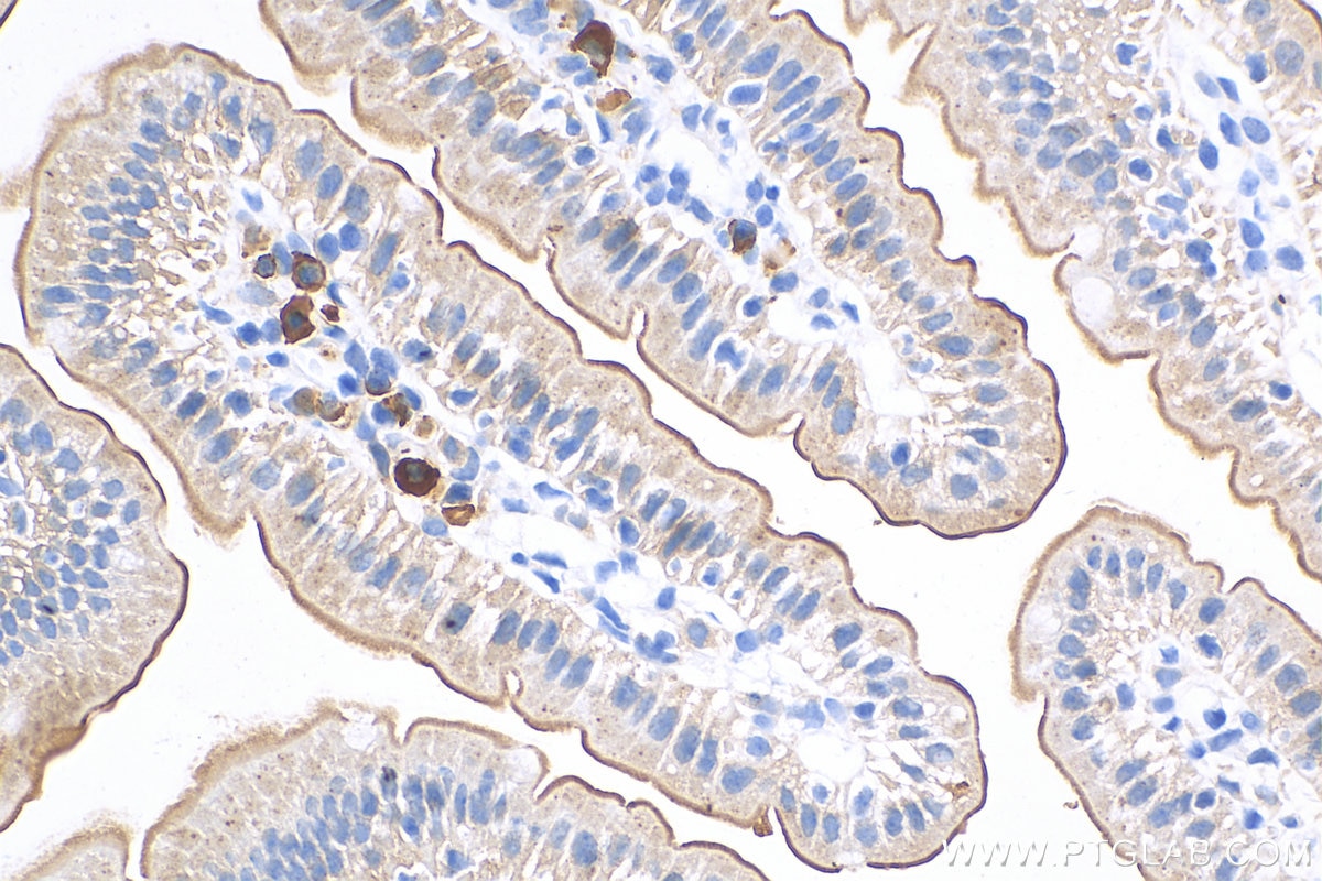
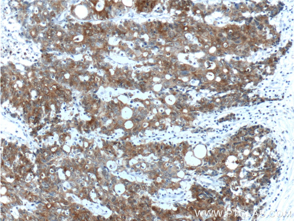
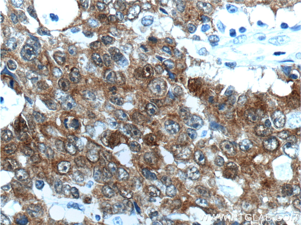
| Résultats positifs en IHC | tissu d'intestin grêle de souris, tissu de cancer du côlon humain il est suggéré de démasquer l'antigène avec un tampon de TE buffer pH 9.0; (*) À défaut, 'le démasquage de l'antigène peut être 'effectué avec un tampon citrate pH 6,0. |
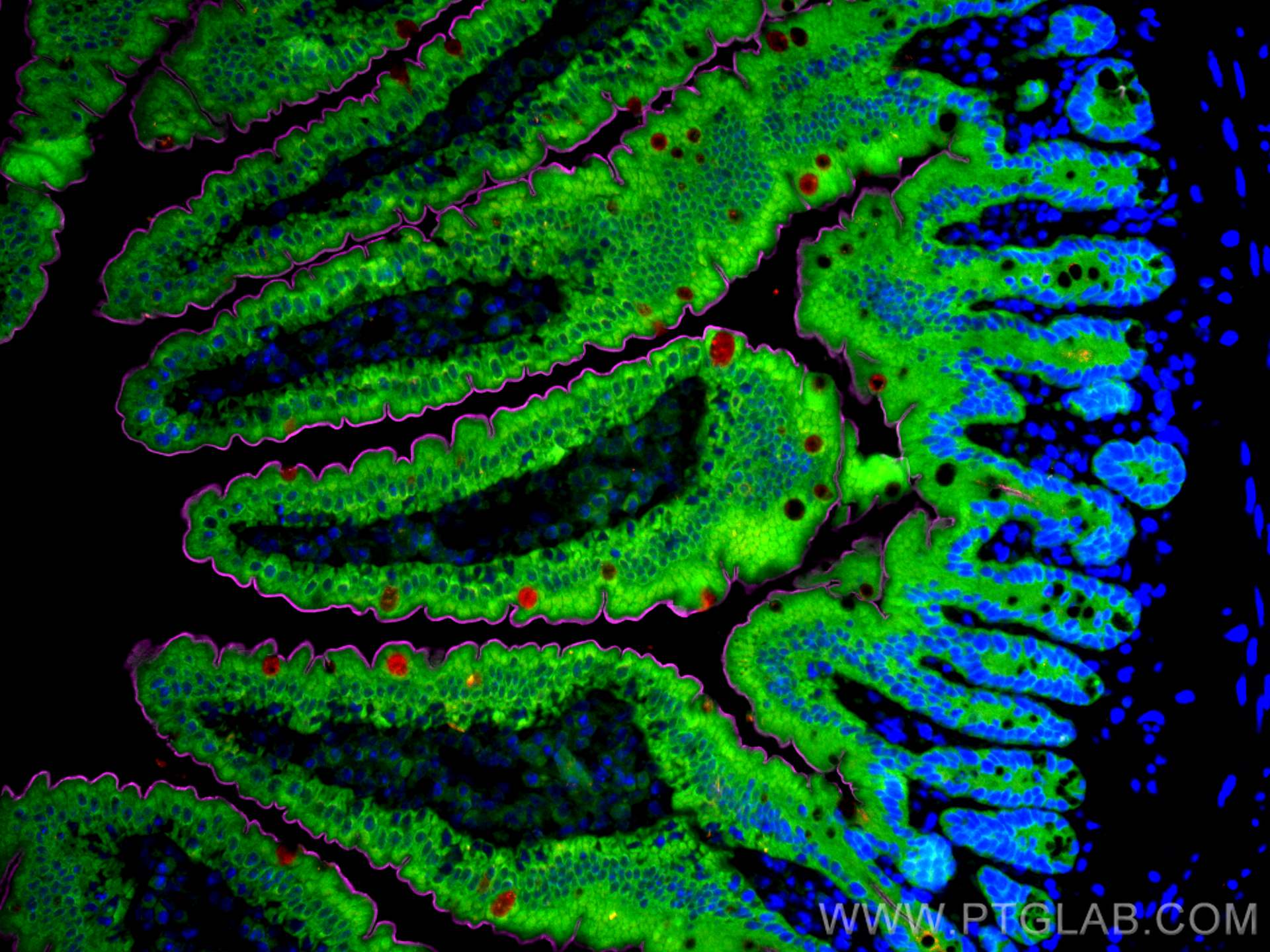
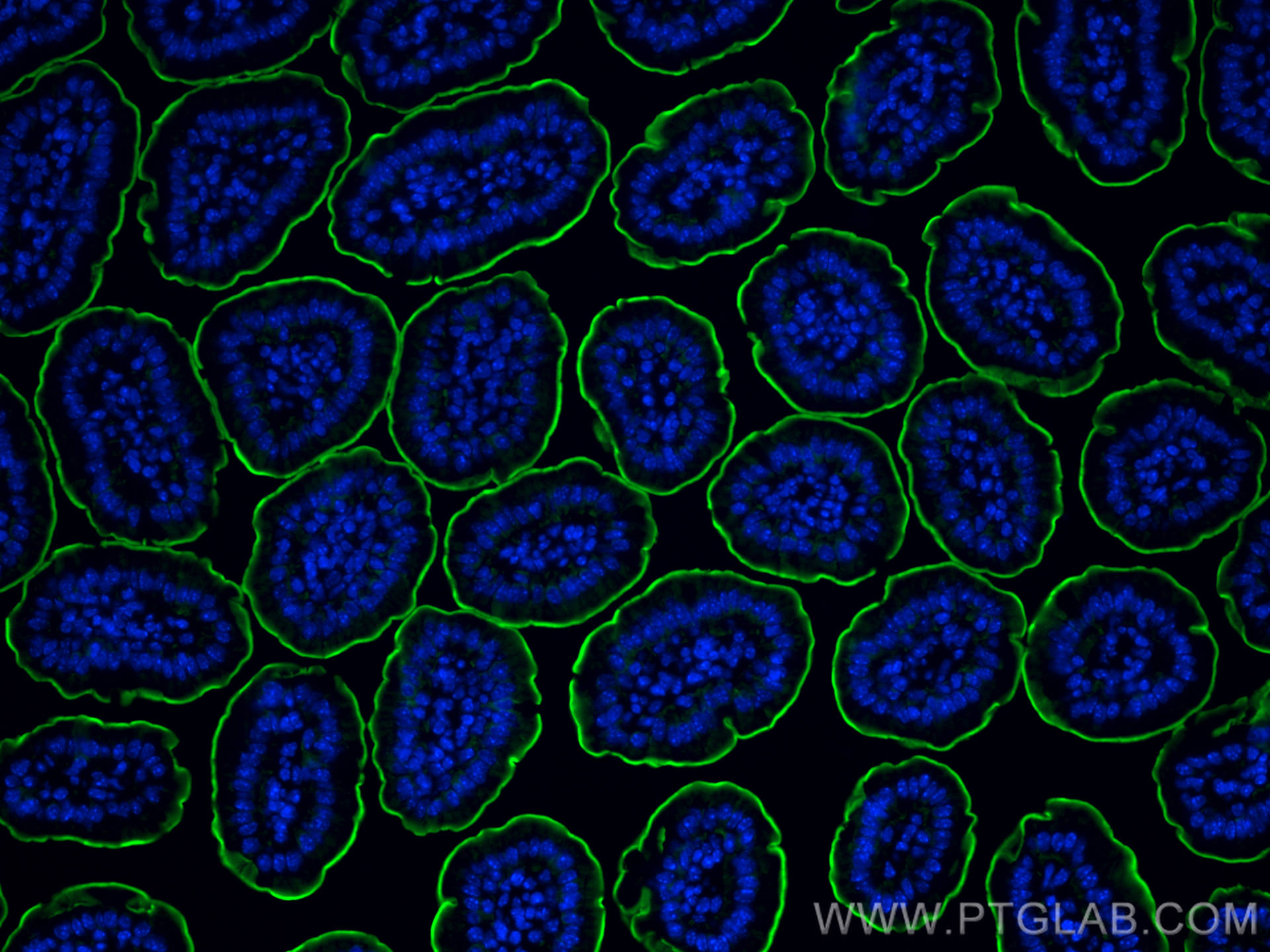
| Résultats positifs en IF-P | tissu d'intestin grêle de souris, |
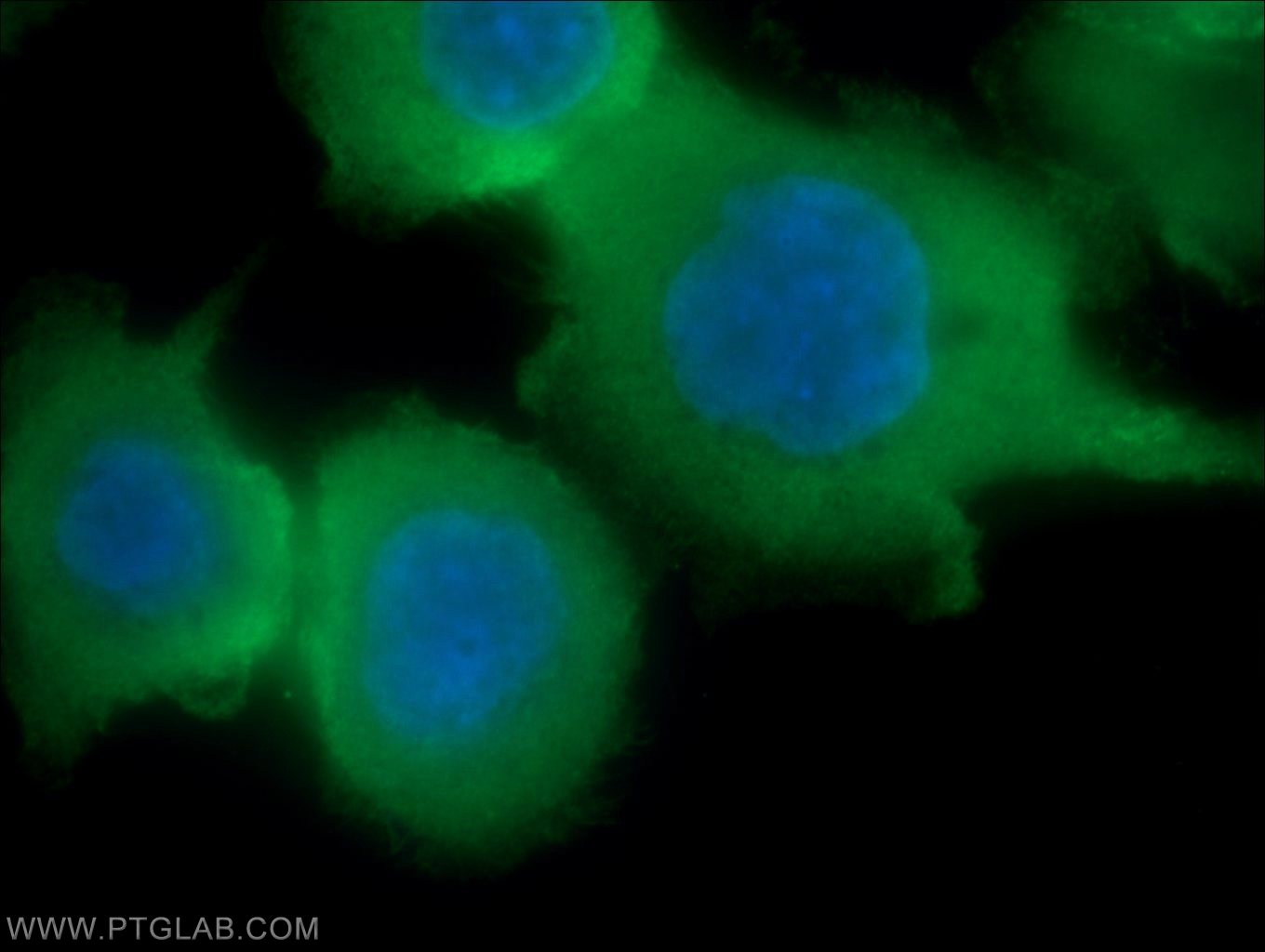
| Résultats positifs en IF/ICC | cellules COLO 320, |
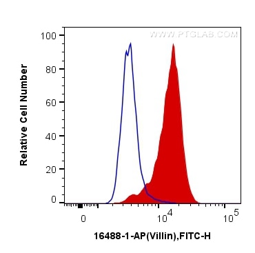
| Résultats positifs en FC (Intra) | cellules HepG2 |
Dilution recommandée
| Application | Dilution |
|---|---|
| Western Blot (WB) | WB : 1:1000-1:4000 |
| Immunoprécipitation (IP) | IP : 0.5-4.0 ug for 1.0-3.0 mg of total protein lysate |
| Immunohistochimie (IHC) | IHC : 1:2500-1:10000 |
| Immunofluorescence (IF)-P | IF-P : 1:50-1:500 |
| Immunofluorescence (IF)/ICC | IF/ICC : 1:50-1:500 |
| Flow Cytometry (FC) (INTRA) | FC (INTRA) : 0.20 ug per 10^6 cells in a 100 µl suspension |
| It is recommended that this reagent should be titrated in each testing system to obtain optimal results. | |
| Sample-dependent, check data in validation data gallery | |
Applications publiées
| WB | See 3 publications below |
| IHC | See 4 publications below |
| IF | See 6 publications below |
Informations sur le produit
16488-1-AP cible Villin dans les applications de WB, IHC, IF/ICC, IF-P, FC (Intra), IP, ELISA et montre une réactivité avec des échantillons Humain, rat, souris
| Réactivité | Humain, rat, souris |
| Réactivité citée | Humain, porc, souris |
| Hôte / Isotype | Lapin / IgG |
| Clonalité | Polyclonal |
| Type | Anticorps |
| Immunogène | Villin Protéine recombinante Ag9610 |
| Nom complet | villin 1 |
| Masse moléculaire calculée | 827aa,93 kDa; 826aa,93 kDa |
| Poids moléculaire observé | 93 kDa |
| Numéro d’acquisition GenBank | BC017303 |
| Symbole du gène | Villin |
| Identification du gène (NCBI) | 7429 |
| Conjugaison | Non conjugué |
| Forme | Liquide |
| Méthode de purification | Purification par affinité contre l'antigène |
| Tampon de stockage | PBS avec azoture de sodium à 0,02 % et glycérol à 50 % pH 7,3 |
| Conditions de stockage | Stocker à -20°C. Stable pendant un an après l'expédition. L'aliquotage n'est pas nécessaire pour le stockage à -20oC Les 20ul contiennent 0,1% de BSA. |
Informations générales
Villin 1 (VIL1) is a 95-kd F-actin bundling and severing protein and its expression is restricted to epithelial cells with a brush border, like epithelial cells of the intestinal mucosa, gall bladder, renal proximal tubules and ductuli efferentes of the testis. VIL1 has been reported to be an epithelial cell-specific anti-apoptotic protein, and to have an important function in regulating actin dynamics, cell morphology, epithelial-to-mesenchymal transitions, cell migration and cell survival. In addition, VIL1 is a useful diagnostic marker for of various cancer, like cervical and endometrial adenocarcinomas, renal cell carcinoma. VIL1 was recently identified as a novel biomarker predictive for postoperative recurrence and poorer prognosis of high serum AFP associated HCC.
Protocole
| Product Specific Protocols | |
|---|---|
| WB protocol for Villin antibody 16488-1-AP | Download protocol |
| IHC protocol for Villin antibody 16488-1-AP | Download protocol |
| IF protocol for Villin antibody 16488-1-AP | Download protocol |
| IP protocol for Villin antibody 16488-1-AP | Download protocol |
| Standard Protocols | |
|---|---|
| Click here to view our Standard Protocols |
Publications
| Species | Application | Title |
|---|---|---|
Proc Natl Acad Sci U S A Organotypic models of type III interferon-mediated protection from Zika virus infections at the maternal-fetal interface. | ||
Clin Transl Med NLRP3 inflammasome of renal tubular epithelial cells induces kidney injury in acute hemolytic transfusion reactions. | ||
PLoS Pathog Bile acids promote the caveolae-associated entry of swine acute diarrhea syndrome coronavirus in porcine intestinal enteroids. | ||
Front Cell Dev Biol Cathelicidin-WA Protects Against LPS-Induced Gut Damage Through Enhancing Survival and Function of Intestinal Stem Cells. | ||
Int J Mol Sci Nrf2-Knockout Protects from Intestinal Injuries in C57BL/6J Mice Following Abdominal Irradiation with γ Rays. | ||
Toxicol In Vitro Survival and cellular heterogeneity of epithelium in cultured mouse and rat precision-cut intestinal slices. |
