- Phare
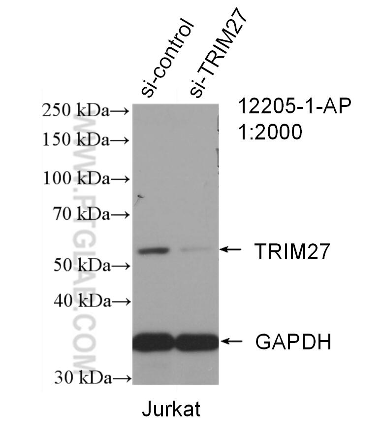
- Validé par KD/KO
Anticorps Polyclonal de lapin anti-TRIM27
TRIM27 Polyclonal Antibody for WB, IP, IHC, ELISA
Hôte / Isotype
Lapin / IgG
Réactivité testée
Humain, rat, souris
Applications
WB, IHC, IP, CoIP, ELISA, IF
Conjugaison
Non conjugué
N° de cat : 12205-1-AP
Synonymes
Galerie de données de validation
Applications testées
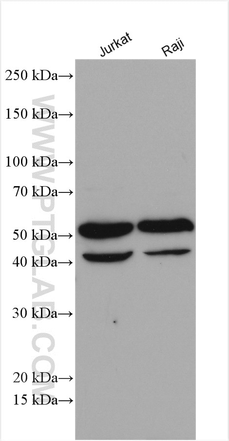
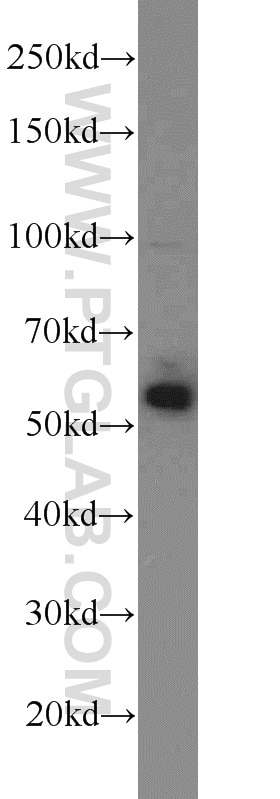
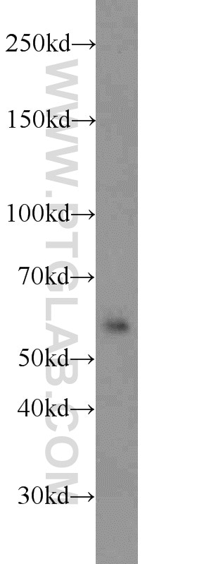
| Résultats positifs en WB | cellules Jurkat, cellules Raji, tissu testiculaire de souris |
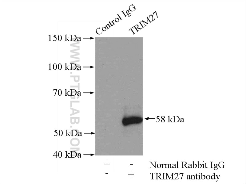
| Résultats positifs en IP | tissu testiculaire de souris |
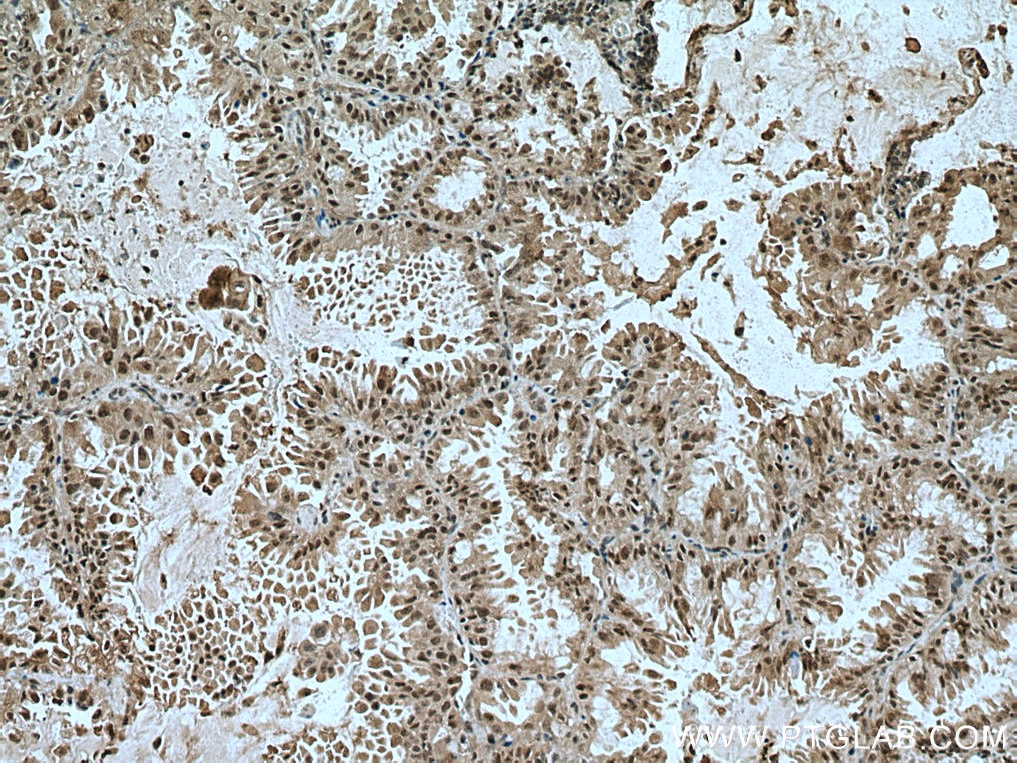
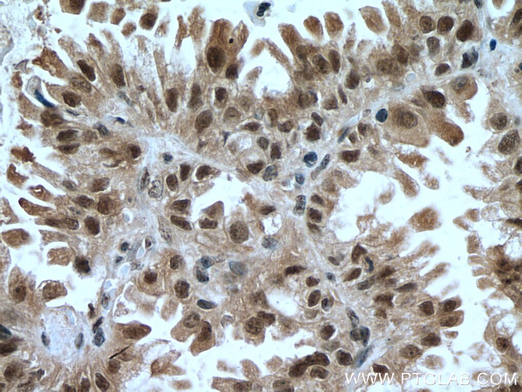
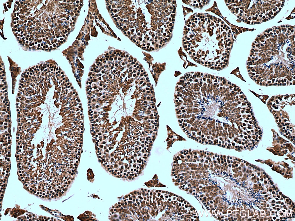
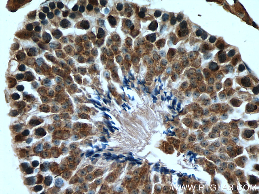
| Résultats positifs en IHC | tissu de cancer du poumon humain, tissu testiculaire de souris il est suggéré de démasquer l'antigène avec un tampon de TE buffer pH 9.0; (*) À défaut, 'le démasquage de l'antigène peut être 'effectué avec un tampon citrate pH 6,0. |
Dilution recommandée
| Application | Dilution |
|---|---|
| Western Blot (WB) | WB : 1:1000-1:6000 |
| Immunoprécipitation (IP) | IP : 0.5-4.0 ug for 1.0-3.0 mg of total protein lysate |
| Immunohistochimie (IHC) | IHC : 1:50-1:500 |
| It is recommended that this reagent should be titrated in each testing system to obtain optimal results. | |
| Sample-dependent, check data in validation data gallery | |
Applications publiées
| KD/KO | See 8 publications below |
| WB | See 13 publications below |
| IHC | See 3 publications below |
| IF | See 7 publications below |
| CoIP | See 3 publications below |
Informations sur le produit
12205-1-AP cible TRIM27 dans les applications de WB, IHC, IP, CoIP, ELISA, IF et montre une réactivité avec des échantillons Humain, rat, souris
| Réactivité | Humain, rat, souris |
| Réactivité citée | Humain, souris |
| Hôte / Isotype | Lapin / IgG |
| Clonalité | Polyclonal |
| Type | Anticorps |
| Immunogène | TRIM27 Protéine recombinante Ag2849 |
| Nom complet | tripartite motif-containing 27 |
| Masse moléculaire calculée | 513 aa, 58 kDa |
| Poids moléculaire observé | 58 kDa, 41 kDa |
| Numéro d’acquisition GenBank | BC013580 |
| Symbole du gène | TRIM27 |
| Identification du gène (NCBI) | 5987 |
| Conjugaison | Non conjugué |
| Forme | Liquide |
| Méthode de purification | Purification par affinité contre l'antigène |
| Tampon de stockage | PBS avec azoture de sodium à 0,02 % et glycérol à 50 % pH 7,3 |
| Conditions de stockage | Stocker à -20°C. Stable pendant un an après l'expédition. L'aliquotage n'est pas nécessaire pour le stockage à -20oC Les 20ul contiennent 0,1% de BSA. |
Protocole
| Product Specific Protocols | |
|---|---|
| WB protocol for TRIM27 antibody 12205-1-AP | Download protocol |
| IHC protocol for TRIM27 antibody 12205-1-AP | Download protocol |
| IP protocol for TRIM27 antibody 12205-1-AP | Download protocol |
| Standard Protocols | |
|---|---|
| Click here to view our Standard Protocols |
Publications
| Species | Application | Title |
|---|---|---|
Elife The testis protein ZNF165 is a SMAD3 cofactor that coordinates oncogenic TGFβ signaling in triple-negative breast cancer. | ||
Mol Ther Nucleic Acids TRIM27 Functions as a Novel Oncogene in Non-Triple-Negative Breast Cancer by Blocking Cellular Senescence through p21 Ubiquitination. | ||
FASEB J TRIM28 and TRIM27 are required for expressions of PDGFRβ and contractile phenotypic genes by vascular smooth muscle cells.
| ||
J Cell Mol Med NT5DC2 promotes leiomyosarcoma tumour cell growth via stabilizing unpalmitoylated TEAD4 and generating a positive feedback loop. | ||
J Leukoc Biol Trim27 confers myeloid hematopoiesis competitiveness by up-regulating myeloid master genes. | ||
FEBS J TRIM27 is an autophagy substrate facilitating mitochondria clustering and mitophagy via phosphorylated TBK1
|
Avis
The reviews below have been submitted by verified Proteintech customers who received an incentive forproviding their feedback.
FH Amy (Verified Customer) (03-18-2024) | Clean bands for U2OS cell lysates 25ug.
|
FH Jason (Verified Customer) (08-17-2022) | Third time purchase. I am satisfied. This antibody works very well in human breast cancer cells using WB at dilution 1:1000, but many non-specific IF singling detected.
|
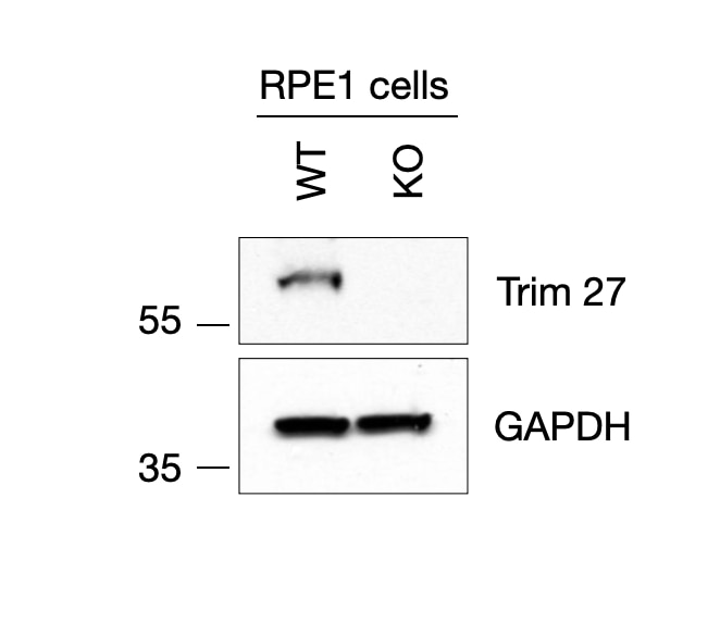
FH Elisa (Verified Customer) (12-21-2021) | HAP1 were lysates with RIPA buffer. 1mg of protein was loaded per condition. Proteins were run on a 12-4% gradient polyacrylamide gel at 180V constant. Gel was transferred on nitrocellulose filter at 300mA constant. Blots were then blocked in 5%milk in TBS-0,1%Tween for 30 min and then incubated with Trim27 primary antibody (1:1000 dilution). Anti-rabbit -HRP conjugated antibody (1:2000) was used to detect TRIM27 signal.
 |
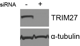
FH Sarah (Verified Customer) (07-03-2019) | Total cell lysate (30 ug) was resolved on a 4-12% Bis-Tris gel and transferred to nitrocellulose membrane. Membrane was incubated in blocking buffer (5% milk/0.1% Tween-20) for 1h. Membrane was incubated with anti-TRIM27 in blocking buffer (1:1000) at 4C overnight. After washing, membrane was incubated in anti-rabbit-HRP in blocking bufffer (1:3000) for 1h at room temperature. Protein was detected using ECL reagent and imaged on a chemiluminescence detection system.
 |
FH Francesca (Verified Customer) (05-16-2019) | Excellent antibody, clean and strong signal.
 |
