- Phare
- Validé par KD/KO
Anticorps Polyclonal de lapin anti-TFAP2A,AP-2
TFAP2A,AP-2 Polyclonal Antibody for WB, IF, ELISA
Hôte / Isotype
Lapin / IgG
Réactivité testée
Humain et plus (1)
Applications
WB, IF/ICC, IP, ChIP, ELISA
Conjugaison
Non conjugué
N° de cat : 13019-3-AP
Synonymes
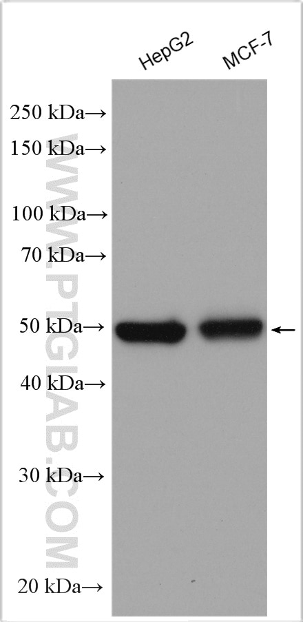
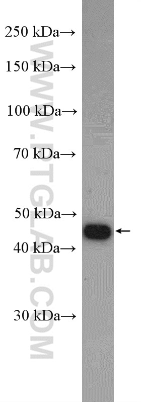
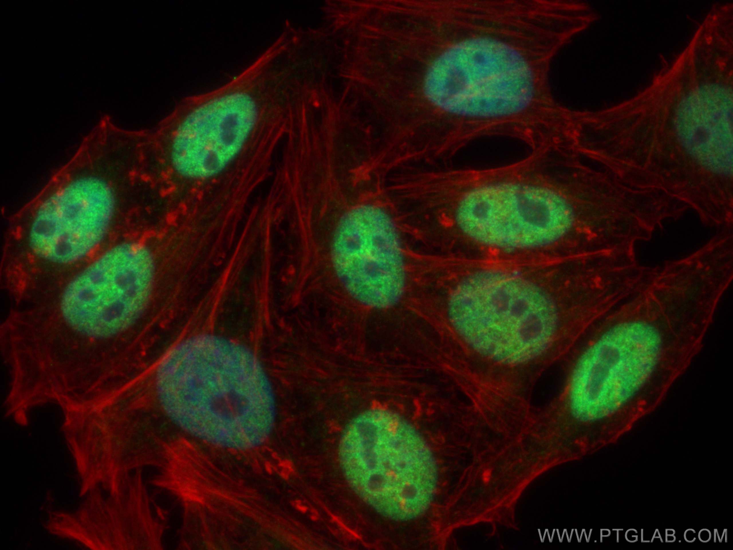
Galerie de données de validation
Applications testées
| Résultats positifs en WB | cellules HepG2, cellules MCF-7, cellules Y79 |
| Résultats positifs en IF/ICC | cellules HepG2, |
Dilution recommandée
| Application | Dilution |
|---|---|
| Western Blot (WB) | WB : 1:2000-1:10000 |
| Immunofluorescence (IF)/ICC | IF/ICC : 1:50-1:500 |
| It is recommended that this reagent should be titrated in each testing system to obtain optimal results. | |
| Sample-dependent, check data in validation data gallery | |
Applications publiées
| KD/KO | See 1 publications below |
| WB | See 3 publications below |
| IP | See 1 publications below |
| ChIP | See 4 publications below |
Informations sur le produit
13019-3-AP cible TFAP2A,AP-2 dans les applications de WB, IF/ICC, IP, ChIP, ELISA et montre une réactivité avec des échantillons Humain
| Réactivité | Humain |
| Réactivité citée | Humain, souris |
| Hôte / Isotype | Lapin / IgG |
| Clonalité | Polyclonal |
| Type | Anticorps |
| Immunogène | TFAP2A,AP-2 Protéine recombinante Ag4112 |
| Nom complet | transcription factor AP-2 alpha (activating enhancer binding protein 2 alpha) |
| Masse moléculaire calculée | 431 aa, 47 kDa |
| Poids moléculaire observé | 47 kDa |
| Numéro d’acquisition GenBank | BC017754 |
| Symbole du gène | TFAP2A |
| Identification du gène (NCBI) | 7020 |
| Conjugaison | Non conjugué |
| Forme | Liquide |
| Méthode de purification | Purification par affinité contre l'antigène |
| Tampon de stockage | PBS avec azoture de sodium à 0,02 % et glycérol à 50 % pH 7,3 |
| Conditions de stockage | Stocker à -20°C. Stable pendant un an après l'expédition. L'aliquotage n'est pas nécessaire pour le stockage à -20oC Les 20ul contiennent 0,1% de BSA. |
Informations générales
The activator protein-2 (AP-2) family of transcription factors comprises five 52-kDa isoforms (AP-2α, AP-2β, AP-2γ, AP-2δ, and AP-2ε), which share a common structure: a proline/glutamine-rich transactivation domain in the N-terminal region and a helix-span-helix domain in the C-terminal region, which mediates dimerization and site-specific DNA binding. Depending on the cellular context, the AP-2 transcription factors are individually associated either with cell differentiation and development or with cancer progression/regression (PMID: 21966377). TFAP2A (AP-2-alpha) is the only AP-2 protein required for early morphogenesis of the lens vesicle. Together with the CITED2 coactivator, stimulates the PITX2 P1 promoter transcription activation (PMID: 11694877).
Protocole
| Product Specific Protocols | |
|---|---|
| WB protocol for TFAP2A,AP-2 antibody 13019-3-AP | Download protocol |
| IF protocol for TFAP2A,AP-2 antibody 13019-3-AP | Download protocol |
| Standard Protocols | |
|---|---|
| Click here to view our Standard Protocols |
Publications
| Species | Application | Title |
|---|---|---|
Oncol Rep Regulation of DEK expression by AP-2α and methylation level of DEK promoter in hepatocellular carcinoma. | ||
Acta Pharmacol Sin Hepatitis B virus X protein promotes human hepatoma cell growth via upregulation of transcription factor AP2α and sphingosine kinase 1.
| ||
Cell Death Dis TFAP2A potentiates lung adenocarcinoma metastasis by a novel miR-16 family/TFAP2A/PSG9/TGF-β signaling pathway. | ||
Cell Death Dis The m6A modification-mediated OGDHL exerts a tumor suppressor role in ccRCC by downregulating FASN to inhibit lipid synthesis and ERK signaling | ||
Int Immunopharmacol miR-1293 suppresses osteosarcoma progression by modulating drug sensitivity in response to cisplatin treatment | ||
Commun Biol Macroscopic inhibition of DNA damage repair pathways by targeting AP-2α with LEI110 eradicates hepatocellular carcinoma |
Avis
The reviews below have been submitted by verified Proteintech customers who received an incentive forproviding their feedback.
FH Alessandro (Verified Customer) (12-09-2023) | good IF antibody. no unspecific staining
|


