- Phare
- Validé par KD/KO
Anticorps Polyclonal de lapin anti-SRX1
SRX1 Polyclonal Antibody for WB, IP, IHC, ELISA
Hôte / Isotype
Lapin / IgG
Réactivité testée
Humain, souris et plus (1)
Applications
WB, IP, IF, IHC, ELISA
Conjugaison
Non conjugué
N° de cat : 14273-1-AP
Synonymes
Galerie de données de validation
Applications testées
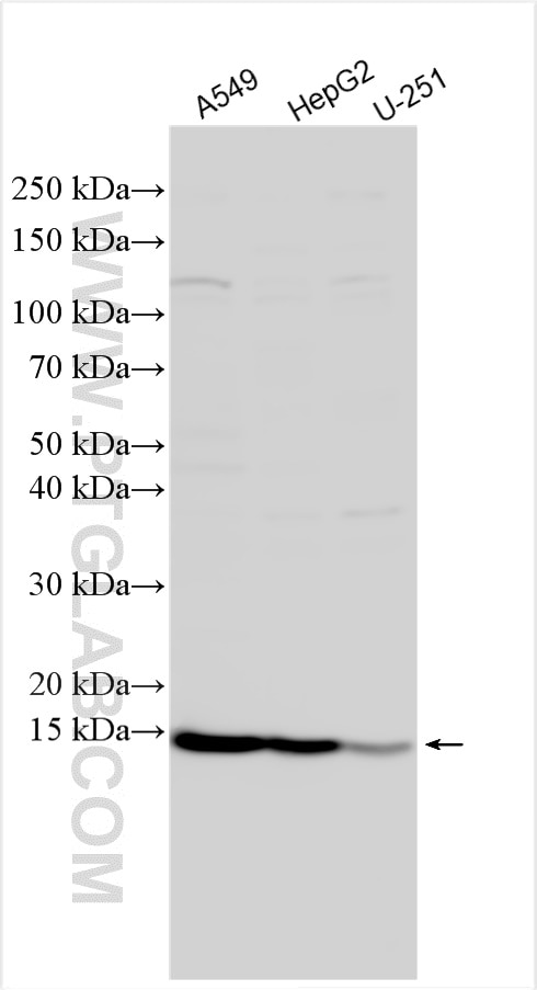
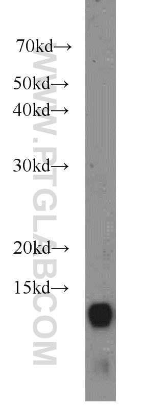
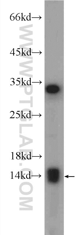
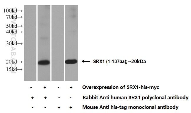
| Résultats positifs en WB | cellules A549, cellules HEK-293 transfectées, cellules HepG2, cellules U-251, tissu hépatique de souris |
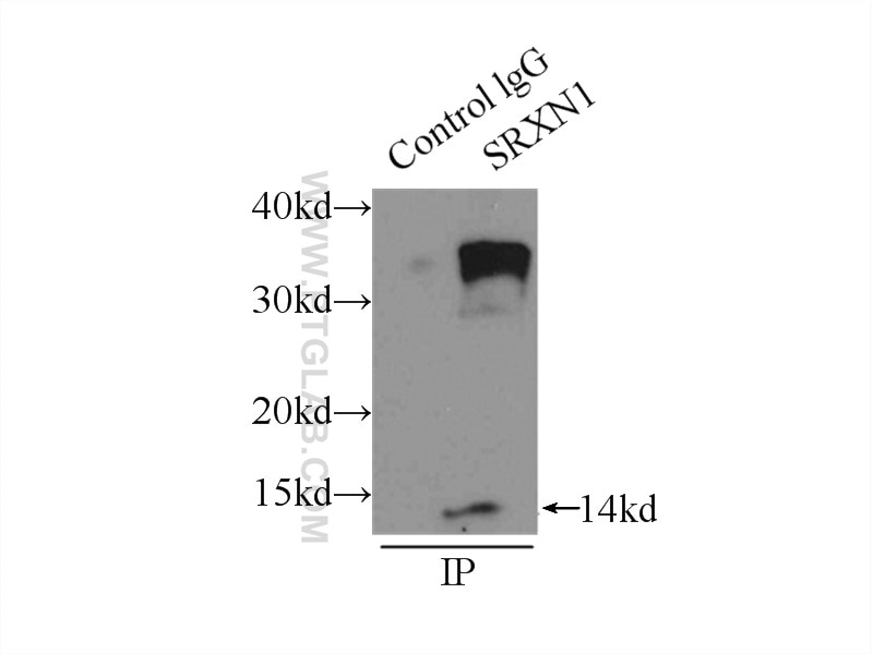
| Résultats positifs en IP | cellules A549, |
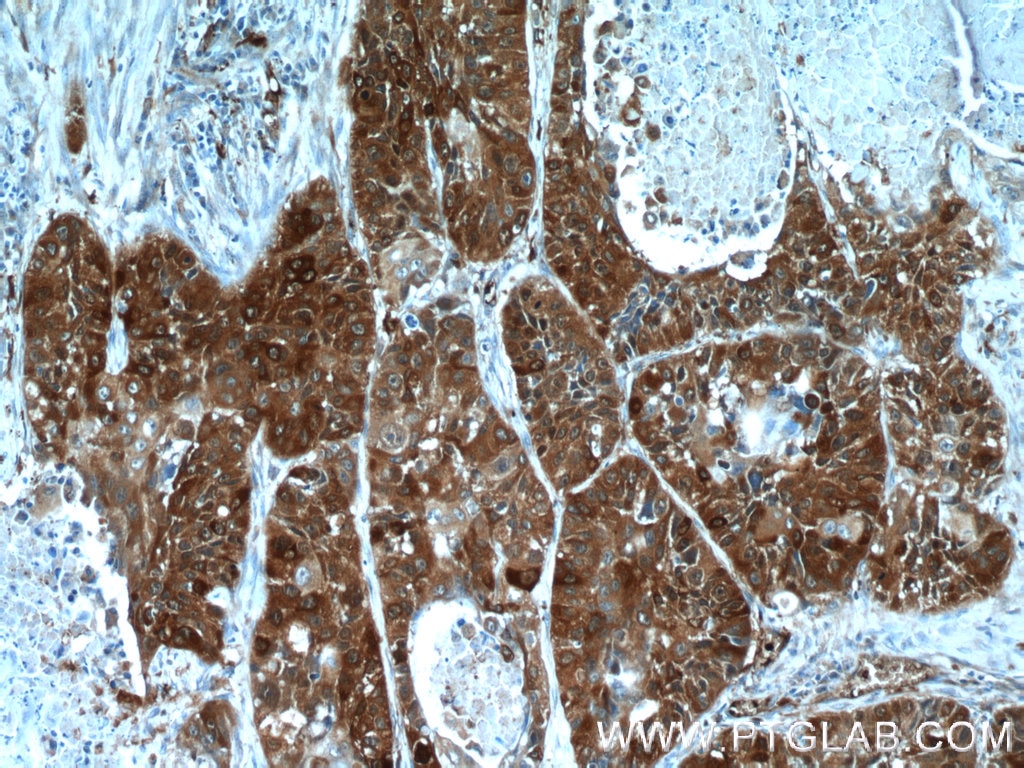
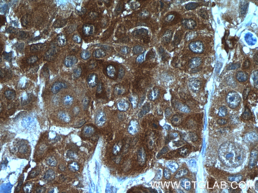
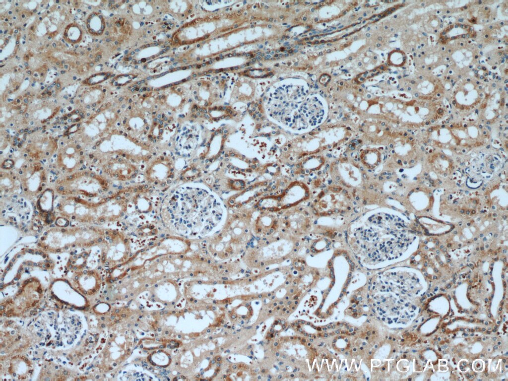
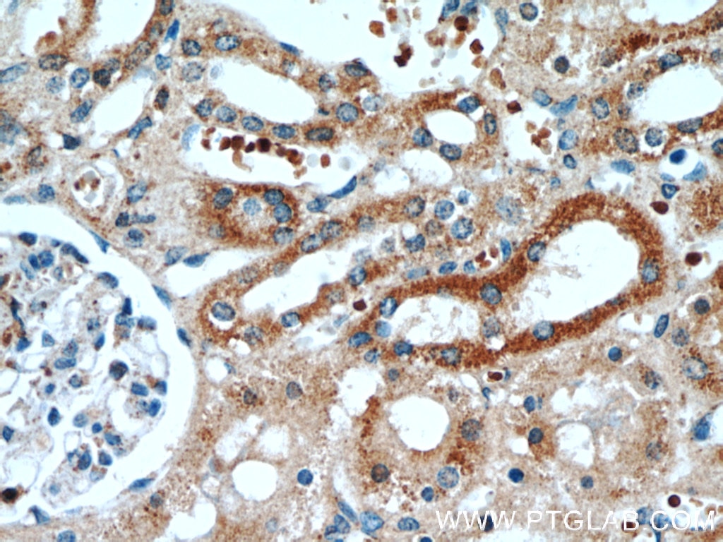
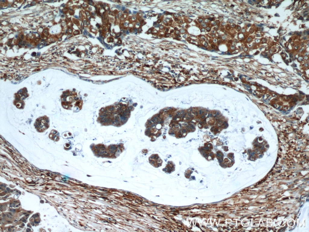
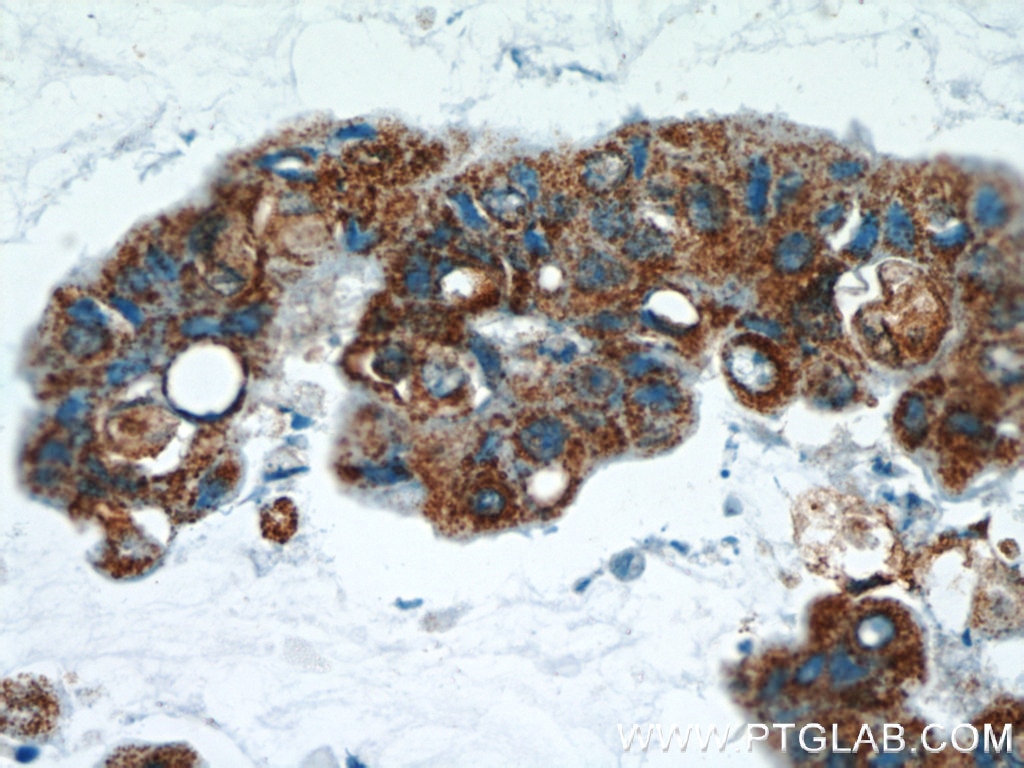
| Résultats positifs en IHC | tissu de cancer du poumon humain, tissu de cancer du sein humain, tissu rénal humain il est suggéré de démasquer l'antigène avec un tampon de TE buffer pH 9.0; (*) À défaut, 'le démasquage de l'antigène peut être 'effectué avec un tampon citrate pH 6,0. |
Dilution recommandée
| Application | Dilution |
|---|---|
| Western Blot (WB) | WB : 1:500-1:3000 |
| Immunoprécipitation (IP) | IP : 0.5-4.0 ug for 1.0-3.0 mg of total protein lysate |
| Immunohistochimie (IHC) | IHC : 1:20-1:200 |
| It is recommended that this reagent should be titrated in each testing system to obtain optimal results. | |
| Sample-dependent, check data in validation data gallery | |
Applications publiées
| KD/KO | See 6 publications below |
| WB | See 25 publications below |
| IHC | See 13 publications below |
| IF | See 2 publications below |
Informations sur le produit
14273-1-AP cible SRX1 dans les applications de WB, IP, IF, IHC, ELISA et montre une réactivité avec des échantillons Humain, souris
| Réactivité | Humain, souris |
| Réactivité citée | rat, Humain, souris |
| Hôte / Isotype | Lapin / IgG |
| Clonalité | Polyclonal |
| Type | Anticorps |
| Immunogène | SRX1 Protéine recombinante Ag5613 |
| Nom complet | sulfiredoxin 1 homolog (S. cerevisiae) |
| Masse moléculaire calculée | 14 kDa |
| Poids moléculaire observé | 14 kDa |
| Numéro d’acquisition GenBank | BC047707 |
| Symbole du gène | SRXN1 |
| Identification du gène (NCBI) | 140809 |
| Conjugaison | Non conjugué |
| Forme | Liquide |
| Méthode de purification | Purification par affinité contre l'antigène |
| Tampon de stockage | PBS avec azoture de sodium à 0,02 % et glycérol à 50 % pH 7,3 |
| Conditions de stockage | Stocker à -20°C. Stable pendant un an après l'expédition. L'aliquotage n'est pas nécessaire pour le stockage à -20oC Les 20ul contiennent 0,1% de BSA. |
Protocole
| Product Specific Protocols | |
|---|---|
| WB protocol for SRX1 antibody 14273-1-AP | Download protocol |
| IHC protocol for SRX1 antibody 14273-1-AP | Download protocol |
| IP protocol for SRX1 antibody 14273-1-AP | Download protocol |
| Standard Protocols | |
|---|---|
| Click here to view our Standard Protocols |
Publications
| Species | Application | Title |
|---|---|---|
Nat Commun Loss of peroxiredoxin-2 exacerbates eccentric contraction-induced force loss in dystrophin-deficient muscle. | ||
Proc Natl Acad Sci U S A Sulfiredoxin-Peroxiredoxin IV axis promotes human lung cancer progression through modulation of specific phosphokinase signaling. | ||
Cancer Res Genetic polymorphisms and protein expression of NRF2 and Sulfiredoxin predict survival outcomes in breast cancer. | ||
Cancer Lett Nrf2-activated expression of sulfiredoxin contributes to urethane-induced lung tumorigenesis.
| ||
Carcinogenesis Tumor Promoter-induced Sulfiredoxin Is Required for Mouse Skin Tumorigenesis. |
Avis
The reviews below have been submitted by verified Proteintech customers who received an incentive forproviding their feedback.
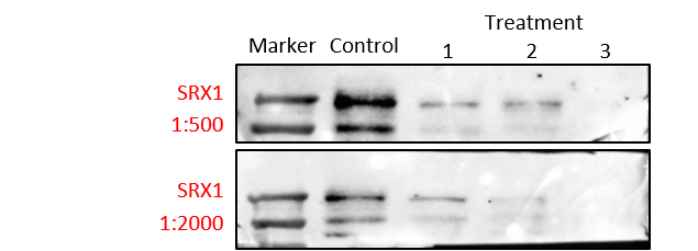
FH Shinford (Verified Customer) (09-23-2023) | Detecting lower molecular weight molecules in gels is relatively difficult, we tried several antibodies from different companies and this worked well at a concentration of 1:500.
 |
FH Christopher (Verified Customer) (12-03-2018) | Primary neurons were lysed in RIPA and 20ug samples (containing 1x Laemmli) were run on Bis-Tris gels in 1xMOPS buffer. This antibody recognised SRXN-1 in HEK293FT cells transfected with Human SRXN1 at 17kDa. In HEK cells, non specific bands were visible at around 28 and 35kDa as well.In primary neurons, the antibody recognised a band at roughly 15kDa however it binds non sepcifically to the Pageruler Prestained protein ladder which is an issue.
|
