- Phare
- Validé par KD/KO
Anticorps Polyclonal de lapin anti-SPRY2
SPRY2 Polyclonal Antibody for WB, IF, IHC, ELISA
Hôte / Isotype
Lapin / IgG
Réactivité testée
Humain, rat, souris
Applications
WB, IHC, IF/ICC, ELISA
Conjugaison
Non conjugué
N° de cat : 11383-1-AP
Synonymes
Galerie de données de validation
Applications testées
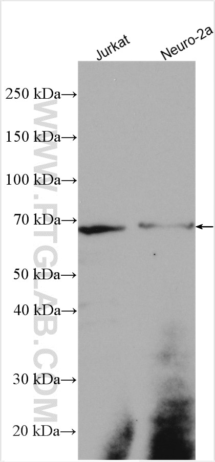
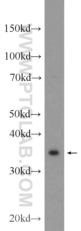
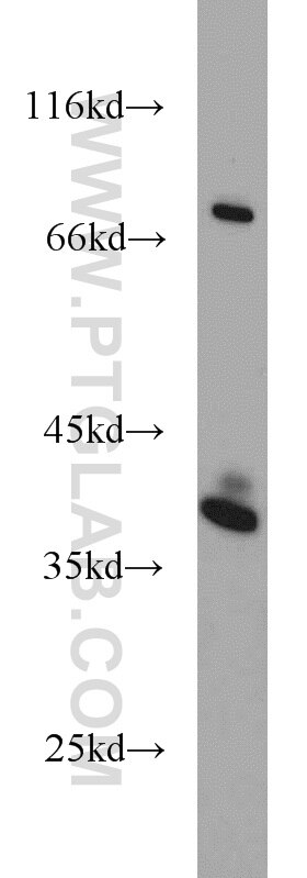
| Résultats positifs en WB | cellules SW-1990, cellules A375, cellules Jurkat, cellules Neuro-2a |
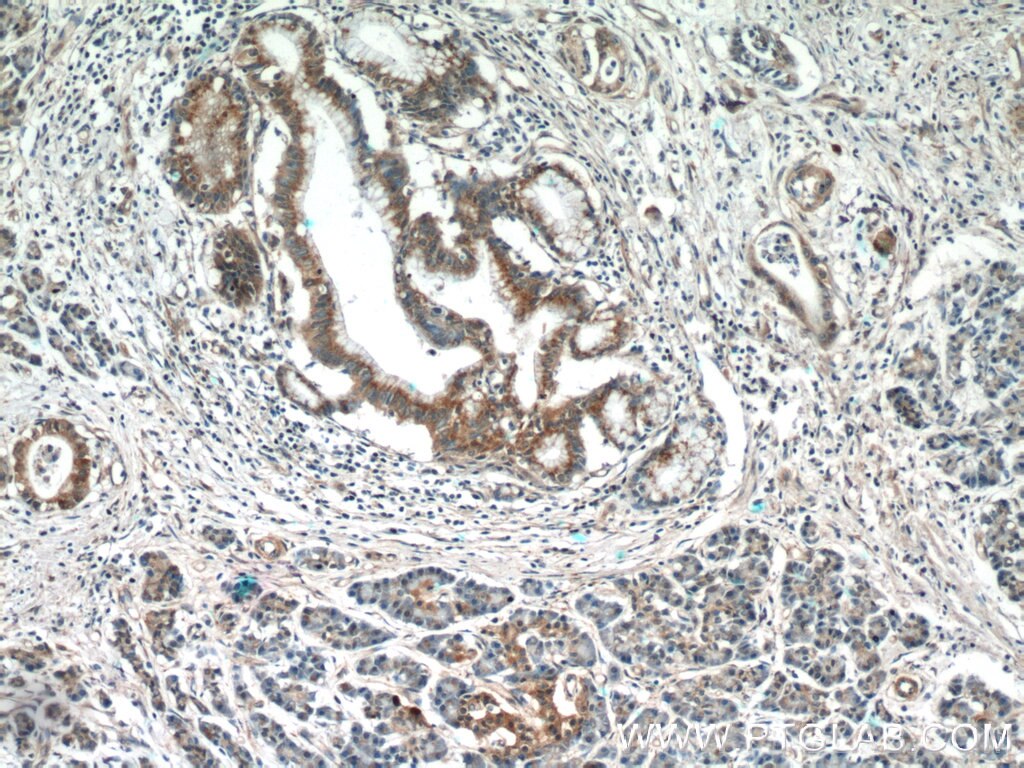
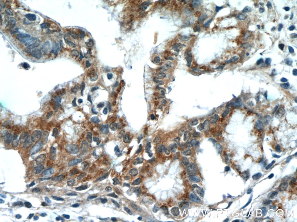
| Résultats positifs en IHC | tissu de cancer du pancréas humain, il est suggéré de démasquer l'antigène avec un tampon de TE buffer pH 9.0; (*) À défaut, 'le démasquage de l'antigène peut être 'effectué avec un tampon citrate pH 6,0. |
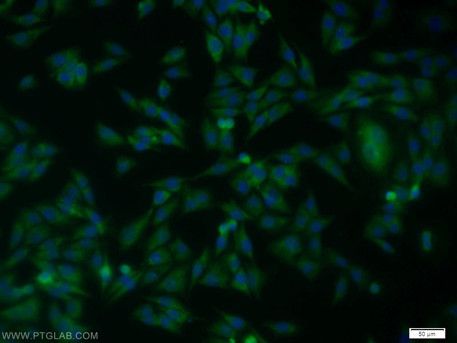
| Résultats positifs en IF/ICC | cellules A375 |
Dilution recommandée
| Application | Dilution |
|---|---|
| Western Blot (WB) | WB : 1:500-1:1000 |
| Immunohistochimie (IHC) | IHC : 1:50-1:500 |
| Immunofluorescence (IF)/ICC | IF/ICC : 1:10-1:100 |
| It is recommended that this reagent should be titrated in each testing system to obtain optimal results. | |
| Sample-dependent, check data in validation data gallery | |
Applications publiées
| KD/KO | See 2 publications below |
| WB | See 6 publications below |
| IHC | See 1 publications below |
| IF | See 1 publications below |
Informations sur le produit
11383-1-AP cible SPRY2 dans les applications de WB, IHC, IF/ICC, ELISA et montre une réactivité avec des échantillons Humain, rat, souris
| Réactivité | Humain, rat, souris |
| Réactivité citée | Humain, souris |
| Hôte / Isotype | Lapin / IgG |
| Clonalité | Polyclonal |
| Type | Anticorps |
| Immunogène | SPRY2 Protéine recombinante Ag1918 |
| Nom complet | sprouty homolog 2 (Drosophila) |
| Masse moléculaire calculée | 35 kDa |
| Poids moléculaire observé | 35-40 kDa, 60~65 kDa |
| Numéro d’acquisition GenBank | BC015745 |
| Symbole du gène | SPRY2 |
| Identification du gène (NCBI) | 10253 |
| Conjugaison | Non conjugué |
| Forme | Liquide |
| Méthode de purification | Purification par affinité contre l'antigène |
| Tampon de stockage | PBS avec azoture de sodium à 0,02 % et glycérol à 50 % pH 7,3 |
| Conditions de stockage | Stocker à -20°C. Stable pendant un an après l'expédition. L'aliquotage n'est pas nécessaire pour le stockage à -20oC Les 20ul contiennent 0,1% de BSA. |
Informations générales
Sprouty(SPRY) genes encode intracellular inhibitors of receptor tyrosine kinase (RTK) signaling pathways, including those triggered by fibroblast growth factors (Fgfs). In addition to its well-reported function as an inhibitor of the RAS/RAF/MEK signalling cascade, SPRY2 exerts its inhibitory role in the PI3K/AKT signalling pathway through PTEN, and inhibits ERK/MAPK activity via EGFR trafficking. SPRY2 was detected 60-65 kDa (PMID:29291435).
Protocole
| Product Specific Protocols | |
|---|---|
| WB protocol for SPRY2 antibody 11383-1-AP | Download protocol |
| IHC protocol for SPRY2 antibody 11383-1-AP | Download protocol |
| IF protocol for SPRY2 antibody 11383-1-AP | Download protocol |
| Standard Protocols | |
|---|---|
| Click here to view our Standard Protocols |
Publications
| Species | Application | Title |
|---|---|---|
Int J Mol Sci Kub3 Deficiency Causes Aberrant Late Embryonic Lung Development in Mice by the FGF Signaling Pathway. | ||
J Cell Mol Med Exosomal miR-27 negatively regulates ROS production and promotes granulosa cells apoptosis by targeting SPRY2 in OHSS.
| ||
Toxicol Appl Pharmacol Hexachlorophene, a selective SHP2 inhibitor, suppresses proliferation and metastasis of KRAS-mutant NSCLC cells by inhibiting RAS/MEK/ERK and PI3K/AKT signaling pathways. | ||
Cell Biol Int Sprouty2 is involved in the control of osteoblast proliferation and differentiation through the FGF and BMP signaling pathways. | ||
Breast Cancer Res Loss of SPRY2 contributes to cancer-associated fibroblasts activation and promotes breast cancer development
| ||
Cancer Cell Int Therapeutic effect and transcriptome-methylome characteristics of METTL3 inhibition in liver hepatocellular carcinoma |
Avis
The reviews below have been submitted by verified Proteintech customers who received an incentive forproviding their feedback.
FH Christine (Verified Customer) (01-27-2023) | Detects two bands on HEK293T lysates. main one between 36 and 48 kDa, second one between 48 and 85 kDa, in accordance with datasheet
|
FH Silvia (Verified Customer) (01-31-2022) | The antibody worked really well and gave me a clear band on Western Blot around 50 kDa.
|
