- Phare
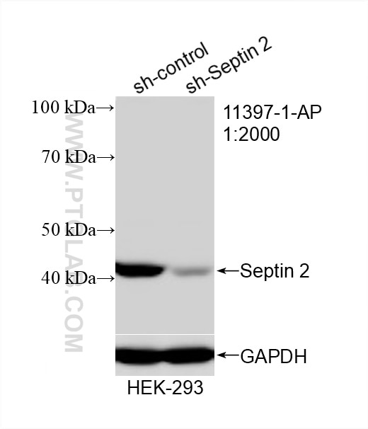
- Validé par KD/KO
Anticorps Polyclonal de lapin anti-Septin 2
Septin 2 Polyclonal Antibody for WB, IHC, IF/ICC, FC (Intra), IP, ELISA
Hôte / Isotype
Lapin / IgG
Réactivité testée
Humain, rat, souris
Applications
WB, IHC, IF/ICC, FC (Intra), IP, ELISA
Conjugaison
Non conjugué
N° de cat : 11397-1-AP
Synonymes
Galerie de données de validation
Applications testées
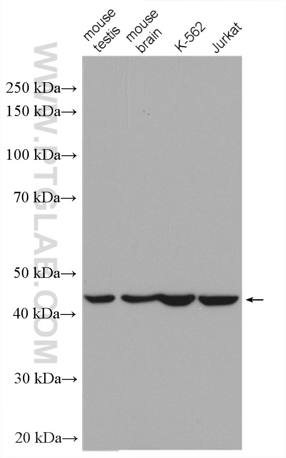
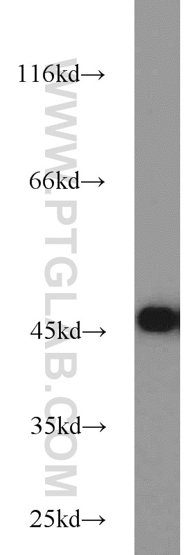
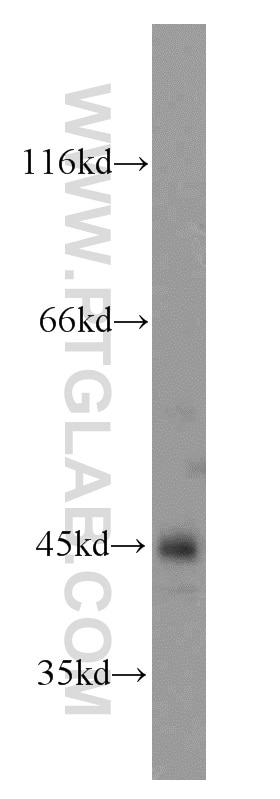
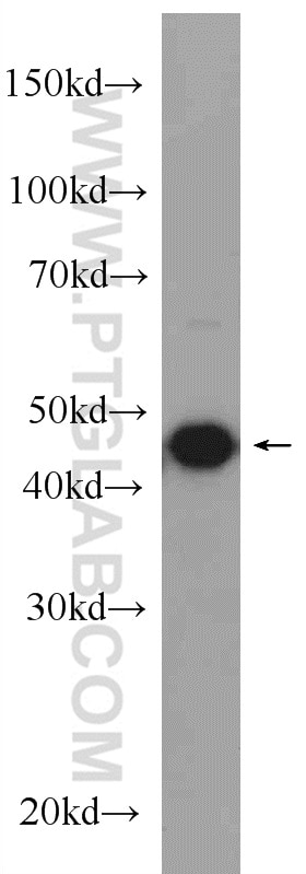
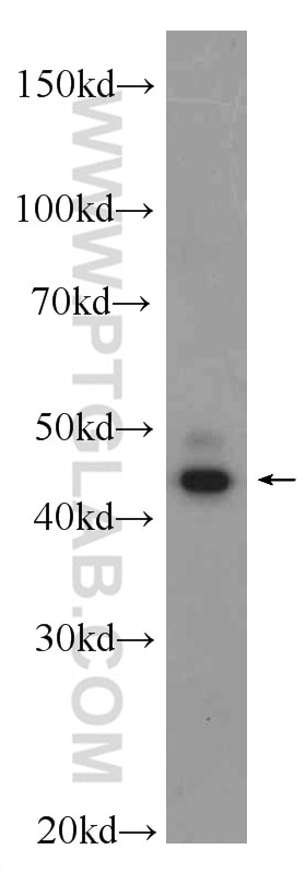
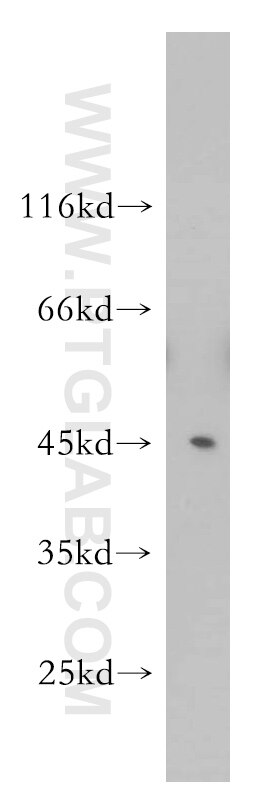
| Résultats positifs en WB | tissu cérébral de souris, cellules C6, cellules HEK-293, cellules HeLa, cellules Jurkat, cellules K-562, tissu cardiaque de souris, tissu cérébral de rat, tissu de muscle squelettique humain |
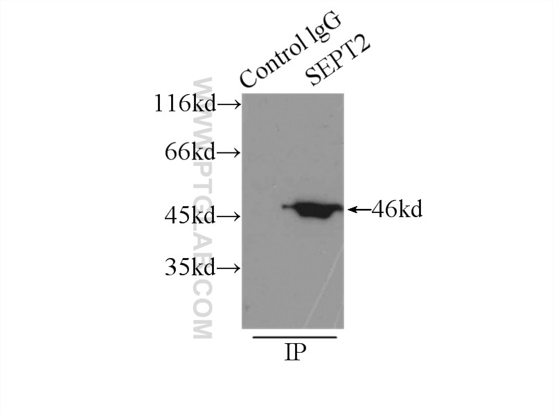
| Résultats positifs en IP | tissu cérébral de souris |
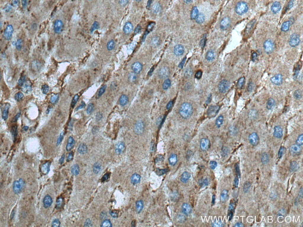
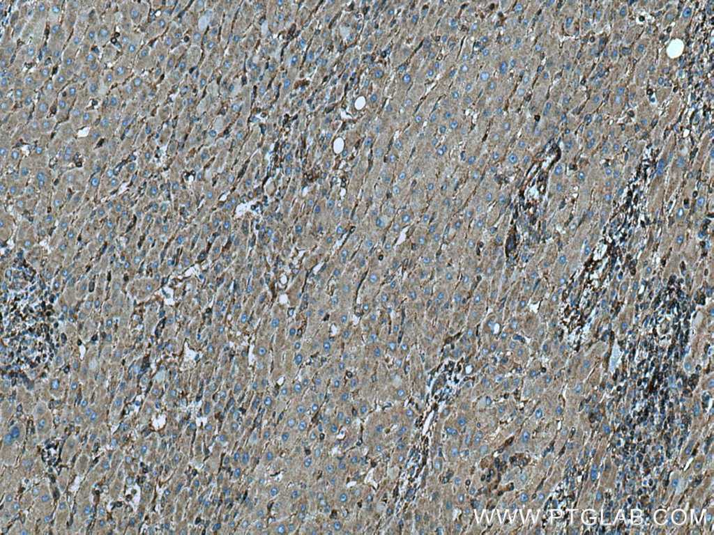
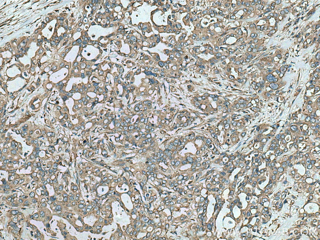
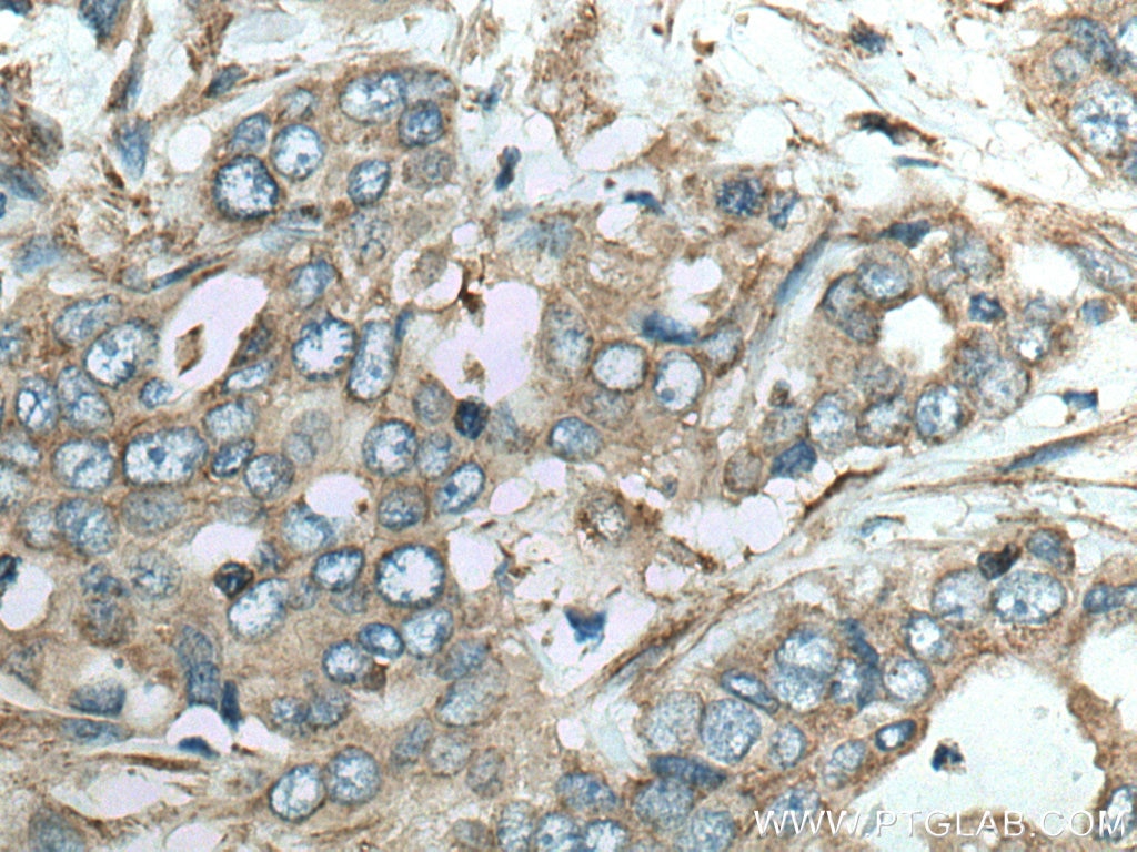
| Résultats positifs en IHC | tissu de cancer du foie humain, tissu de cancer du pancréas humain il est suggéré de démasquer l'antigène avec un tampon de TE buffer pH 9.0; (*) À défaut, 'le démasquage de l'antigène peut être 'effectué avec un tampon citrate pH 6,0. |
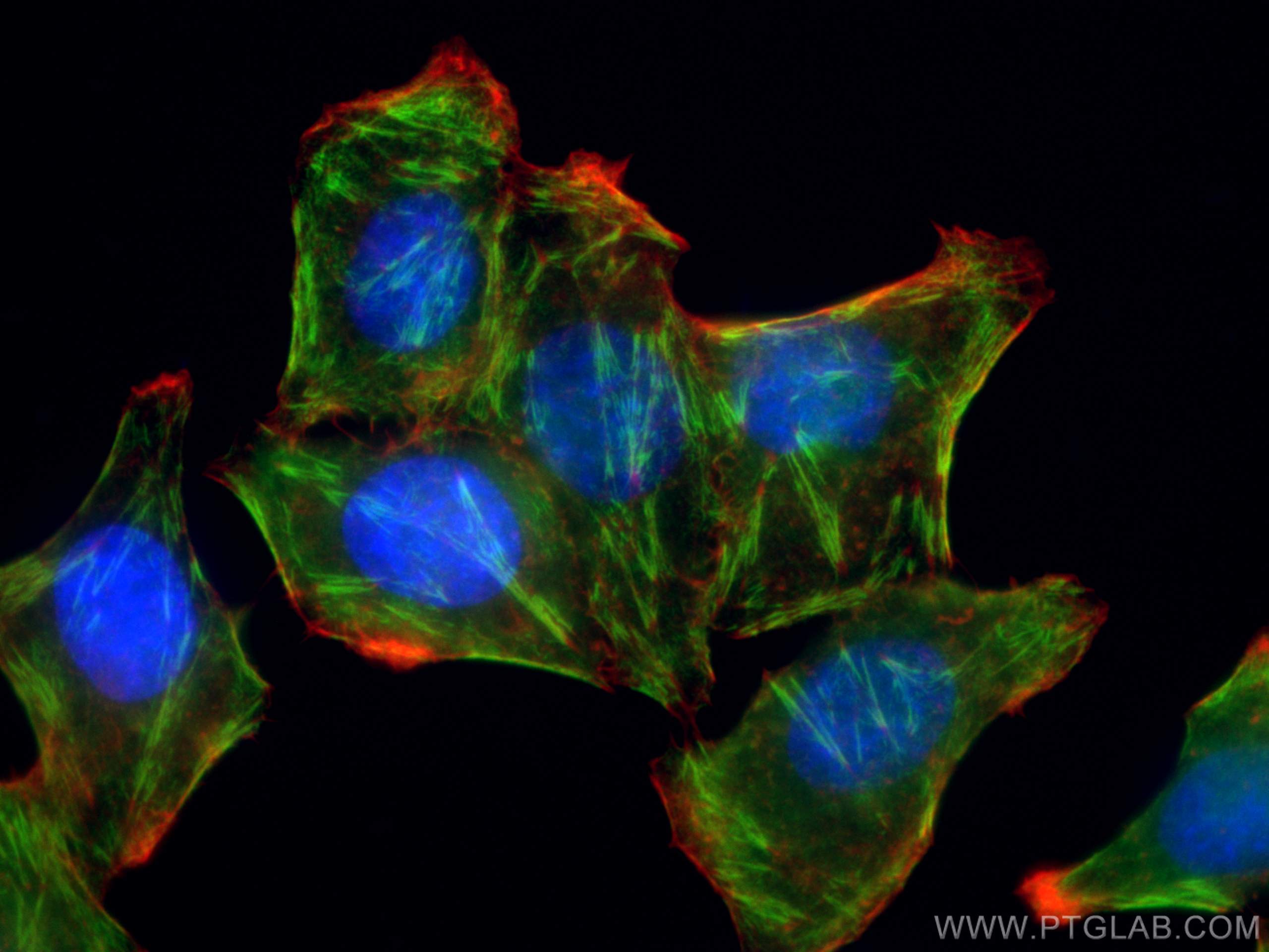
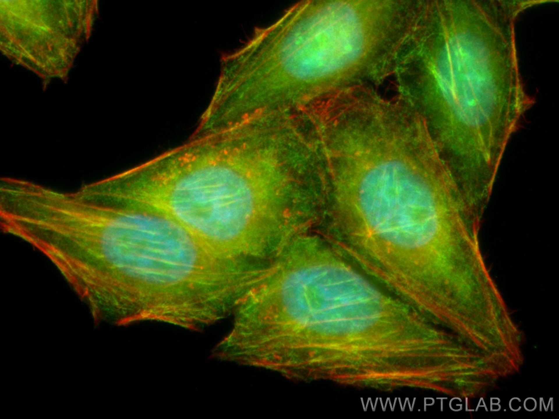
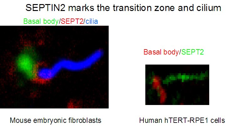
| Résultats positifs en IF/ICC | cellules HepG2, cellules hTERT-RPE1 et fibroblastes embryonnaires de souris |
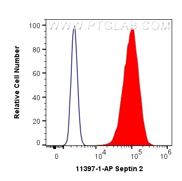
| Résultats positifs en FC (Intra) | cellules MCF-7, |
Dilution recommandée
| Application | Dilution |
|---|---|
| Western Blot (WB) | WB : 1:1000-1:4000 |
| Immunoprécipitation (IP) | IP : 0.5-4.0 ug for 1.0-3.0 mg of total protein lysate |
| Immunohistochimie (IHC) | IHC : 1:50-1:500 |
| Immunofluorescence (IF)/ICC | IF/ICC : 1:50-1:500 |
| Flow Cytometry (FC) (INTRA) | FC (INTRA) : 0.40 ug per 10^6 cells in a 100 µl suspension |
| It is recommended that this reagent should be titrated in each testing system to obtain optimal results. | |
| Sample-dependent, check data in validation data gallery | |
Applications publiées
| KD/KO | See 1 publications below |
| WB | See 16 publications below |
| IF | See 13 publications below |
| IP | See 1 publications below |
Informations sur le produit
11397-1-AP cible Septin 2 dans les applications de WB, IHC, IF/ICC, FC (Intra), IP, ELISA et montre une réactivité avec des échantillons Humain, rat, souris
| Réactivité | Humain, rat, souris |
| Réactivité citée | rat, Humain, souris |
| Hôte / Isotype | Lapin / IgG |
| Clonalité | Polyclonal |
| Type | Anticorps |
| Immunogène | Septin 2 Protéine recombinante Ag1946 |
| Nom complet | septin 2 |
| Masse moléculaire calculée | 41 kDa |
| Poids moléculaire observé | 45 kDa |
| Numéro d’acquisition GenBank | BC014455 |
| Symbole du gène | Septin 2 |
| Identification du gène (NCBI) | 4735 |
| Conjugaison | Non conjugué |
| Forme | Liquide |
| Méthode de purification | Purification par affinité contre l'antigène |
| Tampon de stockage | PBS avec azoture de sodium à 0,02 % et glycérol à 50 % pH 7,3 |
| Conditions de stockage | Stocker à -20°C. Stable pendant un an après l'expédition. L'aliquotage n'est pas nécessaire pour le stockage à -20oC Les 20ul contiennent 0,1% de BSA. |
Informations générales
SEPT2 belongs to a family of septins, GTP-binding proteins that are a component of the cytoskeleton. Septins can assemble into intracellular structures of various shapes and are known to influence membrane stability, forming diffusion barriers and serving as scaffolds.
What is the molecular weight of SEPT2? Is SEPT2 post-translationally modified?
The molecular weight of SEPT2 is 45 kDa. In a cellular context, SEPT2 forms hetero-hexamers and hetero-octamers with other septins. These hexamers and octamers further assemble into high-order structures such as filaments and rings. SEPT2 can be phosphorylated on Ser218 by casein kinase 2 (PMID: 19165576) and can also be SUMOylated (PMID: 29051266).
What is the subcellular localization of SEPT2?
SEPT2 forms a diffusion barrier and localizes to the primary cilium; in some cell types, it has been found at the transition zone/basal body (PMID: 20558667), while in others localized to the axoneme (PMID: 23572511). During cell division, SEPT2 accumulates near the contractile ring from anaphase through telophase, and finally condenses into the midbody (PMID: 9203580). In interphase cells, SEPT2 was found to associate with polyglutamylated microtubules (PMID: 18209106) and can affect microtubule dynamics via interaction with MAP4.
What is the tissue expression pattern of SEPT2?
While some septins have tissue-specific expression profiles, SEPT2 is ubiquitously expressed (PMID: 22314400).
Protocole
| Product Specific Protocols | |
|---|---|
| WB protocol for Septin 2 antibody 11397-1-AP | Download protocol |
| IHC protocol for Septin 2 antibody 11397-1-AP | Download protocol |
| IF protocol for Septin 2 antibody 11397-1-AP | Download protocol |
| IP protocol for Septin 2 antibody 11397-1-AP | Download protocol |
| Standard Protocols | |
|---|---|
| Click here to view our Standard Protocols |
Publications
| Species | Application | Title |
|---|---|---|
Nat Commun Wiskott-Aldrich syndrome protein regulates autophagy and inflammasome activity in innate immune cells. | ||
PLoS Genet Genetic deletion of SEPT7 reveals a cell type-specific role of septins in microtubule destabilization for the completion of cytokinesis. | ||
Cell Death Dis JMJD2C-mediated long non-coding RNA MALAT1/microRNA-503-5p/SEPT2 axis worsens non-small cell lung cancer. | ||
Elife Septin/anillin filaments scaffold central nervous system myelin to accelerate nerve conduction. | ||
PLoS Genet SEPT12 phosphorylation results in loss of the septin ring/sperm annulus, defective sperm motility and poor male fertility. |
Avis
The reviews below have been submitted by verified Proteintech customers who received an incentive forproviding their feedback.
FH Christine (Verified Customer) (01-27-2023) | Incubation for a few hours at room temperature on HEK293T lysates. Detection of a very strong signal just below the 47 kDa marker (expected size of Septin 2) and a fainter signal just above the 47 kDa marker.
|
FH Mohammed (Verified Customer) (08-19-2020) | Some staining at the annulus, there seems to be staining in the acrosome. Overall, really high background (even when the concentration is lowered).
|
