- Phare
- Validé par KD/KO
Anticorps Polyclonal de lapin anti-PKC Alpha
PKC Alpha Polyclonal Antibody for WB, IHC, IF/ICC, FC (Intra), IP, ELISA
Hôte / Isotype
Lapin / IgG
Réactivité testée
Humain, rat, souris et plus (2)
Applications
WB, IHC, IF/ICC, FC (Intra), IP, ELISA
Conjugaison
Non conjugué
N° de cat : 21991-1-AP
Synonymes
Galerie de données de validation
Applications testées
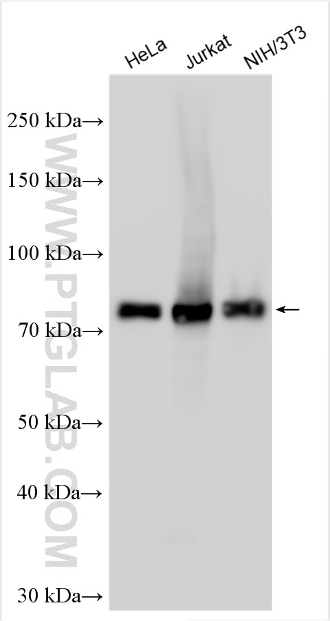
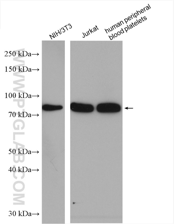
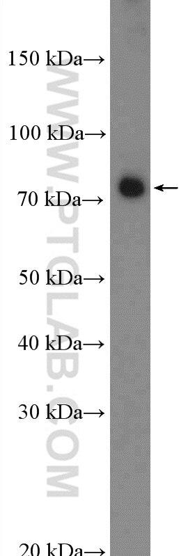
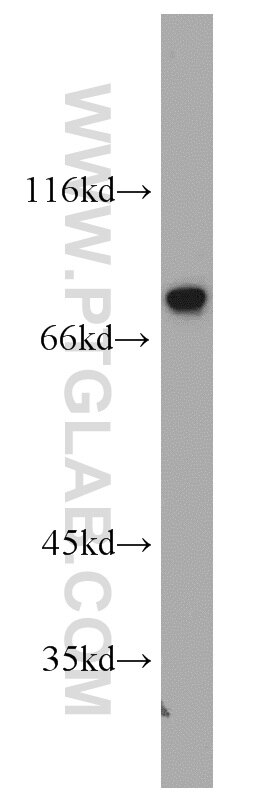
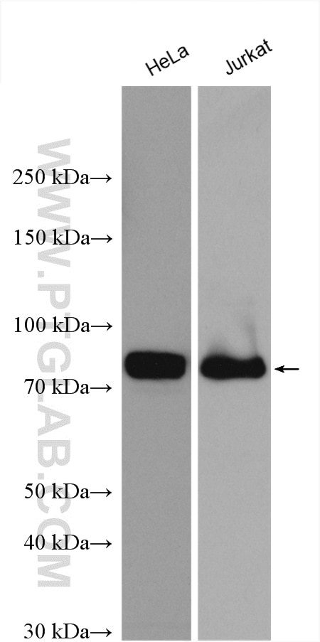
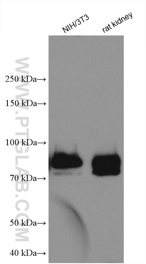
| Résultats positifs en WB | cellules HeLa, cellules HEK-293, cellules Jurkat, cellules NIH/3T3, plaquettes de sang périphérique humain, tissu rénal de rat |
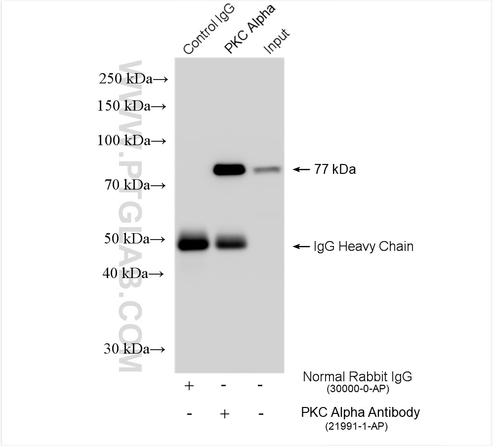
| Résultats positifs en IP | cellules NIH/3T3, |
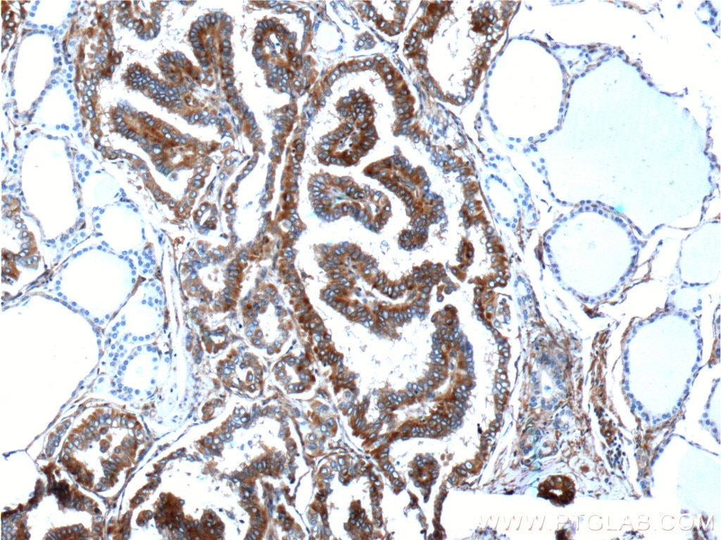
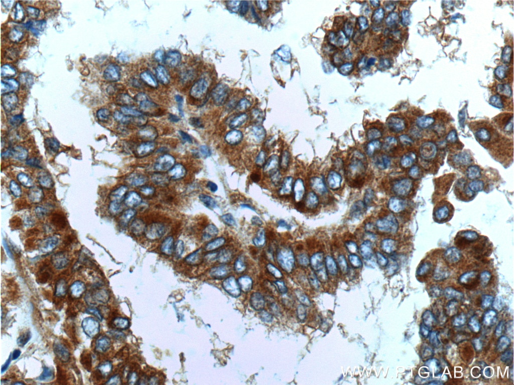
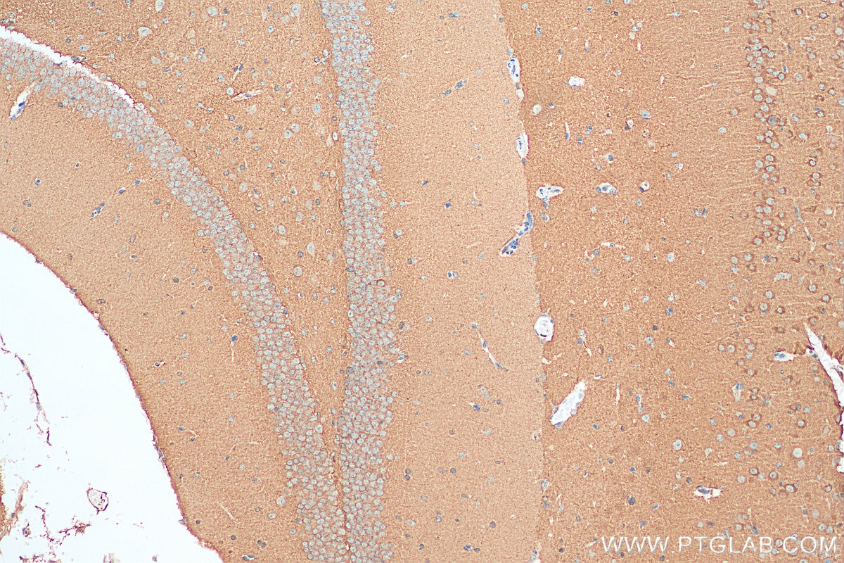
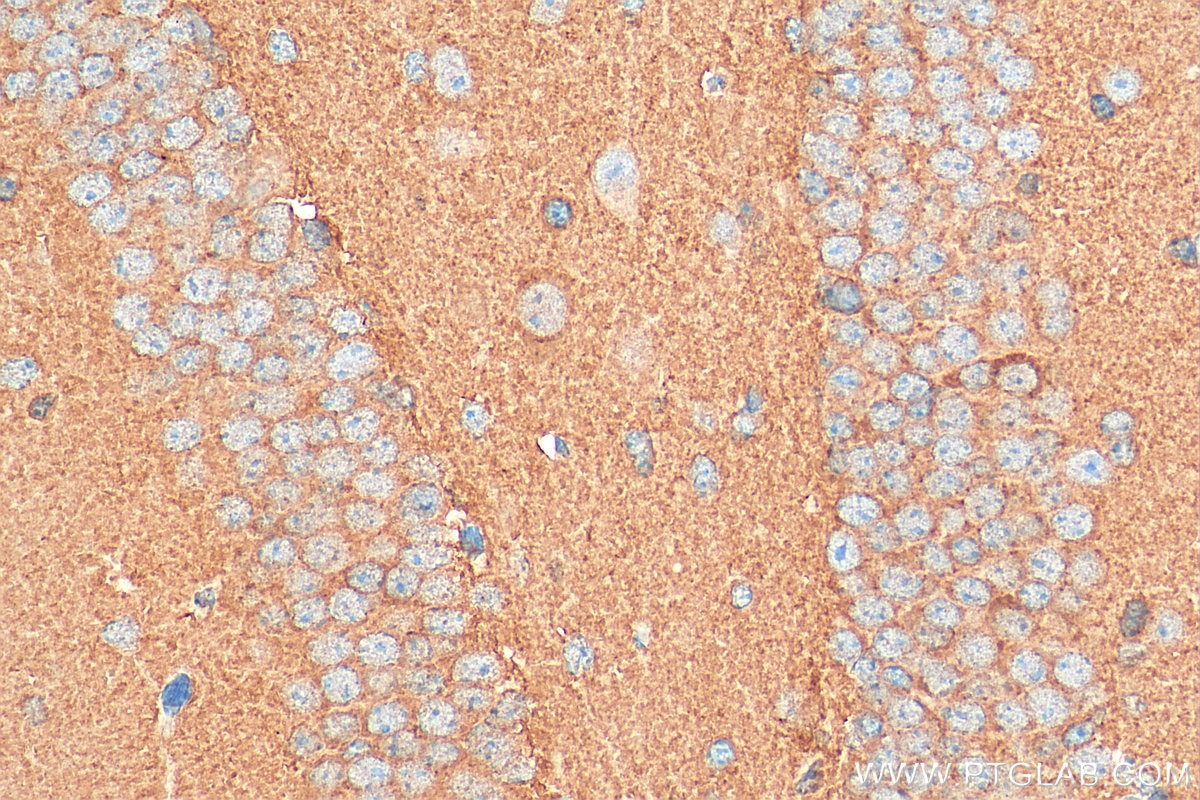
| Résultats positifs en IHC | tissu de cancer de la thyroïde humain, tissu cérébral de souris il est suggéré de démasquer l'antigène avec un tampon de TE buffer pH 9.0; (*) À défaut, 'le démasquage de l'antigène peut être 'effectué avec un tampon citrate pH 6,0. |
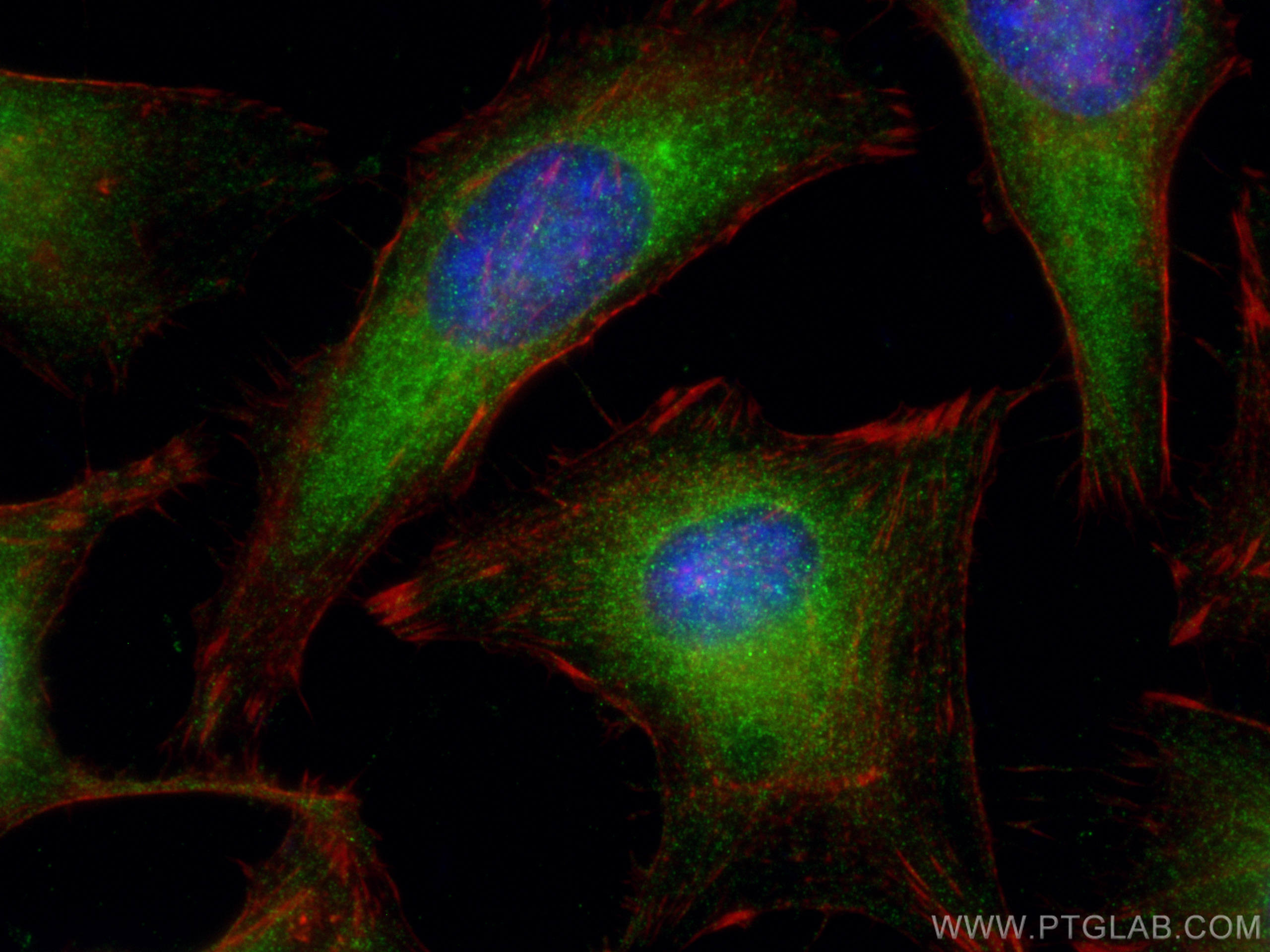
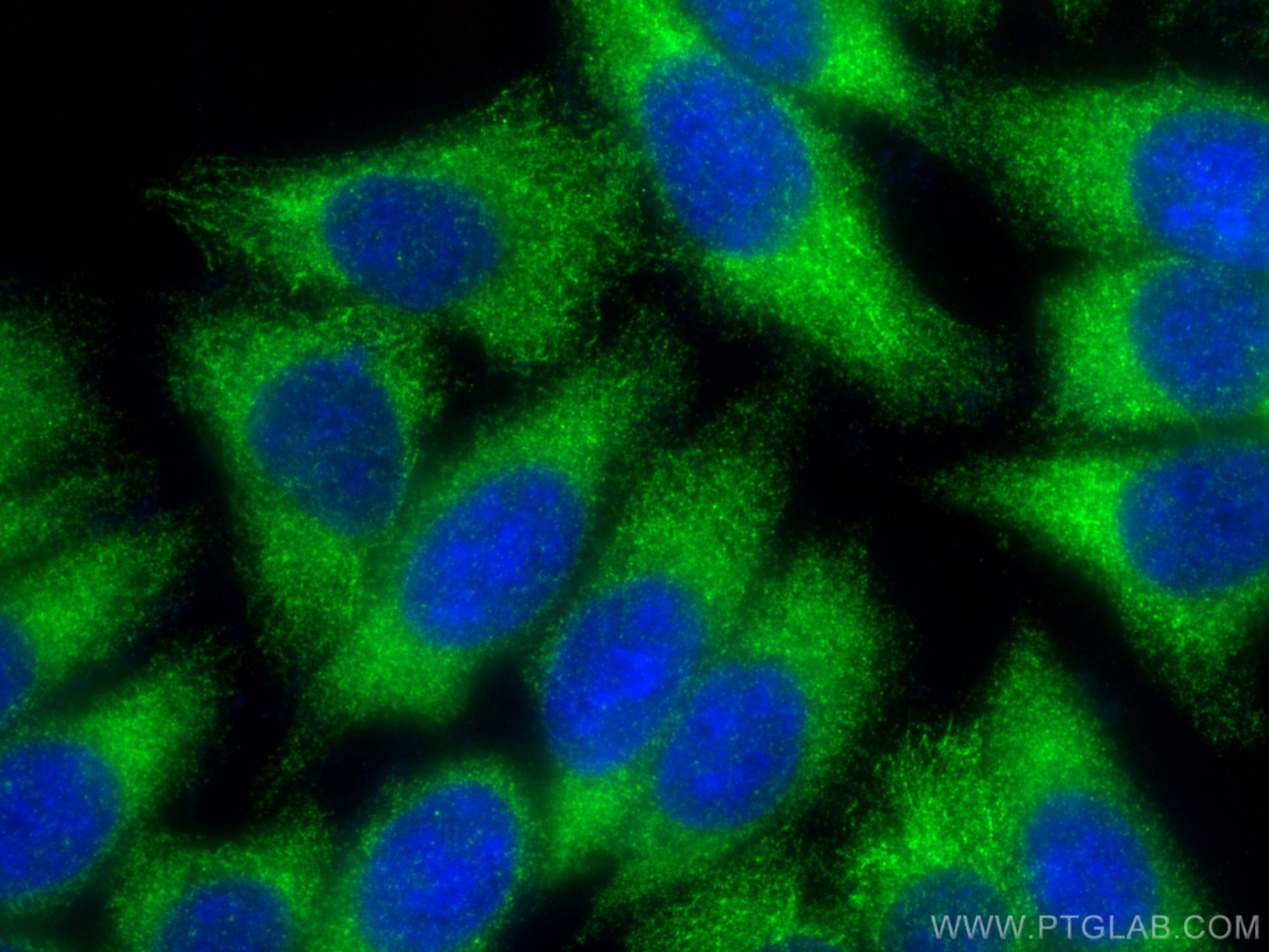
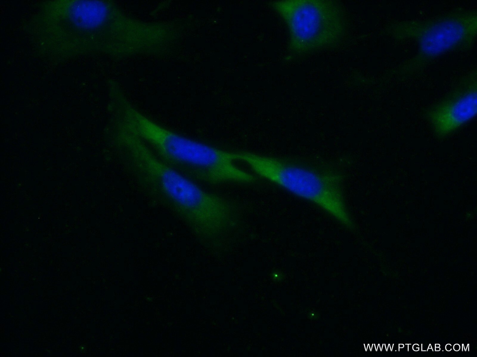
| Résultats positifs en IF/ICC | cellules HeLa, cellules HepG2, cellules NIH/3T3 |
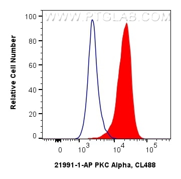
| Résultats positifs en FC (Intra) | cellules HeLa, |
Dilution recommandée
| Application | Dilution |
|---|---|
| Western Blot (WB) | WB : 1:2000-1:12000 |
| Immunoprécipitation (IP) | IP : 0.5-4.0 ug for 1.0-3.0 mg of total protein lysate |
| Immunohistochimie (IHC) | IHC : 1:20-1:200 |
| Immunofluorescence (IF)/ICC | IF/ICC : 1:50-1:500 |
| Flow Cytometry (FC) (INTRA) | FC (INTRA) : 0.40 ug per 10^6 cells in a 100 µl suspension |
| It is recommended that this reagent should be titrated in each testing system to obtain optimal results. | |
| Sample-dependent, check data in validation data gallery | |
Applications publiées
| KD/KO | See 2 publications below |
| WB | See 37 publications below |
| IHC | See 3 publications below |
| IF | See 11 publications below |
Informations sur le produit
21991-1-AP cible PKC Alpha dans les applications de WB, IHC, IF/ICC, FC (Intra), IP, ELISA et montre une réactivité avec des échantillons Humain, rat, souris
| Réactivité | Humain, rat, souris |
| Réactivité citée | rat, Humain, porc, singe, souris |
| Hôte / Isotype | Lapin / IgG |
| Clonalité | Polyclonal |
| Type | Anticorps |
| Immunogène | PKC Alpha Protéine recombinante Ag17275 |
| Nom complet | protein kinase C, alpha |
| Masse moléculaire calculée | 77 kDa |
| Poids moléculaire observé | 77 kDa |
| Numéro d’acquisition GenBank | AK055431 |
| Symbole du gène | PKC Alpha |
| Identification du gène (NCBI) | 5578 |
| Conjugaison | Non conjugué |
| Forme | Liquide |
| Méthode de purification | Purification par affinité contre l'antigène |
| Tampon de stockage | PBS avec azoture de sodium à 0,02 % et glycérol à 50 % pH 7,3 |
| Conditions de stockage | Stocker à -20°C. Stable pendant un an après l'expédition. L'aliquotage n'est pas nécessaire pour le stockage à -20oC Les 20ul contiennent 0,1% de BSA. |
Informations générales
Protein kinase C (PKC) is a family of serine- and threonine-specific protein kinases that can be activated by calcium and the second messenger diacylglycerol. PKC family members phosphorylate a wide variety of protein targets and are known to be involved in diverse cellular signaling pathways. PKC family members also serve as major receptors for phorbol esters, a class of tumor promoters. Each member of the PKC family has a specific expression profile and is believed to play a distinct role in cells. PRKCA is one of the PKC family members. This kinase has been reported to play roles in many different cellular processes, such as cell adhesion, cell transformation, cell cycle checkpoint, and cell volume control. Knockout studies in mice suggest that this kinase may be a fundamental regulator of cardiac contractility and Ca(2+) handling in myocytes.
Protocole
| Product Specific Protocols | |
|---|---|
| WB protocol for PKC Alpha antibody 21991-1-AP | Download protocol |
| IHC protocol for PKC Alpha antibody 21991-1-AP | Download protocol |
| IF protocol for PKC Alpha antibody 21991-1-AP | Download protocol |
| IP protocol for PKC Alpha antibody 21991-1-AP | Download protocol |
| Standard Protocols | |
|---|---|
| Click here to view our Standard Protocols |
Publications
| Species | Application | Title |
|---|---|---|
Theranostics MED13L integrates Mediator-regulated epigenetic control into lung cancer radiosensitivity. | ||
Cell Death Dis ATF4/CEMIP/PKCα promotes anoikis resistance by enhancing protective autophagy in prostate cancer cells. | ||
Cell Death Differ β-Trcp ubiquitin ligase and RSK2 kinase-mediated degradation of FOXN2 promotes tumorigenesis and radioresistance in lung cancer. | ||
Oncogene β-Trcp and CK1δ-mediated degradation of LZTS2 activates PI3K/AKT signaling to drive tumorigenesis and metastasis in hepatocellular carcinoma. | ||
Oncogenesis Blockage of Orai1-Nucleolin interaction meditated calcium influx attenuates breast cancer cells growth | ||
Front Pharmacol Cellular mechanism of action of forsythiaside for the treatment of diabetic kidney disease |
Avis
The reviews below have been submitted by verified Proteintech customers who received an incentive forproviding their feedback.
FH Alessandro (Verified Customer) (11-07-2022) | no unspecific staining, great outcome
|
FH Chiara (Verified Customer) (10-04-2021) | Works very well in Immunoblot
|
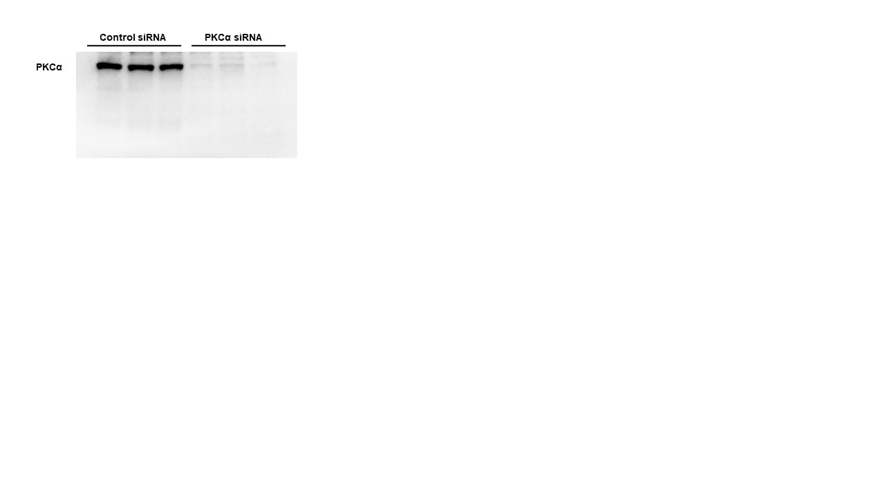
FH Samantha (Verified Customer) (02-24-2020) | Bovine vascular smooth muscle cells were transfected with either control siRNA or PKCalpha siRNA for up to 7 days, with transfections repeated every 48-72 hours. Blocking agent: 5% (w/v) milk in TBST for 1hrPrimary antibody diluted in 5% (w/v) milk in TBST; incubated overnight at 4 degrees.
 |
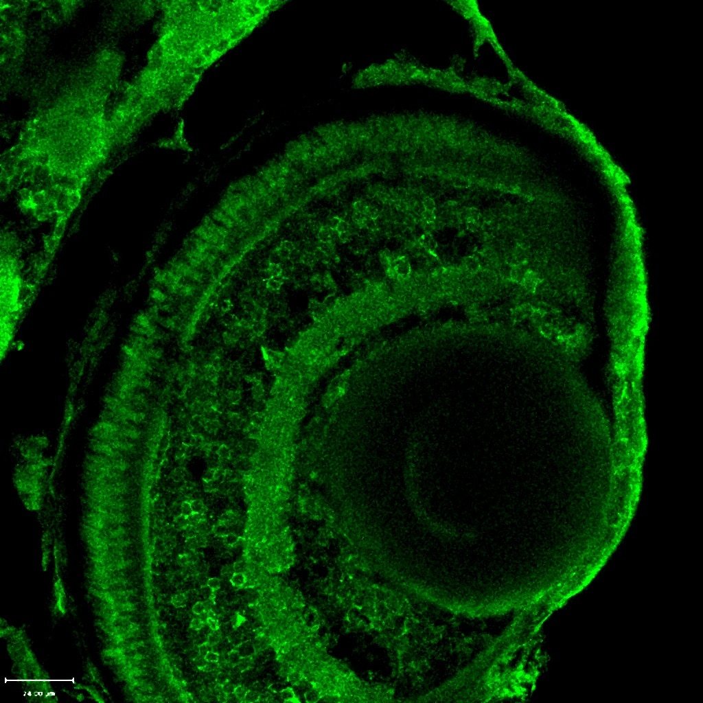
FH Ryan (Verified Customer) (01-10-2018) |
 |
