- Phare
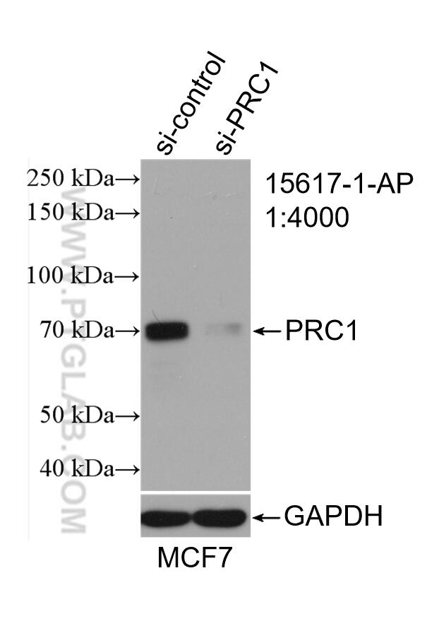
- Validé par KD/KO
Anticorps Polyclonal de lapin anti-PRC1
PRC1 Polyclonal Antibody for WB, IP, IF, IHC, ELISA
Hôte / Isotype
Lapin / IgG
Réactivité testée
Humain, rat et plus (1)
Applications
WB, IHC, IF/ICC, IP, CoIP, ELISA
Conjugaison
Non conjugué
N° de cat : 15617-1-AP
Synonymes
Galerie de données de validation
Applications testées
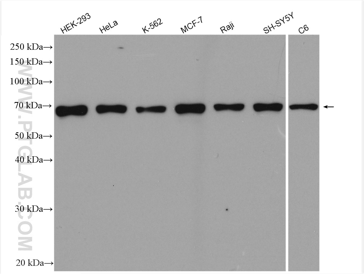
| Résultats positifs en WB | cellules MCF-7, cellules C6, cellules HEK-293, cellules HeLa, cellules K-562, cellules Raji, cellules SH-SY5Y |
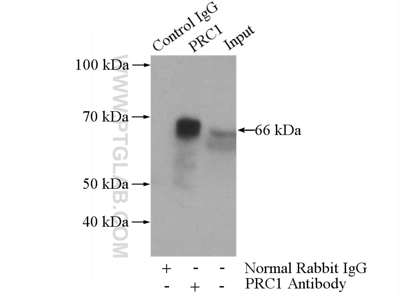
| Résultats positifs en IP | cellules HEK-293 |
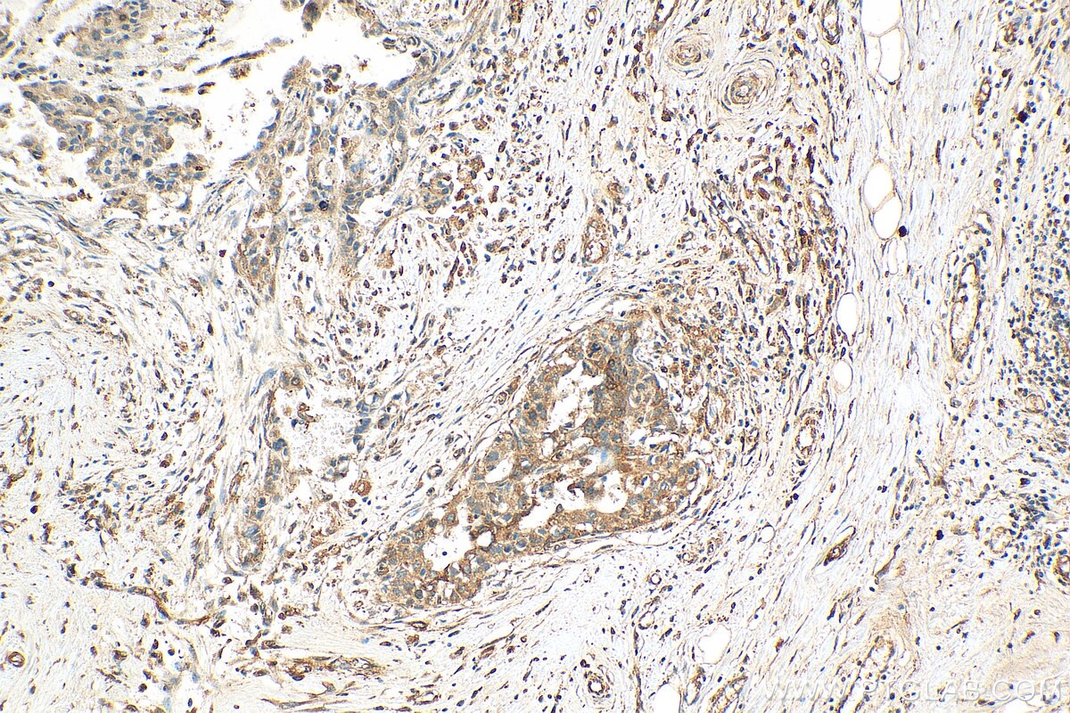
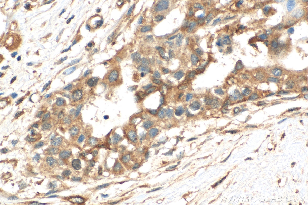
| Résultats positifs en IHC | tissu de cancer du sein humain, il est suggéré de démasquer l'antigène avec un tampon de TE buffer pH 9.0; (*) À défaut, 'le démasquage de l'antigène peut être 'effectué avec un tampon citrate pH 6,0. |
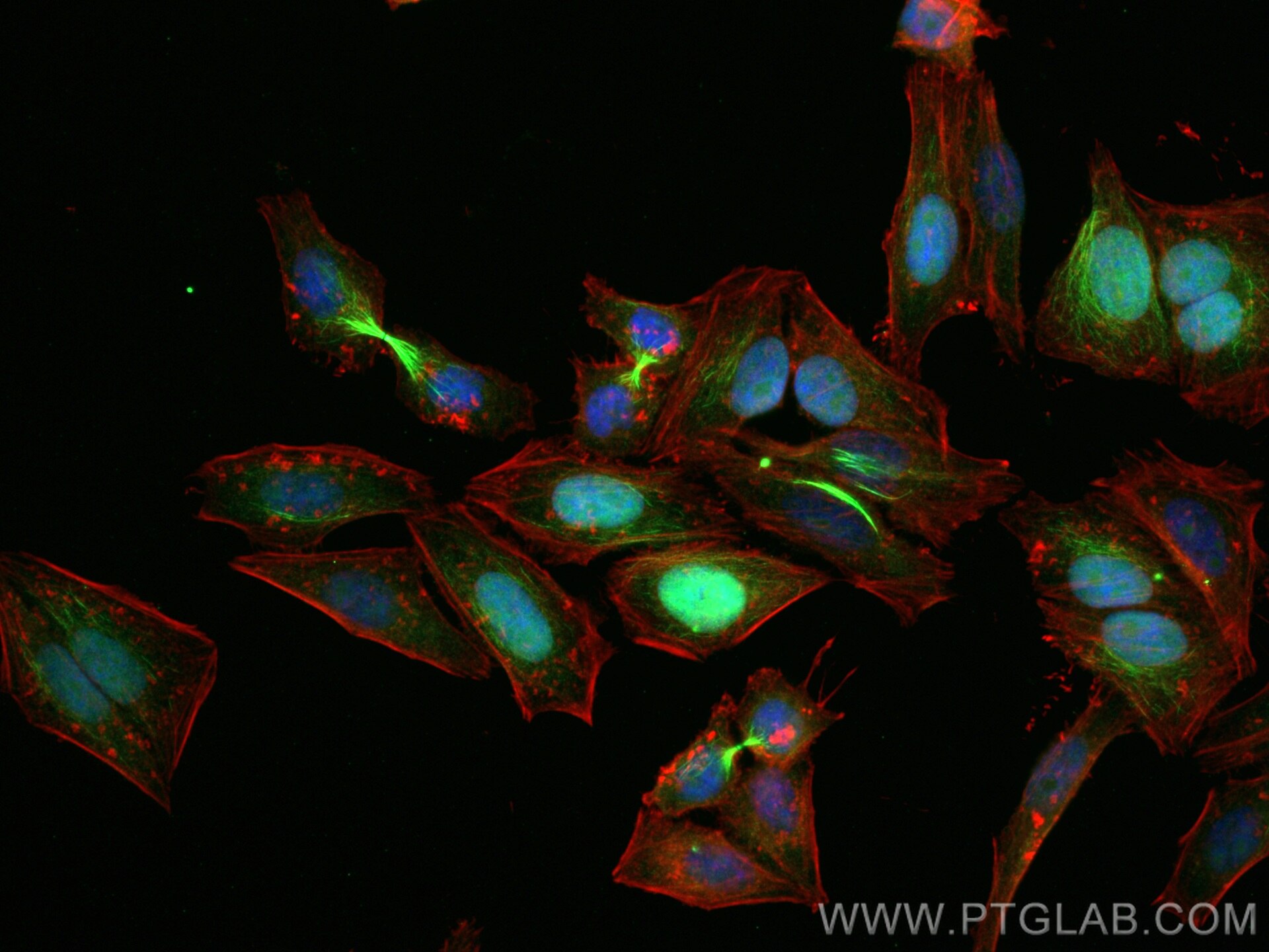
| Résultats positifs en IF/ICC | cellules HepG2, |
Dilution recommandée
| Application | Dilution |
|---|---|
| Western Blot (WB) | WB : 1:1000-1:8000 |
| Immunoprécipitation (IP) | IP : 0.5-4.0 ug for 1.0-3.0 mg of total protein lysate |
| Immunohistochimie (IHC) | IHC : 1:50-1:500 |
| Immunofluorescence (IF)/ICC | IF/ICC : 1:200-1:800 |
| It is recommended that this reagent should be titrated in each testing system to obtain optimal results. | |
| Sample-dependent, check data in validation data gallery | |
Applications publiées
| KD/KO | See 1 publications below |
| WB | See 11 publications below |
| IHC | See 4 publications below |
| IF | See 10 publications below |
| CoIP | See 1 publications below |
Informations sur le produit
15617-1-AP cible PRC1 dans les applications de WB, IHC, IF/ICC, IP, CoIP, ELISA et montre une réactivité avec des échantillons Humain, rat
| Réactivité | Humain, rat |
| Réactivité citée | Humain, souris |
| Hôte / Isotype | Lapin / IgG |
| Clonalité | Polyclonal |
| Type | Anticorps |
| Immunogène | PRC1 Protéine recombinante Ag8028 |
| Nom complet | protein regulator of cytokinesis 1 |
| Masse moléculaire calculée | 72 kDa |
| Poids moléculaire observé | 66 kDa |
| Numéro d’acquisition GenBank | BC003138 |
| Symbole du gène | PRC1 |
| Identification du gène (NCBI) | 9055 |
| Conjugaison | Non conjugué |
| Forme | Liquide |
| Méthode de purification | Purification par affinité contre l'antigène |
| Tampon de stockage | PBS avec azoture de sodium à 0,02 % et glycérol à 50 % pH 7,3 |
| Conditions de stockage | Stocker à -20°C. Stable pendant un an après l'expédition. L'aliquotage n'est pas nécessaire pour le stockage à -20oC Les 20ul contiennent 0,1% de BSA. |
Informations générales
PRC1 (protein regulator of cytokinesis 1) is a microtubule-bundling protein that is involved in cytokinesis. Depletion of PRC1 leads to failed cytokinesis. PRC1 is located in the nucleus during interphase, and becomes associated with mitotic spindles in a highly dynamic manner during mitosis, and localizes to the cell mid-body during cytokinesis. PRC1 is a substrate of CDK1 and it was assumed that CDK1 phosphorylation blocks its activity during metaphase. Three isoforms of PRC1 exist due to the alternative splicing, with predictive molecular weight of 71.6 kDa, 70.2 kDa and 66.2 kDa, respectively. The dual bands between 66-72 kDa detected by this antibody may represent different forms of PRC1.
Protocole
| Product Specific Protocols | |
|---|---|
| WB protocol for PRC1 antibody 15617-1-AP | Download protocol |
| IHC protocol for PRC1 antibody 15617-1-AP | Download protocol |
| IF protocol for PRC1 antibody 15617-1-AP | Download protocol |
| IP protocol for PRC1 antibody 15617-1-AP | Download protocol |
| Standard Protocols | |
|---|---|
| Click here to view our Standard Protocols |
Publications
| Species | Application | Title |
|---|---|---|
Nat Commun Therapeutic targeting of the PLK1-PRC1-axis triggers cell death in genomically silent childhood cancer. | ||
Dev Cell Distinct classes of lagging chromosome underpin age-related oocyte aneuploidy in mouse. | ||
Biomaterials Biocompatible PEGylated Gold nanorods function As cytokinesis inhibitors to suppress angiogenesis. | ||
PLoS Biol The ciliopathy protein CCDC66 controls mitotic progression and cytokinesis by promoting microtubule nucleation and organization. | ||
Eur J Med Chem Identification of naphthalimide-derivatives as novel PBD-targeted polo-like kinase 1 inhibitors with efficacy in drug-resistant lung cancer cells | ||
Development Aurora kinase B inhibits aurora kinase A to control maternal mRNA translation in mouse oocytes. |
