- Phare
- Validé par KD/KO
Anticorps Polyclonal de lapin anti-PIP5K3
PIP5K3 Polyclonal Antibody for WB, IHC, ELISA
Hôte / Isotype
Lapin / IgG
Réactivité testée
Humain, rat, souris et plus (1)
Applications
WB, IHC, ELISA, Dot Blot
Conjugaison
Non conjugué
N° de cat : 13361-1-AP
Synonymes
Galerie de données de validation
Applications testées
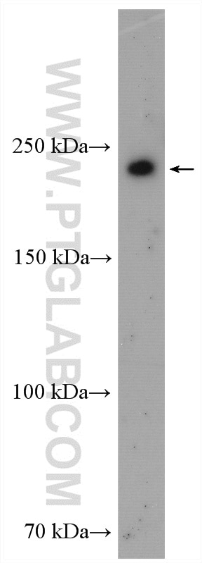
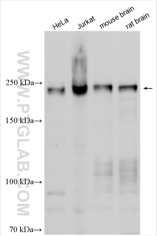
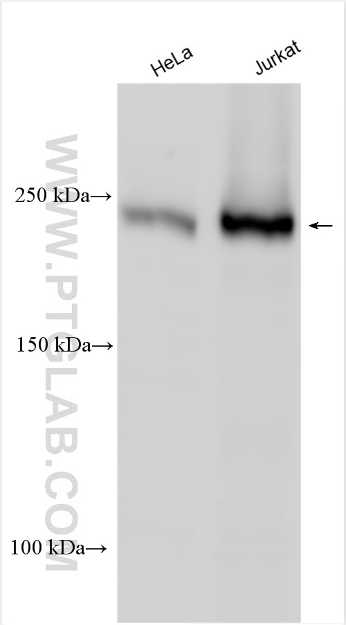
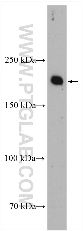
| Résultats positifs en WB | cellules HeLa, cellules Jurkat, tissu cérébral de rat, tissu cérébral de souris, tissu pulmonaire de souris |
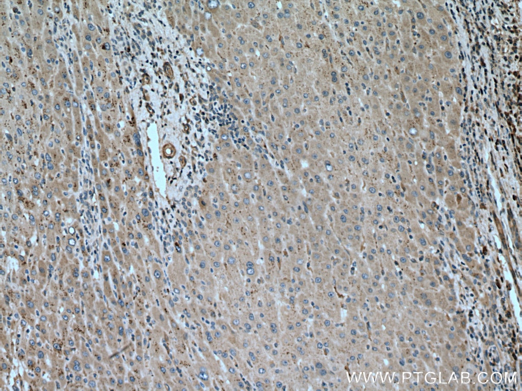
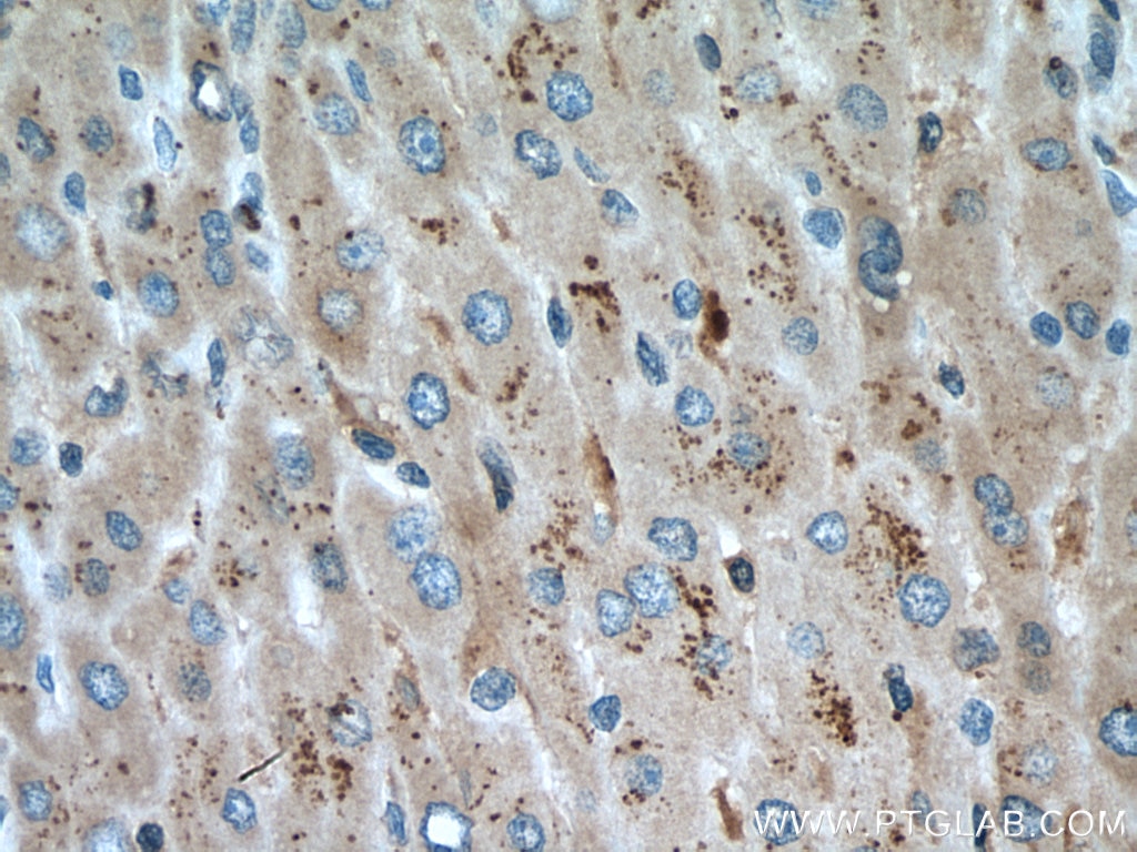
| Résultats positifs en IHC | tissu de cancer du foie humain, il est suggéré de démasquer l'antigène avec un tampon de TE buffer pH 9.0; (*) À défaut, 'le démasquage de l'antigène peut être 'effectué avec un tampon citrate pH 6,0. |
Dilution recommandée
| Application | Dilution |
|---|---|
| Western Blot (WB) | WB : 1:1000-1:4000 |
| Immunohistochimie (IHC) | IHC : 1:50-1:500 |
| It is recommended that this reagent should be titrated in each testing system to obtain optimal results. | |
| Sample-dependent, check data in validation data gallery | |
Applications publiées
| KD/KO | See 1 publications below |
| WB | See 4 publications below |
| IHC | See 1 publications below |
Informations sur le produit
13361-1-AP cible PIP5K3 dans les applications de WB, IHC, ELISA, Dot Blot et montre une réactivité avec des échantillons Humain, rat, souris
| Réactivité | Humain, rat, souris |
| Réactivité citée | Humain, porc, souris |
| Hôte / Isotype | Lapin / IgG |
| Clonalité | Polyclonal |
| Type | Anticorps |
| Immunogène | PIP5K3 Protéine recombinante Ag4177 |
| Nom complet | phosphatidylinositol-3-phosphate/phosphatidylinositol 5-kinase, type III |
| Masse moléculaire calculée | 451 aa, 50 kDa, 237 kDa |
| Poids moléculaire observé | 237 kDa |
| Numéro d’acquisition GenBank | BC032389 |
| Symbole du gène | PIP5K3 |
| Identification du gène (NCBI) | 200576 |
| Conjugaison | Non conjugué |
| Forme | Liquide |
| Méthode de purification | Purification par affinité contre l'antigène |
| Tampon de stockage | PBS avec azoture de sodium à 0,02 % et glycérol à 50 % pH 7,3 |
| Conditions de stockage | Stocker à -20°C. Stable pendant un an après l'expédition. L'aliquotage n'est pas nécessaire pour le stockage à -20oC Les 20ul contiennent 0,1% de BSA. |
Informations générales
PIP5K3 (1-phosphatidylinositol 3-phosphate 5-kinase), also callled PIKFYVE, belongs to a large family of lipid kinases that alter the phosphorylation status of intracellular phosphatidylinositol (PMID: 15902656; 9858586). The content of phosphatidylinositol 3,5-bisphosphate (PtdIns(3,5)P2) in endosomal membranes changes dynamically with fission and fusion events that generate or absorb intracellular transport vesicles. PIKFYVE is the PtdIns(3,5)P2-producing component of a trimolecular complex that tightly regulates the level of PtdIns(3,5)P2. Other components of this complex are the PIKFYVE activator VAC14 and the PtdIns(3,5)P2 phosphatase FIG4 (PMID: 17556371). In addition to its phosphoinositide 5-kinase activity, PIKFYVE also has protein kinase activity (PubMed: 11714711; 17556371).PIP5K3 has four isoforms with molecular weights of 237 kDa, 50 kDa, 51 kDa and 62 kDa. This antibody primarily recognizes the 237 kDa isoform.
Protocole
| Product Specific Protocols | |
|---|---|
| WB protocol for PIP5K3 antibody 13361-1-AP | Download protocol |
| IHC protocol for PIP5K3 antibody 13361-1-AP | Download protocol |
| Standard Protocols | |
|---|---|
| Click here to view our Standard Protocols |
Publications
| Species | Application | Title |
|---|---|---|
Mol Cell TSC1 binding to lysosomal PIPs is required for TSC complex translocation and mTORC1 regulation.
| ||
Elife Lipid kinases VPS34 and PIKfyve coordinate a phosphoinositide cascade to regulate retriever-mediated recycling on endosomes. | ||
PLoS Pathog Pseudorabies virus inhibits progesterone-induced inactivation of TRPML1 to facilitate viral entry | ||
Front Oncol TRIM68, PIKFYVE, and DYNLL2: The Possible Novel Autophagy- and Immunity-Associated Gene Biomarkers for Osteosarcoma Prognosis. | ||
Eur J Pharmacol Inhibition of c-Rel DNA binding is critical for the anti-inflammatory effects of novel PIKfyve inhibitor. | ||
BMC Immunol miR32-5p promoted vascular smooth muscle cell calcification by upregulating TNFα in the microenvironment. |
Avis
The reviews below have been submitted by verified Proteintech customers who received an incentive forproviding their feedback.
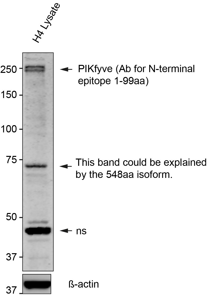
FH Kincaid (Verified Customer) (05-21-2024) | This antibody works very well. I use it 1:200 - overnight incubation at 4°C (3% BSA in TBST). I have validated with H4 cell lysate.
 |
