- Phare
- Validé par KD/KO
Anticorps Polyclonal de lapin anti-PDE1A
PDE1A Polyclonal Antibody for WB, IP, IHC, ELISA
Hôte / Isotype
Lapin / IgG
Réactivité testée
Humain, rat, souris
Applications
WB, IP, IHC, ELISA
Conjugaison
Non conjugué
N° de cat : 12442-2-AP
Synonymes
Galerie de données de validation
Applications testées
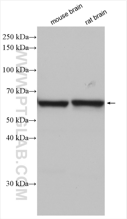
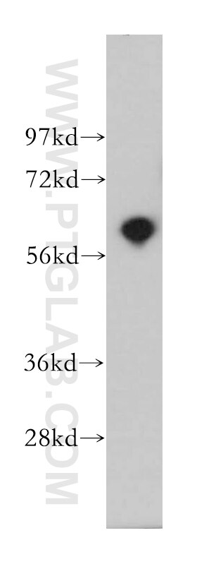
| Résultats positifs en WB | tissu cérébral de souris, tissu cérébral de rat, tissu cérébral humain |
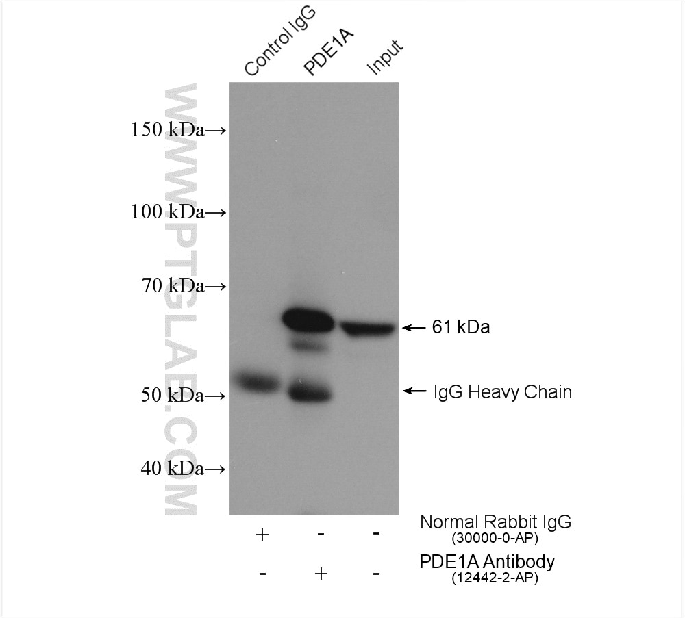
| Résultats positifs en IP | tissu cérébral de souris, |
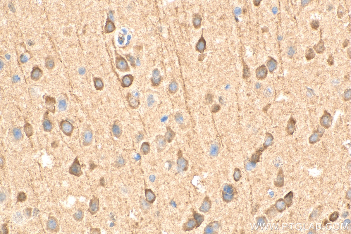
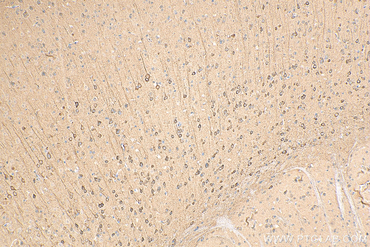
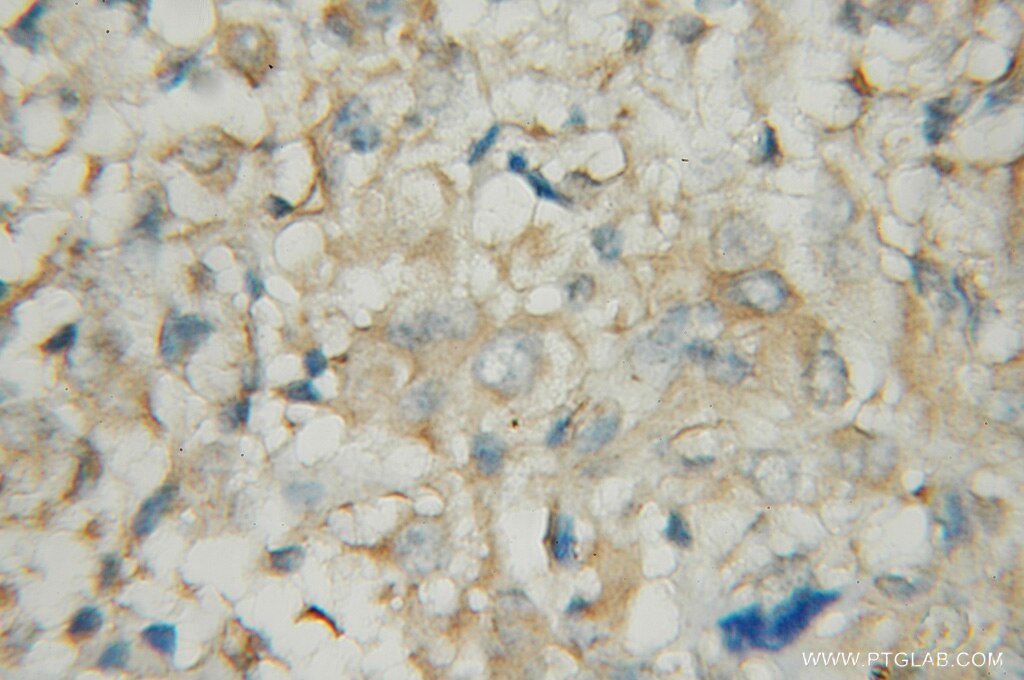
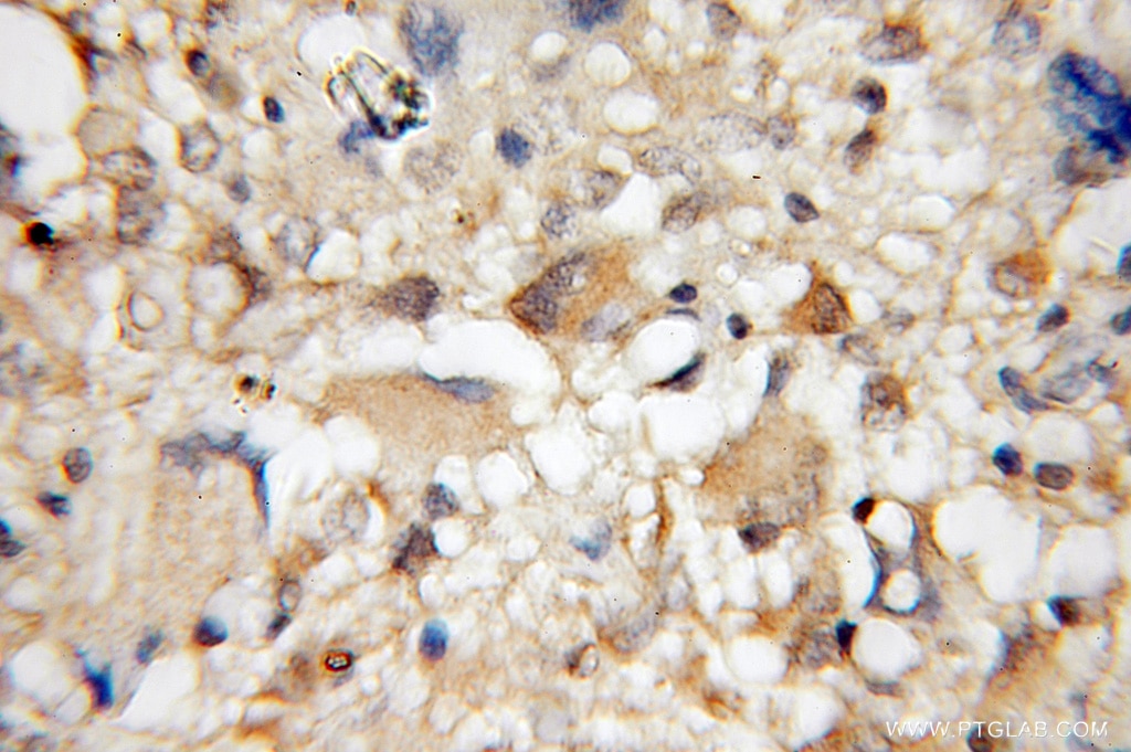
| Résultats positifs en IHC | tissu cérébral de souris, tissu de gliome humain il est suggéré de démasquer l'antigène avec un tampon de TE buffer pH 9.0; (*) À défaut, 'le démasquage de l'antigène peut être 'effectué avec un tampon citrate pH 6,0. |
Dilution recommandée
| Application | Dilution |
|---|---|
| Western Blot (WB) | WB : 1:1000-1:6000 |
| Immunoprécipitation (IP) | IP : 0.5-4.0 ug for 1.0-3.0 mg of total protein lysate |
| Immunohistochimie (IHC) | IHC : 1:50-1:500 |
| It is recommended that this reagent should be titrated in each testing system to obtain optimal results. | |
| Sample-dependent, check data in validation data gallery | |
Applications publiées
| KD/KO | See 1 publications below |
| WB | See 2 publications below |
| IHC | See 1 publications below |
Informations sur le produit
12442-2-AP cible PDE1A dans les applications de WB, IP, IHC, ELISA et montre une réactivité avec des échantillons Humain, rat, souris
| Réactivité | Humain, rat, souris |
| Réactivité citée | Humain, souris |
| Hôte / Isotype | Lapin / IgG |
| Clonalité | Polyclonal |
| Type | Anticorps |
| Immunogène | PDE1A Protéine recombinante Ag3157 |
| Nom complet | phosphodiesterase 1A, calmodulin-dependent |
| Masse moléculaire calculée | 545 aa, 61 kDa |
| Poids moléculaire observé | 61 kDa |
| Numéro d’acquisition GenBank | BC022480 |
| Symbole du gène | PDE1A |
| Identification du gène (NCBI) | 5136 |
| Conjugaison | Non conjugué |
| Forme | Liquide |
| Méthode de purification | Purification par affinité contre l'antigène |
| Tampon de stockage | PBS avec azoture de sodium à 0,02 % et glycérol à 50 % pH 7,3 |
| Conditions de stockage | Stocker à -20°C. Stable pendant un an après l'expédition. L'aliquotage n'est pas nécessaire pour le stockage à -20oC Les 20ul contiennent 0,1% de BSA. |
Informations générales
PDE1A(Calcium/calmodulin-dependent 3',5'-cyclic nucleotide phosphodiesterase 1A) is also named as Cam-PDE 1A, 61 kDa Cam-PDE, hCam-1 and belongs to the cyclic nucleotide phosphodiesterase (PDEs) family. The PDEs play a role in signal transduction by regulating intracellular cyclic nucleotide concentrations through hydrolysis of cAMP and/or cGMP to their respective nucleoside 5-prime monophosphates. This protein has 9 isoforms produced by alternative splicing with the molecular weight from 57 kDa to 63 kDa. There is a report showing the major 67-kD PDE1A isoform identified in human sperm associates tightly with calmodulin and is not activated by Ca(2+)/calmodulin(PMID:12135876).
Protocole
| Product Specific Protocols | |
|---|---|
| WB protocol for PDE1A antibody 12442-2-AP | Download protocol |
| IHC protocol for PDE1A antibody 12442-2-AP | Download protocol |
| IP protocol for PDE1A antibody 12442-2-AP | Download protocol |
| Standard Protocols | |
|---|---|
| Click here to view our Standard Protocols |
Publications
| Species | Application | Title |
|---|---|---|
Biol Reprod Luteinizing Hormone Causes Phosphorylation and Activation of the cGMP Phosphodiesterase PDE5 in Rat Ovarian Follicles, Contributing, Together with PDE1 Activity, to the Resumption of Meiosis. | ||
Biol Reprod Follicle-stimulating hormone and luteinizing hormone increase Ca2+ in the granulosa cells of mouse ovarian follicles†. | ||
Cell Rep Single-Cell Analysis of Foxp1-Driven Mechanisms Essential for Striatal Development. | ||
Front Genet Transcriptome profiling of intact bowel wall reveals that PDE1A and SEMA3D are possible markers with roles in enteric smooth muscle apoptosis, proliferative disorders, and dysautonomia in Crohn's disease |
