- Phare
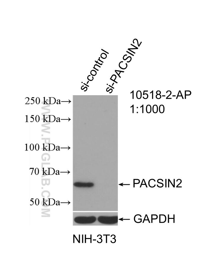
- Validé par KD/KO
Anticorps Polyclonal de lapin anti-PACSIN2
PACSIN2 Polyclonal Antibody for WB, IP, IF, IHC, ELISA
Hôte / Isotype
Lapin / IgG
Réactivité testée
Humain, souris et plus (1)
Applications
WB, IHC, IF/ICC, IP, ELISA
Conjugaison
Non conjugué
N° de cat : 10518-2-AP
Synonymes
Galerie de données de validation
Applications testées
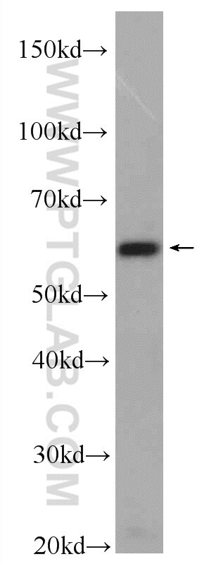
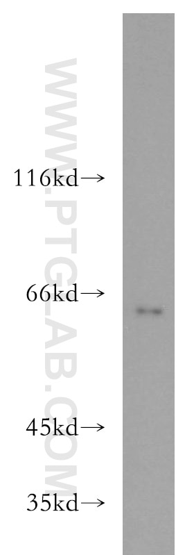
| Résultats positifs en WB | cellules NIH/3T3, tissu cardiaque de souris, tissu pulmonaire de souris |
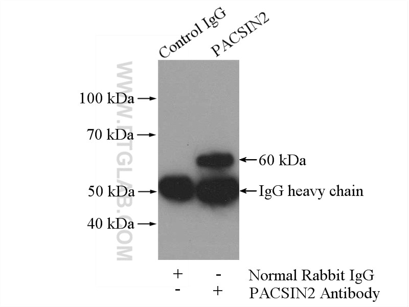
| Résultats positifs en IP | cellules NIH/3T3 |
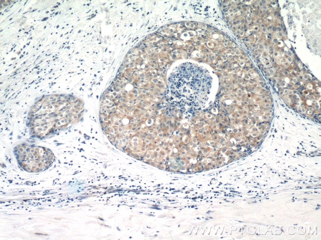
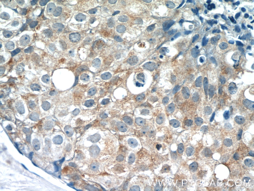
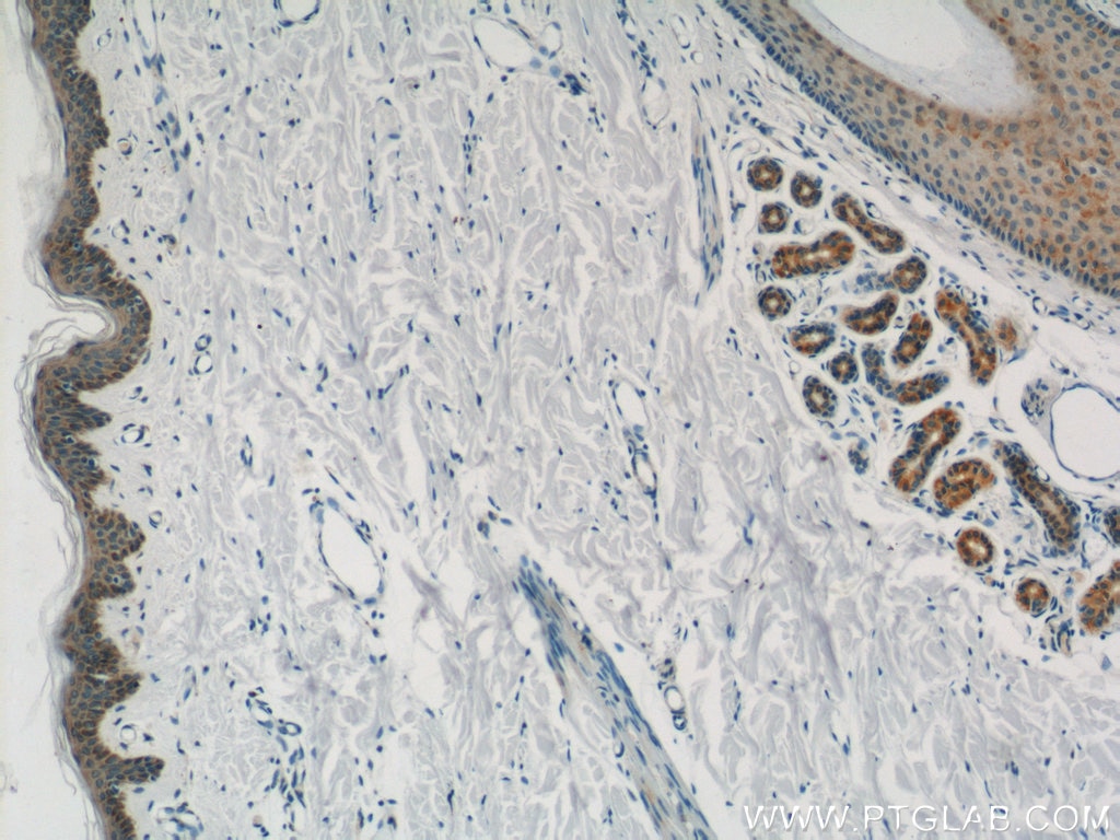
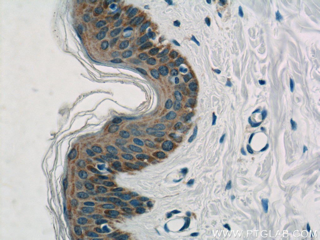
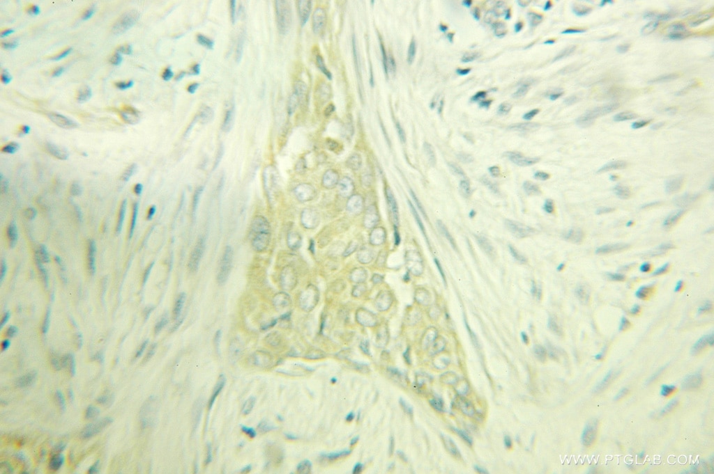
| Résultats positifs en IHC | tissu de cancer du sein humain, tissu cutané humain il est suggéré de démasquer l'antigène avec un tampon de TE buffer pH 9.0; (*) À défaut, 'le démasquage de l'antigène peut être 'effectué avec un tampon citrate pH 6,0. |
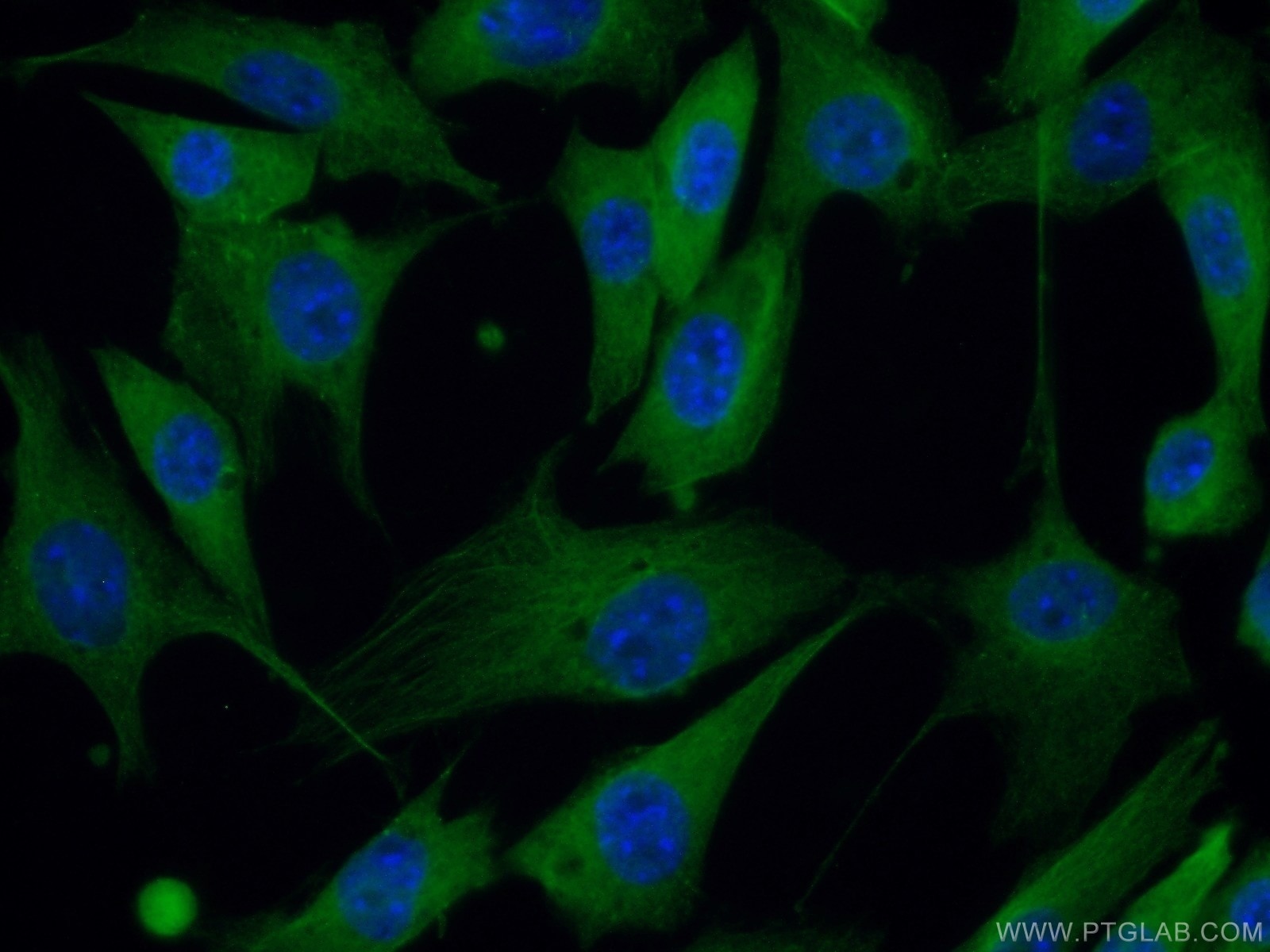
| Résultats positifs en IF/ICC | cellules NIH/3T3 |
Dilution recommandée
| Application | Dilution |
|---|---|
| Western Blot (WB) | WB : 1:500-1:1000 |
| Immunoprécipitation (IP) | IP : 0.5-4.0 ug for 1.0-3.0 mg of total protein lysate |
| Immunohistochimie (IHC) | IHC : 1:20-1:200 |
| Immunofluorescence (IF)/ICC | IF/ICC : 1:50-1:500 |
| It is recommended that this reagent should be titrated in each testing system to obtain optimal results. | |
| Sample-dependent, check data in validation data gallery | |
Applications publiées
| KD/KO | See 3 publications below |
| WB | See 7 publications below |
| IF | See 4 publications below |
Informations sur le produit
10518-2-AP cible PACSIN2 dans les applications de WB, IHC, IF/ICC, IP, ELISA et montre une réactivité avec des échantillons Humain, souris
| Réactivité | Humain, souris |
| Réactivité citée | Humain, poisson-zèbre, souris |
| Hôte / Isotype | Lapin / IgG |
| Clonalité | Polyclonal |
| Type | Anticorps |
| Immunogène | PACSIN2 Protéine recombinante Ag0809 |
| Nom complet | protein kinase C and casein kinase substrate in neurons 2 |
| Masse moléculaire calculée | 56 kDa |
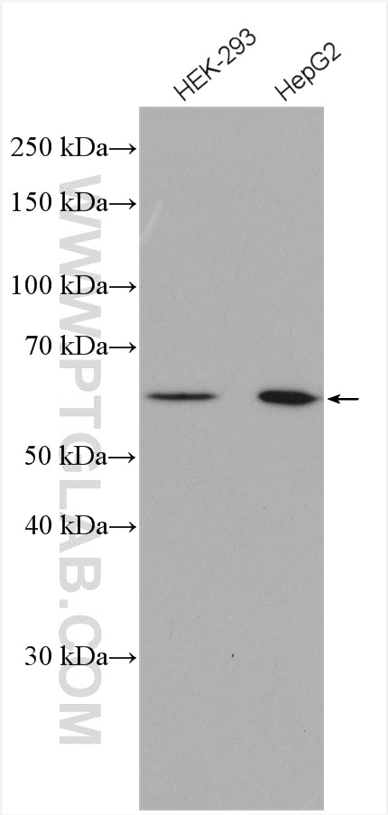
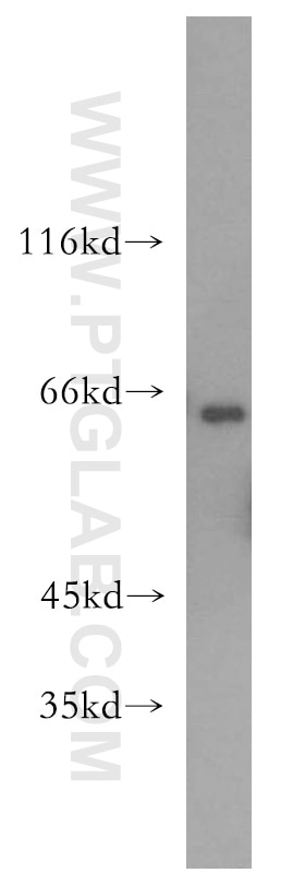
| Poids moléculaire observé | 60-65 kDa |
| Numéro d’acquisition GenBank | BC008037 |
| Symbole du gène | PACSIN2 |
| Identification du gène (NCBI) | 11252 |
| Conjugaison | Non conjugué |
| Forme | Liquide |
| Méthode de purification | Purification par affinité contre l'antigène |
| Tampon de stockage | PBS avec azoture de sodium à 0,02 % et glycérol à 50 % pH 7,3 |
| Conditions de stockage | Stocker à -20°C. Stable pendant un an après l'expédition. L'aliquotage n'est pas nécessaire pour le stockage à -20oC Les 20ul contiennent 0,1% de BSA. |
Informations générales
Peripheral membrane proteins of the Bin/amphiphysin/Rvs (BAR) and Fer-CIP4 homology-BAR (F-BAR) family participate in cellular membrane trafficking (PMID: 19549836). PACSIN2 (also known as syndapin-2) is a member of the protein kinase C and casein kinase substrate in neurons (PACSIN) family, which are membrane-binding proteins characterized by an amino-terminal F-BAR domain. PACSIN2 plays a role in intracellular vesicle-mediated transport. It is involved in the endocytosis of cell-surface receptors like the EGF receptor, contributing to its internalization (PMID: 23129763). PACSIN2 also has a role in caveolae membrane sculpting (PMID: 21610094).
Protocole
| Product Specific Protocols | |
|---|---|
| WB protocol for PACSIN2 antibody 10518-2-AP | Download protocol |
| IHC protocol for PACSIN2 antibody 10518-2-AP | Download protocol |
| IF protocol for PACSIN2 antibody 10518-2-AP | Download protocol |
| IP protocol for PACSIN2 antibody 10518-2-AP | Download protocol |
| Standard Protocols | |
|---|---|
| Click here to view our Standard Protocols |
Publications
| Species | Application | Title |
|---|---|---|
Mol Biol Cell Cooperation of MICAL-L1, syndapin2, and phosphatidic acid in tubular recycling endosome biogenesis.
| ||
PLoS One Role of Phosphatidylinositol 4,5-Bisphosphate in Regulating EHD2 Plasma Membrane Localization.
| ||
J Bacteriol Application of a C. trachomatis expression system to identify C. pneumoniae proteins translocated into host cells. | ||
Cell Rep Endogenous Cyclin D1 Promotes the Rate of Onset and Magnitude of Mitogenic Signaling via Akt1 Ser473 Phosphorylation. | ||
Nat Commun The molecular organization of differentially curved caveolae indicates bendable structural units at the plasma membrane |
Avis
The reviews below have been submitted by verified Proteintech customers who received an incentive forproviding their feedback.
FH Herve (Verified Customer) (11-13-2019) | This antibody recognizes a 55-kDa band that is present in both Pacsin2 WT and KO platelet lysates. PACSIN2 runs normally at 65 kDa in human and mouse platelet lysates and is absent in mouse Pacsin2 KO samples.
|
