Anticorps Polyclonal de lapin anti-NPHP3
NPHP3 Polyclonal Antibody for WB, IHC, IF/ICC, IP, ELISA
Hôte / Isotype
Lapin / IgG
Réactivité testée
canin, Humain, rat, souris
Applications
WB, IHC, IF/ICC, IP, ELISA
Conjugaison
Non conjugué
N° de cat : 22026-1-AP
Synonymes
Galerie de données de validation
Applications testées
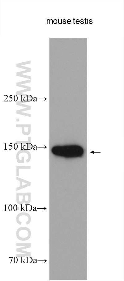
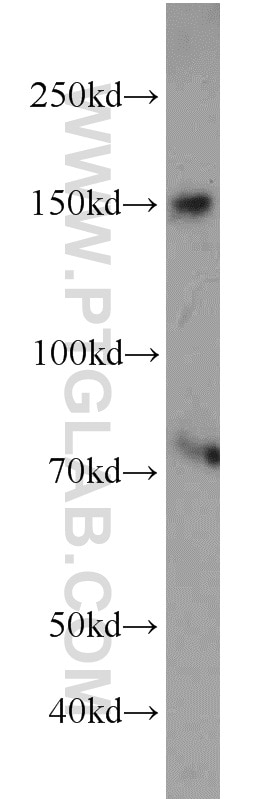
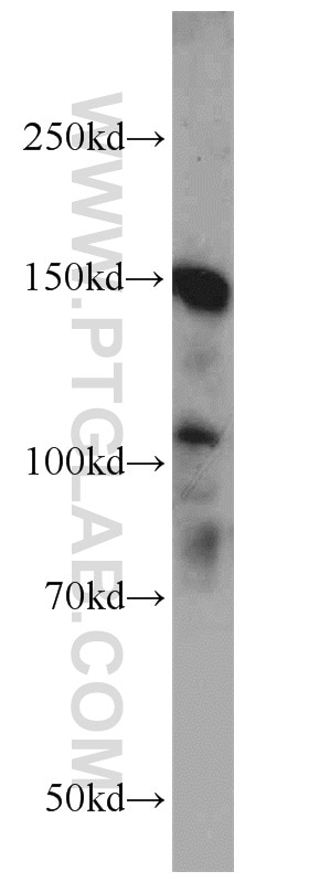
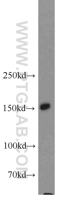
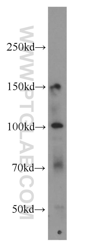
| Résultats positifs en WB | tissu testiculaire de souris, cellules HEK-293, cellules HepG2, tissu hépatique de souris, tissu testiculaire humain |
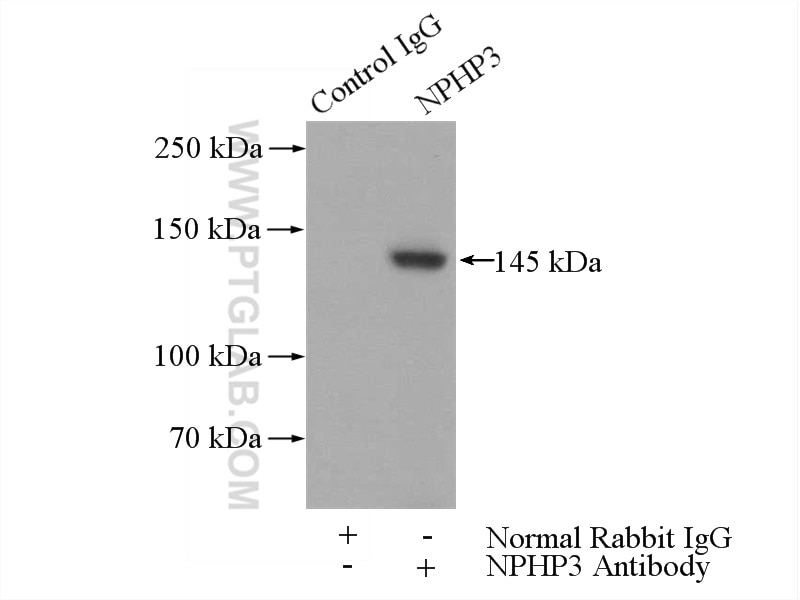
| Résultats positifs en IP | tissu testiculaire de souris |
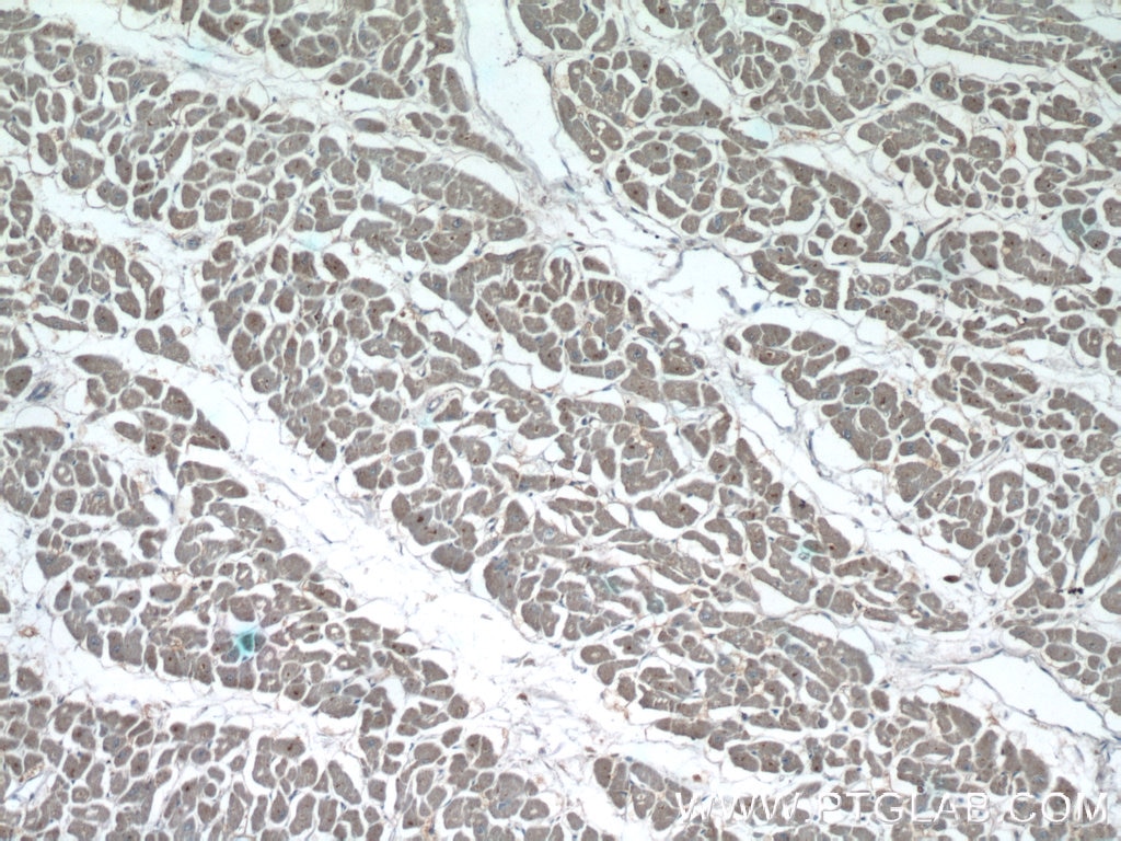
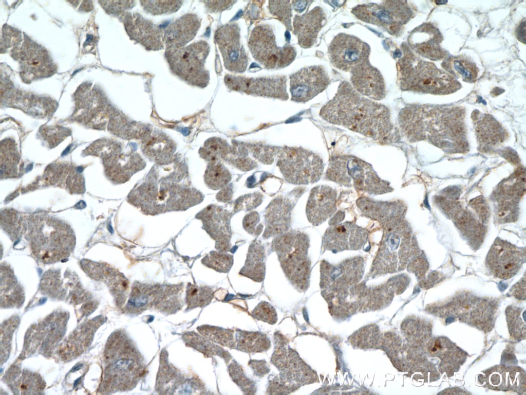
| Résultats positifs en IHC | tissu cardiaque humain il est suggéré de démasquer l'antigène avec un tampon de TE buffer pH 9.0; (*) À défaut, 'le démasquage de l'antigène peut être 'effectué avec un tampon citrate pH 6,0. |
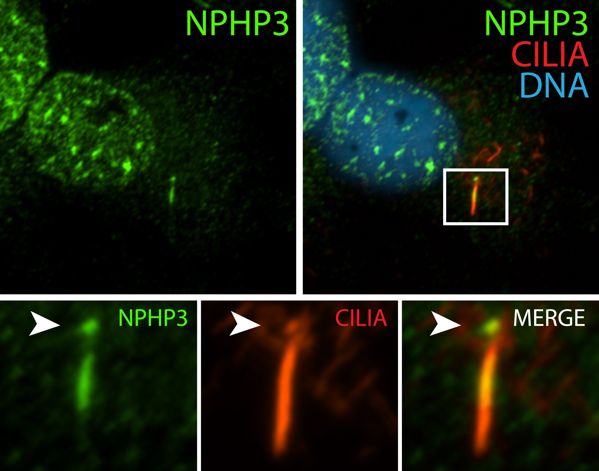
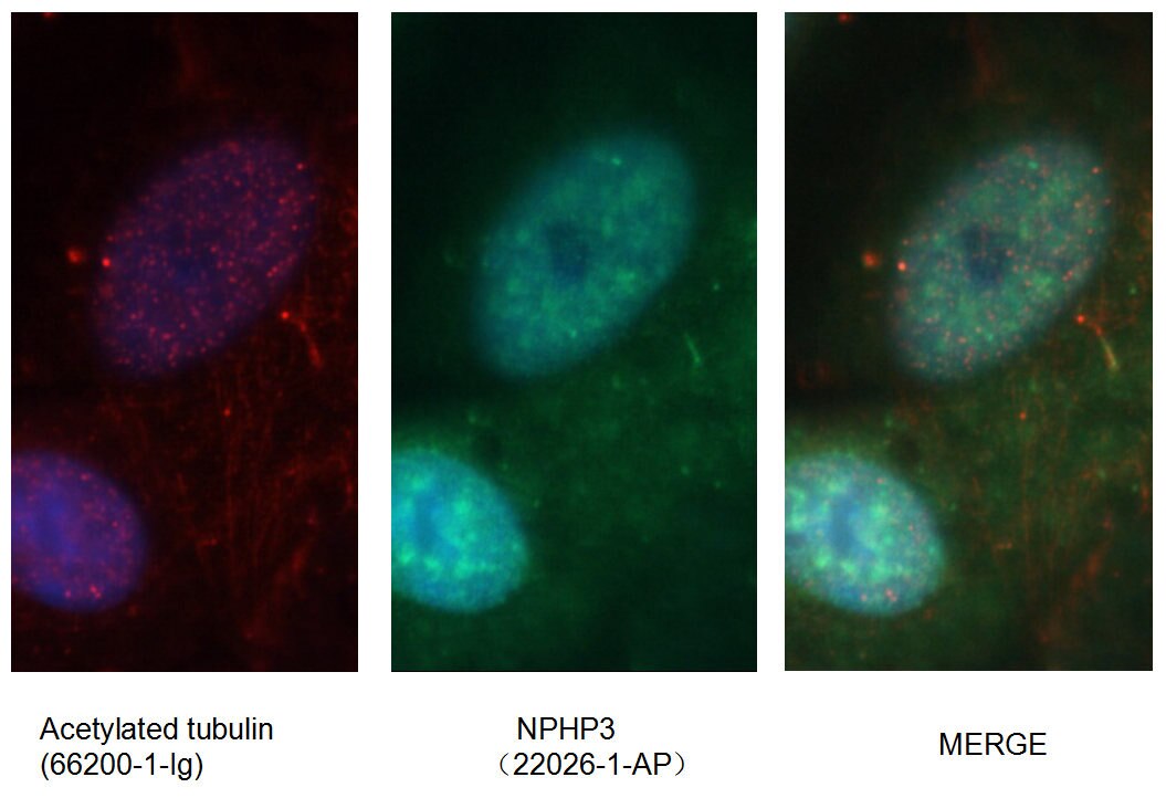
| Résultats positifs en IF/ICC | cellules hTERT-RPE1, cellules MDCK |
Dilution recommandée
| Application | Dilution |
|---|---|
| Western Blot (WB) | WB : 1:500-1:2000 |
| Immunoprécipitation (IP) | IP : 0.5-4.0 ug for 1.0-3.0 mg of total protein lysate |
| Immunohistochimie (IHC) | IHC : 1:20-1:200 |
| Immunofluorescence (IF)/ICC | IF/ICC : 1:20-1:200 |
| It is recommended that this reagent should be titrated in each testing system to obtain optimal results. | |
| Sample-dependent, check data in validation data gallery | |
Applications publiées
| WB | See 1 publications below |
| IHC | See 1 publications below |
| IF | See 5 publications below |
Informations sur le produit
22026-1-AP cible NPHP3 dans les applications de WB, IHC, IF/ICC, IP, ELISA et montre une réactivité avec des échantillons canin, Humain, rat, souris
| Réactivité | canin, Humain, rat, souris |
| Réactivité citée | Humain, souris |
| Hôte / Isotype | Lapin / IgG |
| Clonalité | Polyclonal |
| Type | Anticorps |
| Immunogène | NPHP3 Protéine recombinante Ag16888 |
| Nom complet | nephronophthisis 3 (adolescent) |
| Masse moléculaire calculée | 1330 aa, 151 kDa |
| Poids moléculaire observé | 145-151 kDa |
| Numéro d’acquisition GenBank | BC068082 |
| Symbole du gène | NPHP3 |
| Identification du gène (NCBI) | 27031 |
| Conjugaison | Non conjugué |
| Forme | Liquide |
| Méthode de purification | Purification par affinité contre l'antigène |
| Tampon de stockage | PBS with 0.02% sodium azide and 50% glycerol |
| Conditions de stockage | Stocker à -20°C. Stable pendant un an après l'expédition. L'aliquotage n'est pas nécessaire pour le stockage à -20oC Les 20ul contiennent 0,1% de BSA. |
Protocole
| Product Specific Protocols | |
|---|---|
| WB protocol for NPHP3 antibody 22026-1-AP | Download protocol |
| IHC protocol for NPHP3 antibody 22026-1-AP | Download protocol |
| IF protocol for NPHP3 antibody 22026-1-AP | Download protocol |
| IP protocol for NPHP3 antibody 22026-1-AP | Download protocol |
| Standard Protocols | |
|---|---|
| Click here to view our Standard Protocols |
Publications
| Species | Application | Title |
|---|---|---|
Nat Methods Genetically encoded calcium indicator illuminates calcium dynamics in primary cilia. | ||
Am J Hum Genet ARL3 Mutations Cause Joubert Syndrome by Disrupting Ciliary Protein Composition. | ||
Hum Mol Genet Translational read-through of the RP2 Arg120stop mutation in patient iPSC-derived retinal pigment epithelial cells. | ||
Int J Mol Sci Ciliary Proteins Repurposed by the Synaptic Ribbon: Trafficking Myristoylated Proteins at Rod Photoreceptor Synapses. | ||
Mol Biol Cell Interactions between TULP3 tubby domain and ARL13B amphipathic helix promote lipidated protein transport to cilia | ||
Avis
The reviews below have been submitted by verified Proteintech customers who received an incentive for providing their feedback.
FH Lelongt (Verified Customer) (12-10-2021) | Immunofluorescence is observed in mice cilia but is not restricted to the inversin compartment. Dilution has to be adjusted for WB since there are few bands.
|
