Anticorps Polyclonal de lapin anti-MYO7A
MYO7A Polyclonal Antibody for WB, IF, IHC, ELISA
Hôte / Isotype
Lapin / IgG
Réactivité testée
Humain, rat, souris et plus (1)
Applications
WB, IHC, IF/ICC, ELISA
Conjugaison
Non conjugué
N° de cat : 20720-1-AP
Synonymes
Galerie de données de validation
Applications testées
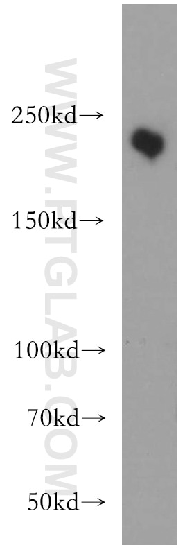
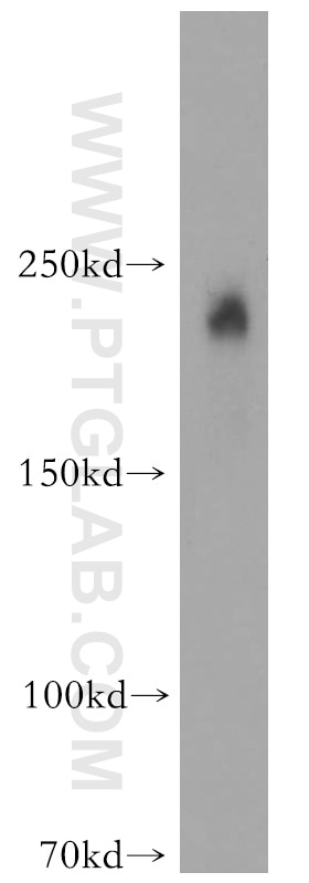
| Résultats positifs en WB | cellules L02, cellules A431 |
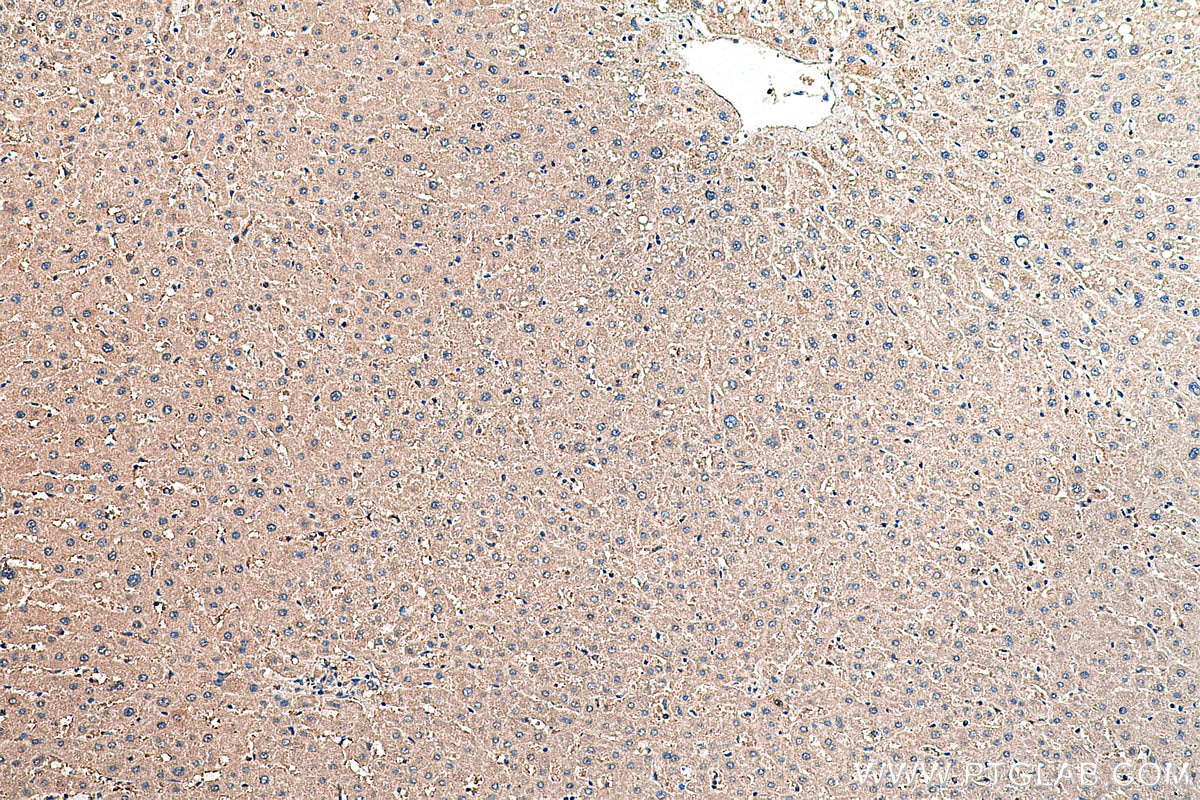
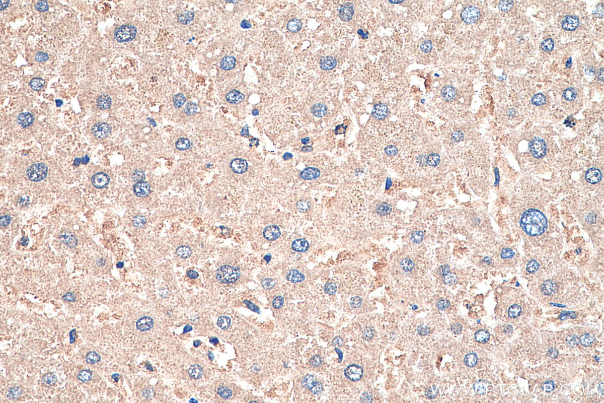
| Résultats positifs en IHC | tissu hépatique humain, il est suggéré de démasquer l'antigène avec un tampon de TE buffer pH 9.0; (*) À défaut, 'le démasquage de l'antigène peut être 'effectué avec un tampon citrate pH 6,0. |
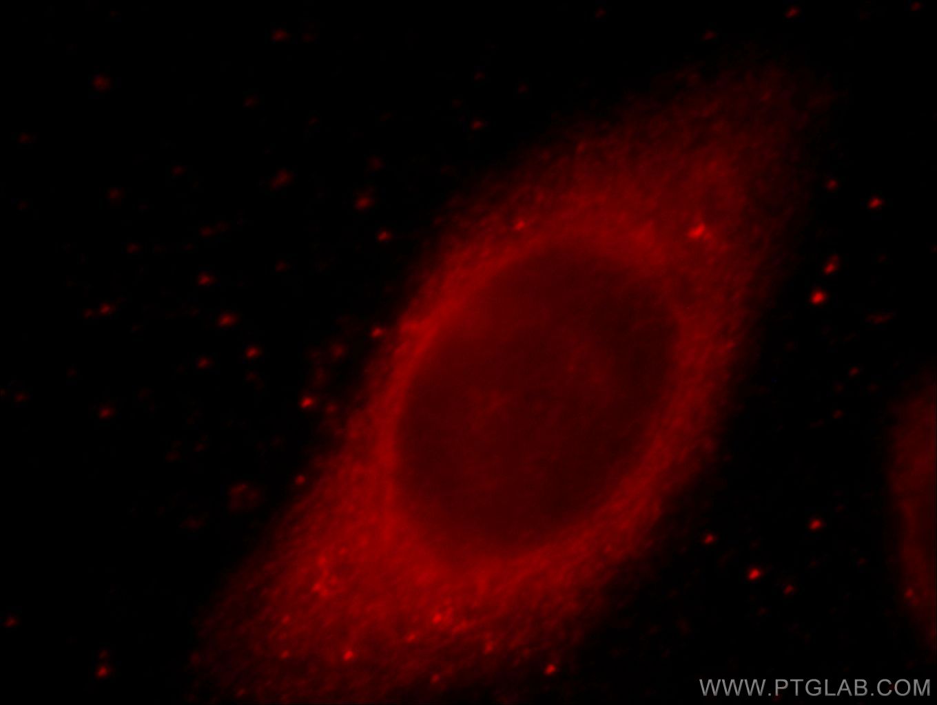
| Résultats positifs en IF/ICC | cellules HepG2 |
Dilution recommandée
| Application | Dilution |
|---|---|
| Western Blot (WB) | WB : 1:500-1:1000 |
| Immunohistochimie (IHC) | IHC : 1:500-1:2000 |
| Immunofluorescence (IF)/ICC | IF/ICC : 1:10-1:100 |
| It is recommended that this reagent should be titrated in each testing system to obtain optimal results. | |
| Sample-dependent, check data in validation data gallery | |
Applications publiées
| WB | See 1 publications below |
| IF | See 3 publications below |
Informations sur le produit
20720-1-AP cible MYO7A dans les applications de WB, IHC, IF/ICC, ELISA et montre une réactivité avec des échantillons Humain, rat, souris
| Réactivité | Humain, rat, souris |
| Réactivité citée | Humain, poisson-zèbre |
| Hôte / Isotype | Lapin / IgG |
| Clonalité | Polyclonal |
| Type | Anticorps |
| Immunogène | Peptide |
| Nom complet | myosin VIIA |
| Masse moléculaire calculée | 254 kDa |
| Poids moléculaire observé | 240-250 kDa |
| Numéro d’acquisition GenBank | NM_000260 |
| Symbole du gène | MYO7A |
| Identification du gène (NCBI) | 4647 |
| Conjugaison | Non conjugué |
| Forme | Liquide |
| Méthode de purification | Purification par affinité contre l'antigène |
| Tampon de stockage | PBS avec azoture de sodium à 0,02 % et glycérol à 50 % pH 7,3 |
| Conditions de stockage | Stocker à -20°C. Stable pendant un an après l'expédition. L'aliquotage n'est pas nécessaire pour le stockage à -20oC Les 20ul contiennent 0,1% de BSA. |
Informations générales
MYO7A, also named a USH1B, is one of myosins protein which are actin-based motor molecules with ATPase activity. Unconventional myosins serve in intracellular movements. Their highly divergent tails are presumed to bind to membranous compartments, which would be moved relative to actin filaments. In retina, MYO7A might play a role in trafficking of ribbon-synaptic vesicle complexes and renewal of the outer photoreceptors disks. In inner ear, it might maintain the rigidity of stereocilia during the dynamic movements of the bundle. It is involved in hair-cell vesicle trafficking of aminoglycosides, which are known to induce ototoxicity. Defects in MYO7A are the cause of Usher syndrome type 1B (USH1B). Defects in MYO7A are the cause of deafness autosomal recessive type 2 (DFNB2). Defects in MYO7A are the cause of deafness autosomal dominant type 11 (DFNA11). The antibody is specific to MYO7A.
Protocole
| Product Specific Protocols | |
|---|---|
| WB protocol for MYO7A antibody 20720-1-AP | Download protocol |
| IHC protocol for MYO7A antibody 20720-1-AP | Download protocol |
| IF protocol for MYO7A antibody 20720-1-AP | Download protocol |
| Standard Protocols | |
|---|---|
| Click here to view our Standard Protocols |
Publications
| Species | Application | Title |
|---|---|---|
ACS Biomater Sci Eng Microenvironment Can Induce Development of Auditory Progenitor Cells from Human Gingival Mesenchymal Stem Cells. | ||
Biomedicines Knockout of mafba Causes Inner-Ear Developmental Defects in Zebrafish via the Impairment of Proliferation and Differentiation of Ionocyte Progenitor Cells. | ||
Mol Biol Cell Comparative analysis of the MyTH4-FERM myosins reveals insights into the determinants of actin track selection in polarized epithelia. |
