Anticorps Polyclonal de lapin anti-MYL3
MYL3 Polyclonal Antibody for WB, IP, IF, IHC, ELISA, FC (Intra)
Hôte / Isotype
Lapin / IgG
Réactivité testée
Humain, rat, souris
Applications
WB, IHC, IF/ICC, FC (Intra), IP, ELISA
Conjugaison
Non conjugué
N° de cat : 10913-1-AP
Synonymes
Galerie de données de validation
Applications testées
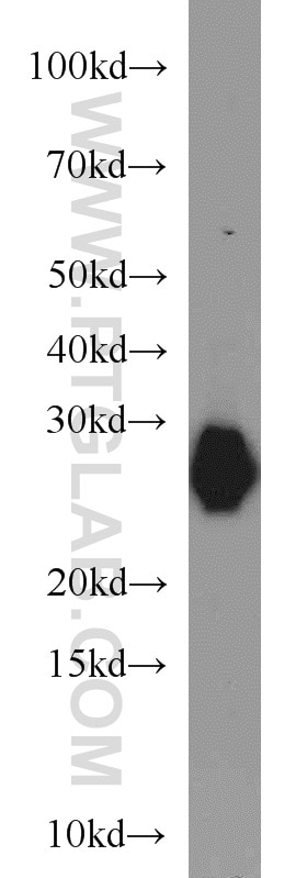
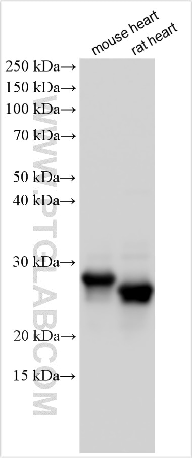
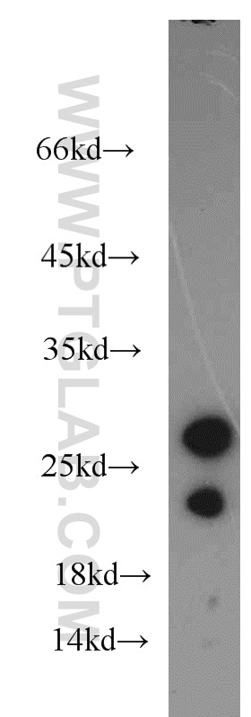
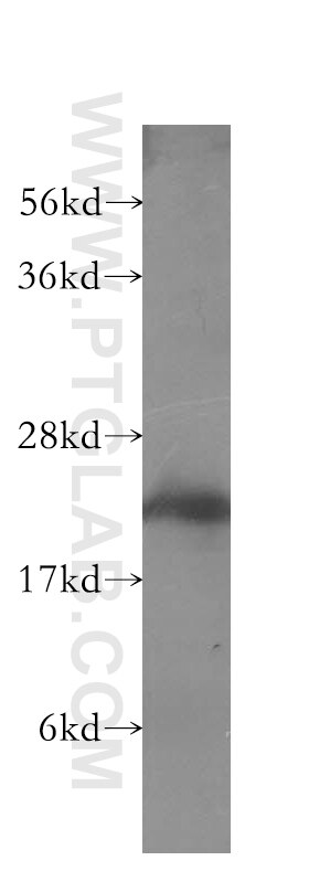
| Résultats positifs en WB | tissu cardiaque de souris, tissu cardiaque de rat, tissu cardiaque humain, tissu de muscle squelettique de souris |
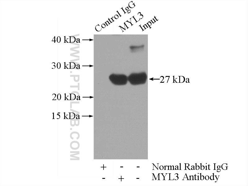
| Résultats positifs en IP | tissu cardiaque de souris |
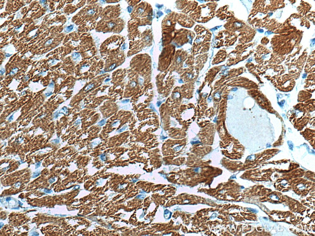
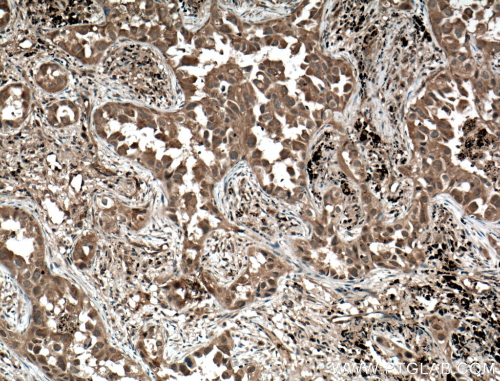
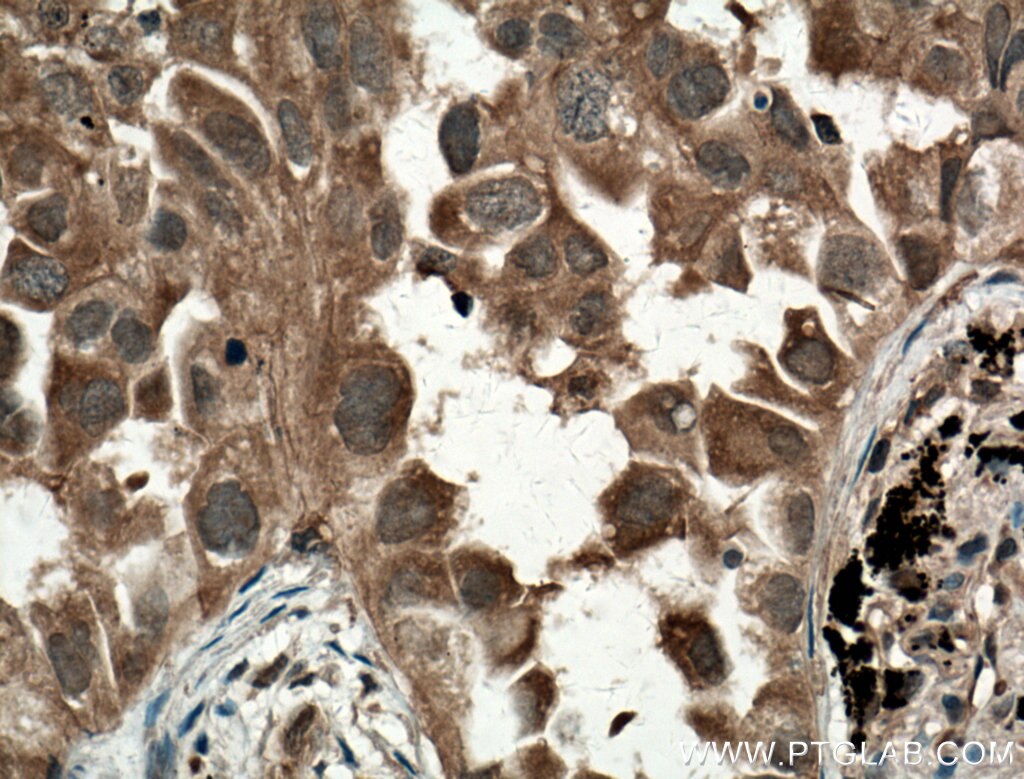
| Résultats positifs en IHC | tissu cardiaque humain, tissu de cancer du poumon humain il est suggéré de démasquer l'antigène avec un tampon de TE buffer pH 9.0; (*) À défaut, 'le démasquage de l'antigène peut être 'effectué avec un tampon citrate pH 6,0. |
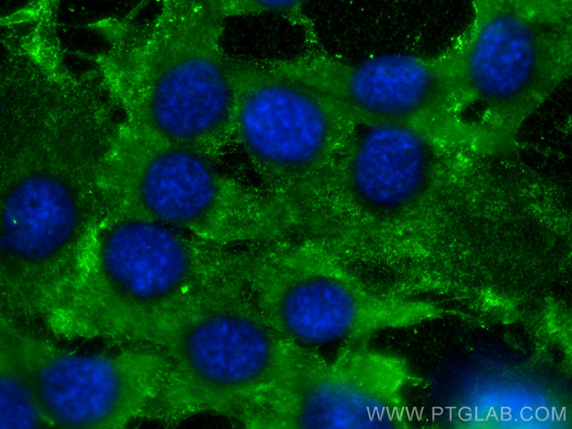
| Résultats positifs en IF/ICC | cellules C2C12, |
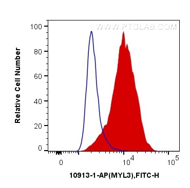
| Résultats positifs en FC (Intra) | cellules C2C12 |
| Résultats positifs en cytométrie | cellules C2C12 |
Dilution recommandée
| Application | Dilution |
|---|---|
| Western Blot (WB) | WB : 1:5000-1:50000 |
| Immunoprécipitation (IP) | IP : 0.5-4.0 ug for 1.0-3.0 mg of total protein lysate |
| Immunohistochimie (IHC) | IHC : 1:500-1:2000 |
| Immunofluorescence (IF)/ICC | IF/ICC : 1:50-1:500 |
| Flow Cytometry (FC) (INTRA) | FC (INTRA) : 0.40 ug per 10^6 cells in a 100 µl suspension |
| Flow Cytometry (FC) | FC : 0.40 ug per 10^6 cells in a 100 µl suspension |
| It is recommended that this reagent should be titrated in each testing system to obtain optimal results. | |
| Sample-dependent, check data in validation data gallery | |
Applications publiées
| WB | See 4 publications below |
| IHC | See 1 publications below |
| IF | See 1 publications below |
Informations sur le produit
10913-1-AP cible MYL3 dans les applications de WB, IHC, IF/ICC, FC (Intra), IP, ELISA et montre une réactivité avec des échantillons Humain, rat, souris
| Réactivité | Humain, rat, souris |
| Réactivité citée | Humain, souris |
| Hôte / Isotype | Lapin / IgG |
| Clonalité | Polyclonal |
| Type | Anticorps |
| Immunogène | MYL3 Protéine recombinante Ag1364 |
| Nom complet | myosin, light chain 3, alkali; ventricular, skeletal, slow |
| Masse moléculaire calculée | 22 kDa |
| Poids moléculaire observé | 22-27 kDa |
| Numéro d’acquisition GenBank | BC009790 |
| Symbole du gène | MYL3 |
| Identification du gène (NCBI) | 4634 |
| Conjugaison | Non conjugué |
| Forme | Liquide |
| Méthode de purification | Purification par affinité contre l'antigène |
| Tampon de stockage | PBS avec azoture de sodium à 0,02 % et glycérol à 50 % pH 7,3 |
| Conditions de stockage | Stocker à -20°C. Stable pendant un an après l'expédition. L'aliquotage n'est pas nécessaire pour le stockage à -20oC Les 20ul contiennent 0,1% de BSA. |
Informations générales
MYL3, also named as MLC1v, is an essential light chain of myosin that is associated with muscle contraction. It is expressed in ventricular and slow skeletal muscle. MYL3 may serve as a target for caspase-3 in dying cardiomyocytes. Mutations of MYL3 gene cause hypertrophic cardiomyopathy. MYL3 has been identified as potential serum biomarker for drug induced myotoxicity. Great increase in MYL3 serum concentration has been observed in rats with cardiac and skeletal muscle injury. (PMID:21685905)
Protocole
| Product Specific Protocols | |
|---|---|
| WB protocol for MYL3 antibody 10913-1-AP | Download protocol |
| IHC protocol for MYL3 antibody 10913-1-AP | Download protocol |
| IF protocol for MYL3 antibody 10913-1-AP | Download protocol |
| IP protocol for MYL3 antibody 10913-1-AP | Download protocol |
| Standard Protocols | |
|---|---|
| Click here to view our Standard Protocols |
Publications
| Species | Application | Title |
|---|---|---|
Nat Biotechnol A pipeline that integrates the discovery and verification of plasma protein biomarkers reveals candidate markers for cardiovascular disease. | ||
J Proteome Res Multi-Proteomic Analysis Reveals the Effect of Protein Lactylation on Matrix and Cholesterol Metabolism in Tendinopathy | ||
Nat Commun MYL3 protects chondrocytes from senescence by inhibiting clathrin-mediated endocytosis and activating of Notch signaling | ||
Nat Commun Systematic HOIP interactome profiling reveals critical roles of linear ubiquitination in tissue homeostasis |
