- Phare
- Validé par KD/KO
Anticorps Polyclonal de lapin anti-MTA3
MTA3 Polyclonal Antibody for WB, IHC, IF/ICC, IP, ELISA
Hôte / Isotype
Lapin / IgG
Réactivité testée
Humain, rat, souris
Applications
WB, IHC, IF/ICC, IP, ELISA
Conjugaison
Non conjugué
N° de cat : 14682-1-AP
Synonymes
Galerie de données de validation
Applications testées
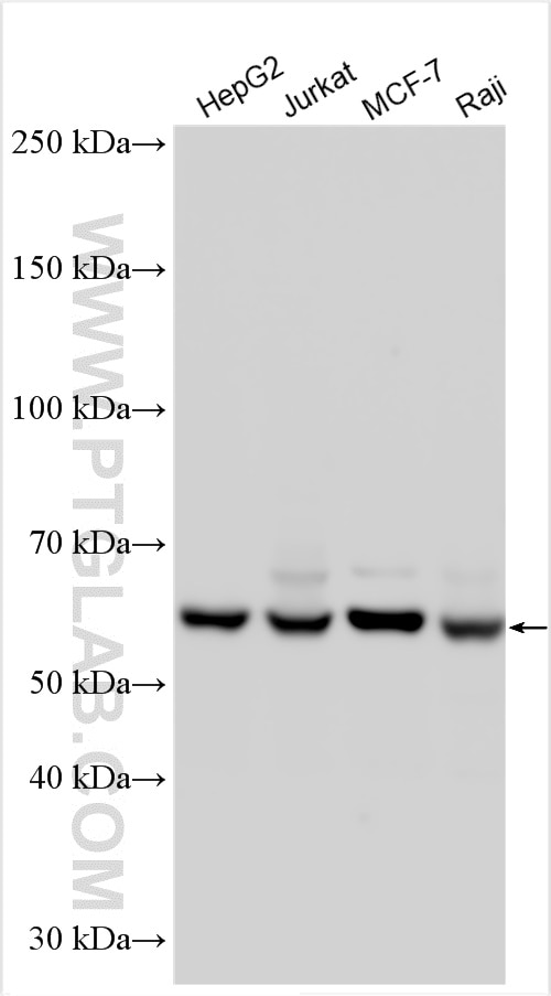
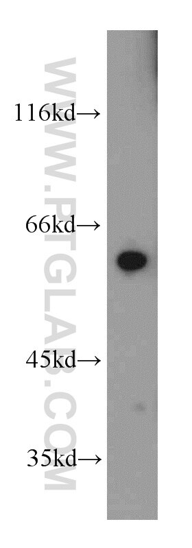
| Résultats positifs en WB | cellules HepG2, cellules Jurkat, cellules MCF-7, cellules Raji, tissu de thymus de souris |
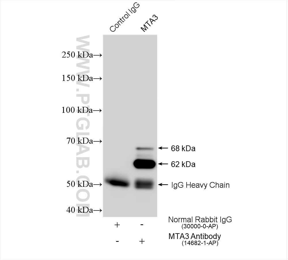
| Résultats positifs en IP | cellules Raji, |
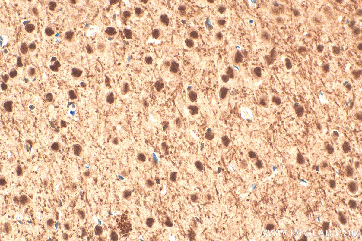
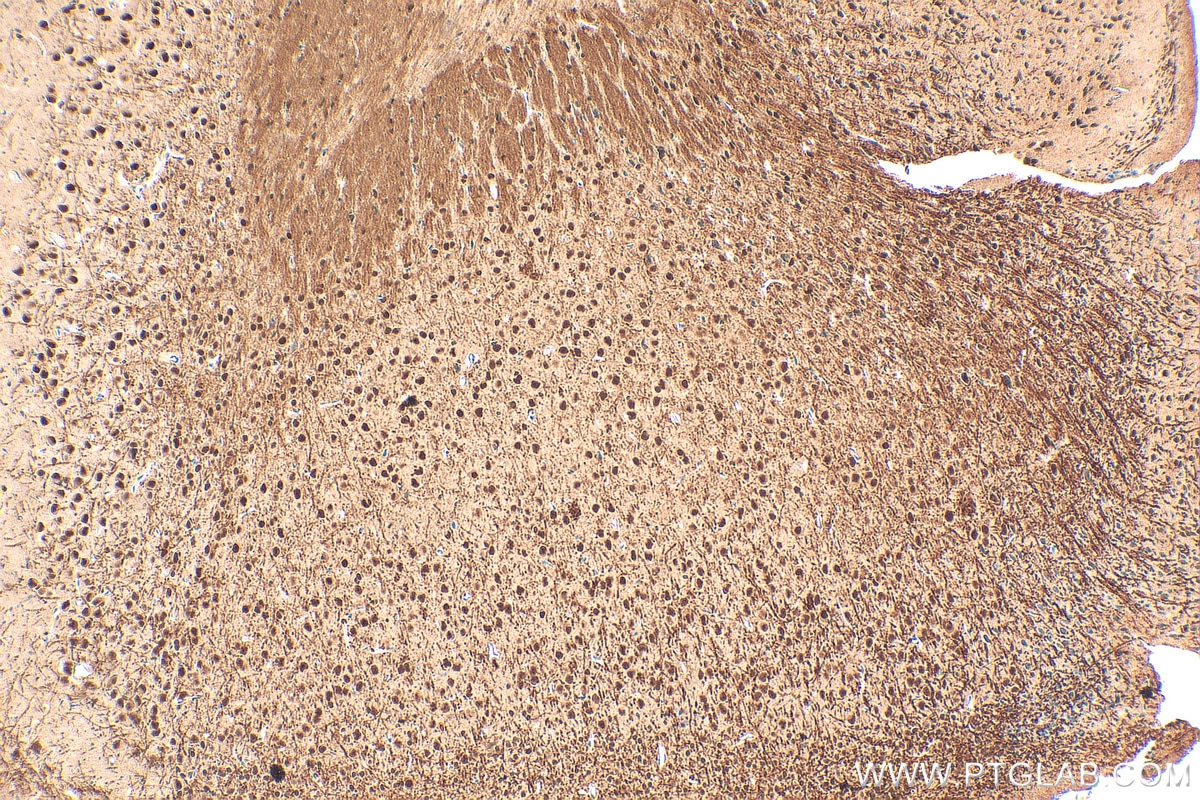
| Résultats positifs en IHC | tissu cérébral de souris, il est suggéré de démasquer l'antigène avec un tampon de TE buffer pH 9.0; (*) À défaut, 'le démasquage de l'antigène peut être 'effectué avec un tampon citrate pH 6,0. |
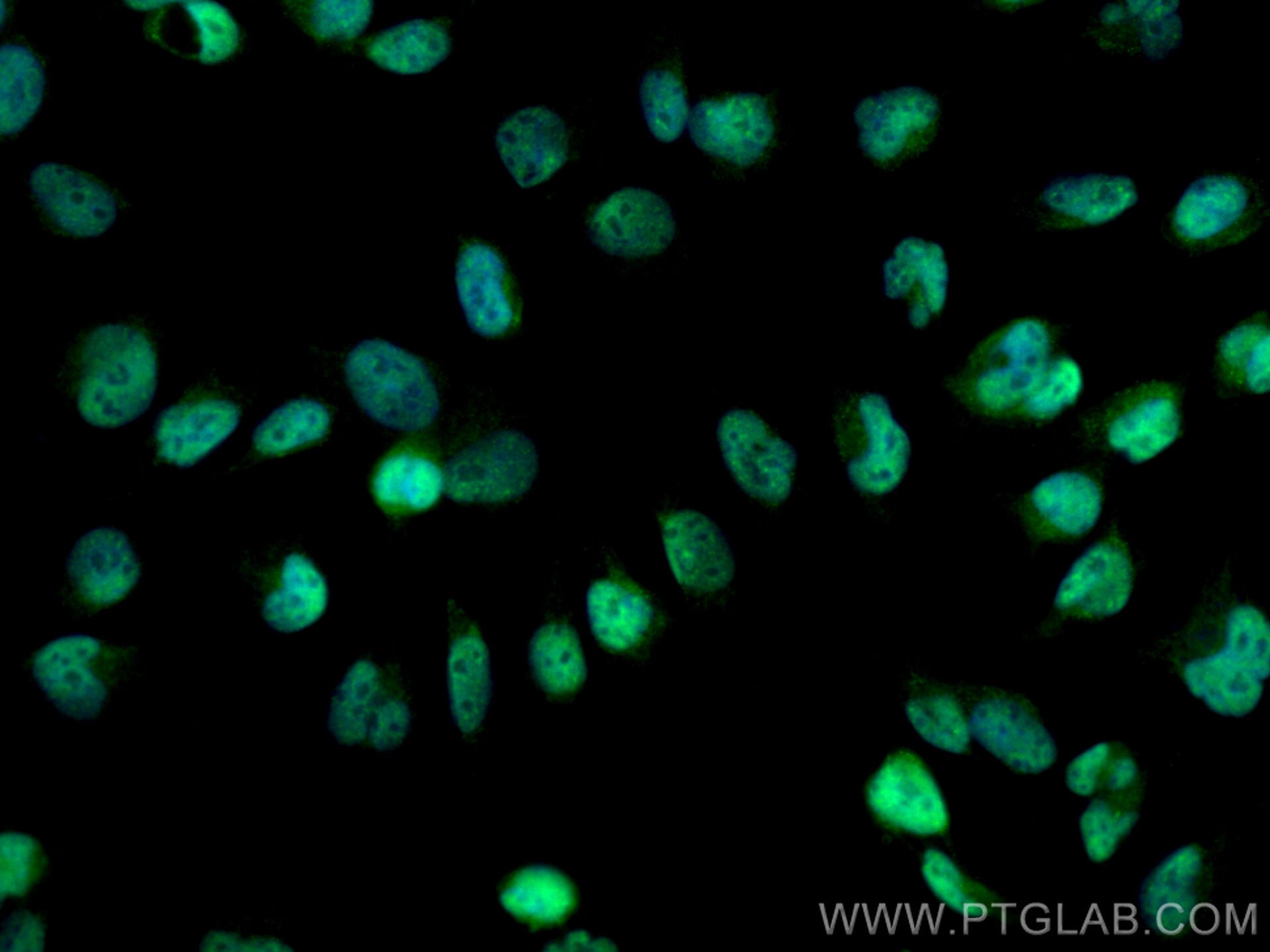
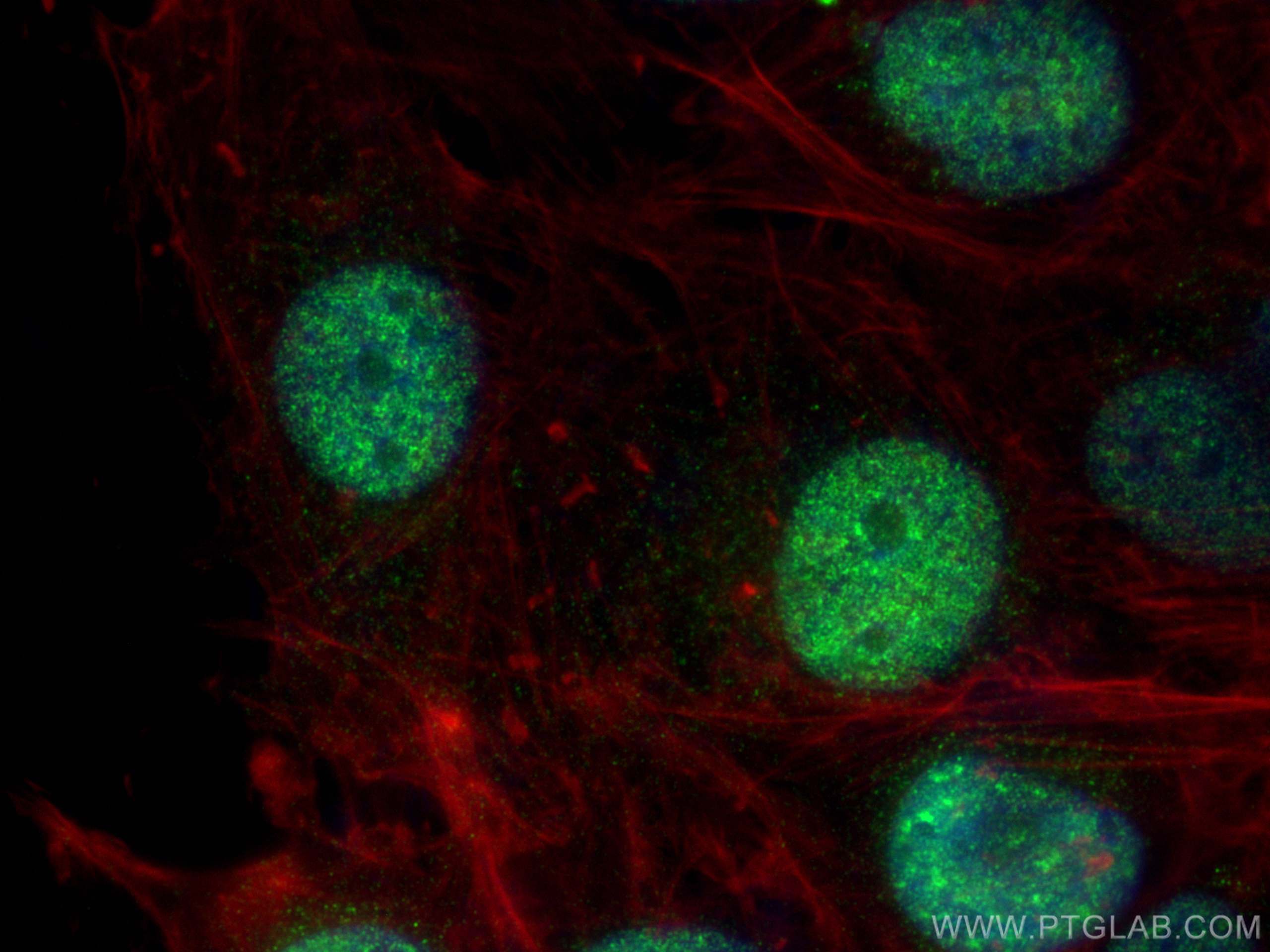
| Résultats positifs en IF/ICC | cellules U-251, |
Dilution recommandée
| Application | Dilution |
|---|---|
| Western Blot (WB) | WB : 1:2000-1:10000 |
| Immunoprécipitation (IP) | IP : 0.5-4.0 ug for 1.0-3.0 mg of total protein lysate |
| Immunohistochimie (IHC) | IHC : 1:250-1:1000 |
| Immunofluorescence (IF)/ICC | IF/ICC : 1:200-1:800 |
| It is recommended that this reagent should be titrated in each testing system to obtain optimal results. | |
| Sample-dependent, check data in validation data gallery | |
Applications publiées
| KD/KO | See 2 publications below |
| WB | See 8 publications below |
| IHC | See 10 publications below |
| IP | See 1 publications below |
Informations sur le produit
14682-1-AP cible MTA3 dans les applications de WB, IHC, IF/ICC, IP, ELISA et montre une réactivité avec des échantillons Humain, rat, souris
| Réactivité | Humain, rat, souris |
| Réactivité citée | Humain, souris |
| Hôte / Isotype | Lapin / IgG |
| Clonalité | Polyclonal |
| Type | Anticorps |
| Immunogène | MTA3 Protéine recombinante Ag6400 |
| Nom complet | metastasis associated 1 family, member 3 |
| Masse moléculaire calculée | 68 kDa |
| Poids moléculaire observé | 62 kDa, 68 kDa |
| Numéro d’acquisition GenBank | BC053631 |
| Symbole du gène | MTA3 |
| Identification du gène (NCBI) | 57504 |
| Conjugaison | Non conjugué |
| Forme | Liquide |
| Méthode de purification | Purification par affinité contre l'antigène |
| Tampon de stockage | PBS with 0.02% sodium azide and 50% glycerol |
| Conditions de stockage | Stocker à -20°C. Stable pendant un an après l'expédition. L'aliquotage n'est pas nécessaire pour le stockage à -20oC Les 20ul contiennent 0,1% de BSA. |
Informations générales
The metastasis-associated (MTA) family contains three members, MTA1, MTA2 and MTA3 (PMID:12167865).Metastasis-associated protein 3 (MTA3) is a cell-type specific component of the Mi-2-NURD transcriptional co-repressor complex. It has a role in maintenance of the normal epithelial architecture through the repression of SNAI1 transcription in a histone deacetylase-dependent manner, and thus the regulation of E-cadherin levels.
Protocole
| Product Specific Protocols | |
|---|---|
| WB protocol for MTA3 antibody 14682-1-AP | Download protocol |
| IHC protocol for MTA3 antibody 14682-1-AP | Download protocol |
| IF protocol for MTA3 antibody 14682-1-AP | Download protocol |
| IP protocol for MTA3 antibody 14682-1-AP | Download protocol |
| Standard Protocols | |
|---|---|
| Click here to view our Standard Protocols |
Publications
| Species | Application | Title |
|---|---|---|
Cell Stem Cell The polycomb group protein L3mbtl2 assembles an atypical PRC1-family complex that is essential in pluripotent stem cells and early development. | ||
Mol Cell The Nucleosome Remodeling and Deacetylation Complex Modulates Chromatin Structure at Sites of Active Transcription to Fine-Tune Gene Expression. | ||
Oncotarget Metastasis-associated protein 3 in colorectal cancer determines tumor recurrence and prognosis. | ||
PLoS One Overexpression of MTA3 Correlates with Tumor Progression in Non-Small Cell Lung Cancer.
| ||
J Gastroenterol Hepatol Overexpression of the metastasis-associated gene MTA3 correlates with tumor progression and poor prognosis in hepatocellular carcinoma. | ||
Tumour Biol MTA3 regulates malignant progression of colorectal cancer through Wnt signaling pathway. |
