Anticorps Polyclonal de lapin anti-MSH3
MSH3 Polyclonal Antibody for WB, IP, IF, IHC, ELISA
Hôte / Isotype
Lapin / IgG
Réactivité testée
Humain et plus (1)
Applications
WB, IHC, IF/ICC, IP, ELISA
Conjugaison
Non conjugué
N° de cat : 22393-1-AP
Synonymes
Galerie de données de validation
Applications testées
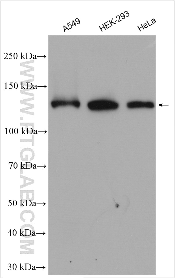
| Résultats positifs en WB | cellules A549, cellules HEK-293, cellules HeLa |
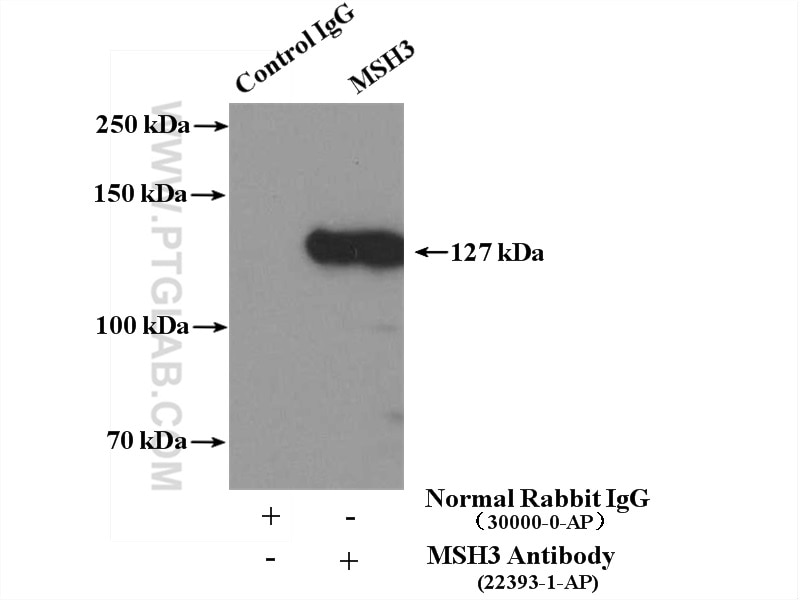
| Résultats positifs en IP | cellules HeLa |
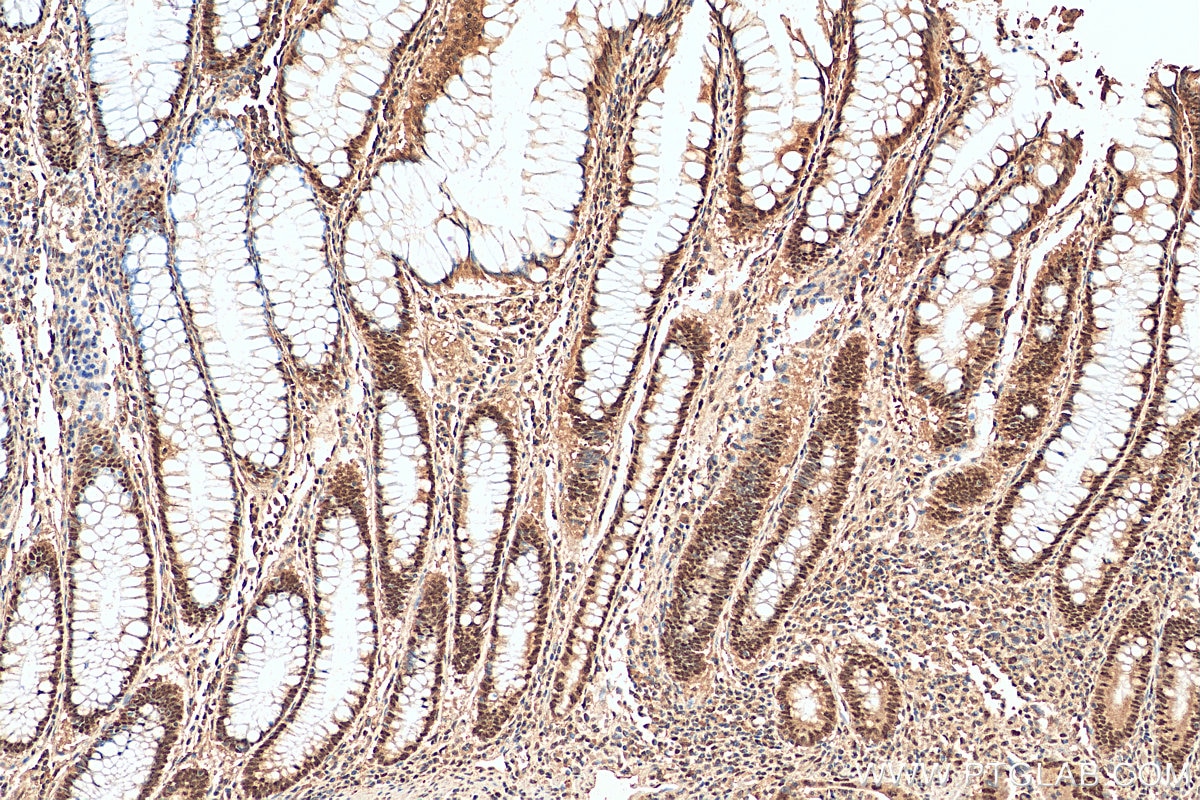
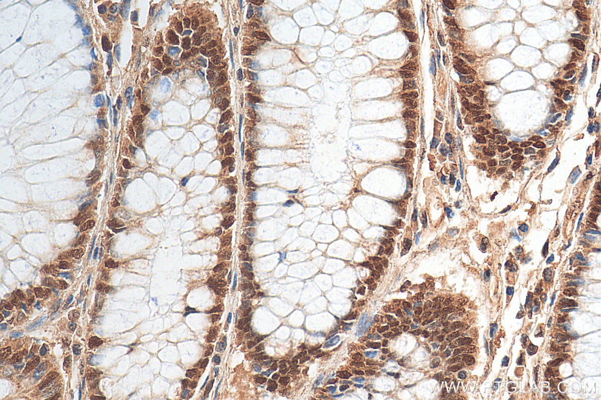
| Résultats positifs en IHC | tissu de cancer du côlon humain, il est suggéré de démasquer l'antigène avec un tampon de TE buffer pH 9.0; (*) À défaut, 'le démasquage de l'antigène peut être 'effectué avec un tampon citrate pH 6,0. |
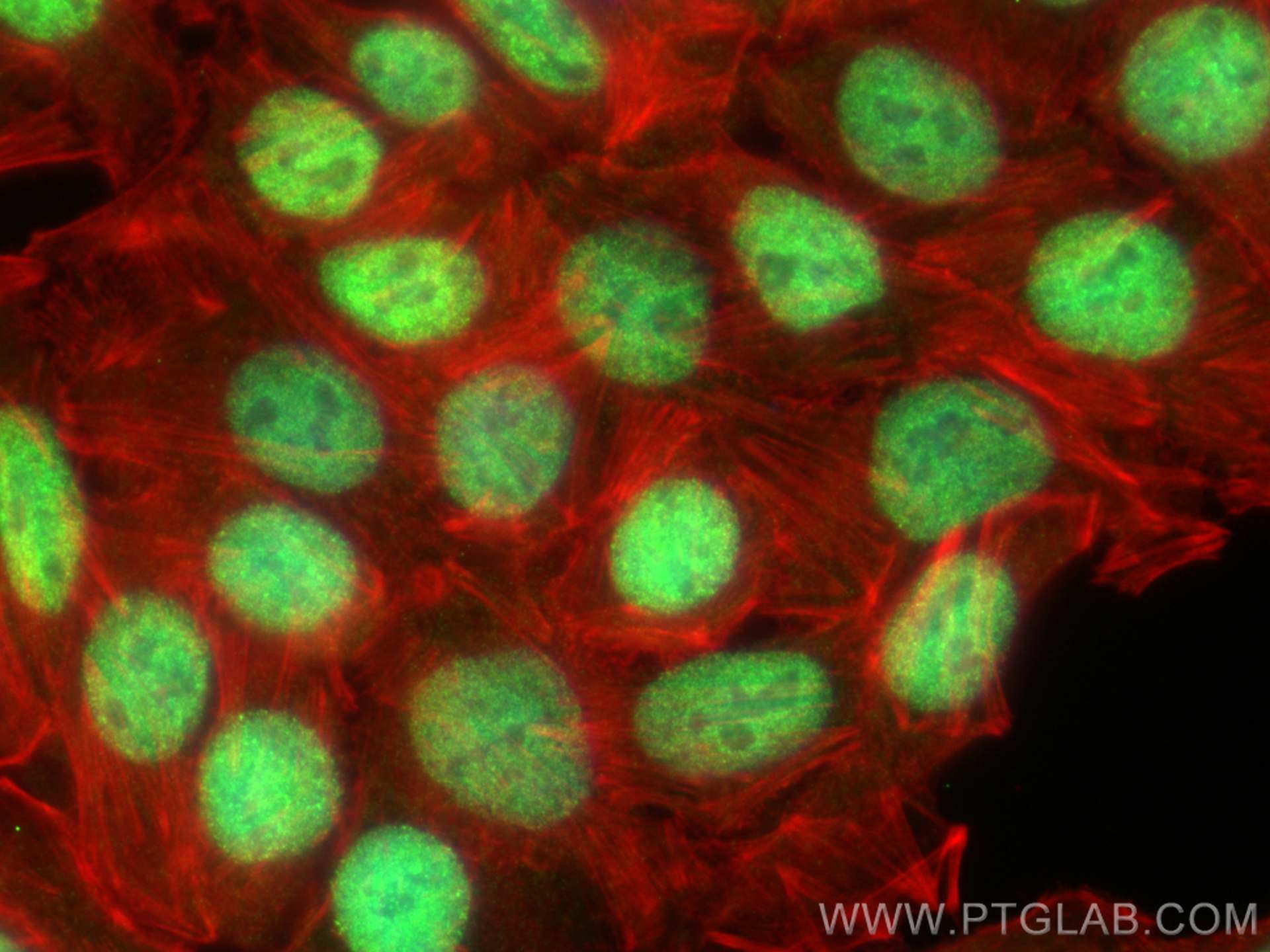
| Résultats positifs en IF/ICC | cellules HT-29, |
Dilution recommandée
| Application | Dilution |
|---|---|
| Western Blot (WB) | WB : 1:5000-1:50000 |
| Immunoprécipitation (IP) | IP : 0.5-4.0 ug for 1.0-3.0 mg of total protein lysate |
| Immunohistochimie (IHC) | IHC : 1:50-1:500 |
| Immunofluorescence (IF)/ICC | IF/ICC : 1:50-1:500 |
| It is recommended that this reagent should be titrated in each testing system to obtain optimal results. | |
| Sample-dependent, check data in validation data gallery | |
Applications publiées
| KD/KO | See 1 publications below |
| WB | See 6 publications below |
Informations sur le produit
22393-1-AP cible MSH3 dans les applications de WB, IHC, IF/ICC, IP, ELISA et montre une réactivité avec des échantillons Humain
| Réactivité | Humain |
| Réactivité citée | Humain, souris |
| Hôte / Isotype | Lapin / IgG |
| Clonalité | Polyclonal |
| Type | Anticorps |
| Immunogène | MSH3 Protéine recombinante Ag18060 |
| Nom complet | mutS homolog 3 (E. coli) |
| Masse moléculaire calculée | 1137 aa, 127 kDa |
| Poids moléculaire observé | 127 kDa |
| Numéro d’acquisition GenBank | BC130434 |
| Symbole du gène | MSH3 |
| Identification du gène (NCBI) | 4437 |
| Conjugaison | Non conjugué |
| Forme | Liquide |
| Méthode de purification | Purification par affinité contre l'antigène |
| Tampon de stockage | PBS avec azoture de sodium à 0,02 % et glycérol à 50 % pH 7,3 |
| Conditions de stockage | Stocker à -20°C. Stable pendant un an après l'expédition. L'aliquotage n'est pas nécessaire pour le stockage à -20oC Les 20ul contiennent 0,1% de BSA. |
Informations générales
MSH3 is a DNA mismatch repair (MMR) gene that heterodimerizes with MSH2 to form MutS beta which binds to DNA mismatches thereby initiating DNA repair. It undergoes frequent somatic mutation in colorectal cancers (CRCs) with MMR deficiency. MSH3 mismatch repair protein regulates sensitivity to cytotoxic drugs and a histone deacetylase inhibitor in human colon carcinoma cells (PMID: 23724141, PMID:32671096).
Protocole
| Product Specific Protocols | |
|---|---|
| WB protocol for MSH3 antibody 22393-1-AP | Download protocol |
| IHC protocol for MSH3 antibody 22393-1-AP | Download protocol |
| IF protocol for MSH3 antibody 22393-1-AP | Download protocol |
| IP protocol for MSH3 antibody 22393-1-AP | Download protocol |
| Standard Protocols | |
|---|---|
| Click here to view our Standard Protocols |
Publications
| Species | Application | Title |
|---|---|---|
Mol Cancer CircLIFR synergizes with MSH2 to attenuate chemoresistance via MutSα/ATM-p73 axis in bladder cancer. | ||
Sci Adv Deficiency in mammalian STN1 promotes colon cancer development via inhibiting DNA repair | ||
Hum Mol Genet Lack of association of somatic CAG repeat expansion with striatal neurodegeneration in HD knock-in animal models. | ||
bioRxiv D-type cyclins regulate DNA mismatch repair in the G1 and S phases of the cell cycle, maintaining genome stability | ||
Prog Neurobiol Loss of TDP-43 promotes somatic CAG repeat expansion in Huntington's disease knock-in mice
|
Avis
The reviews below have been submitted by verified Proteintech customers who received an incentive forproviding their feedback.
FH Daniela (Verified Customer) (03-17-2022) | In western blot we observed multiple bands, but none at the expected molecular weight. In immunofluorescene, staining was citoplasmatic, while MSH3 is expected to be in the nucleus.
 |
