- Phare
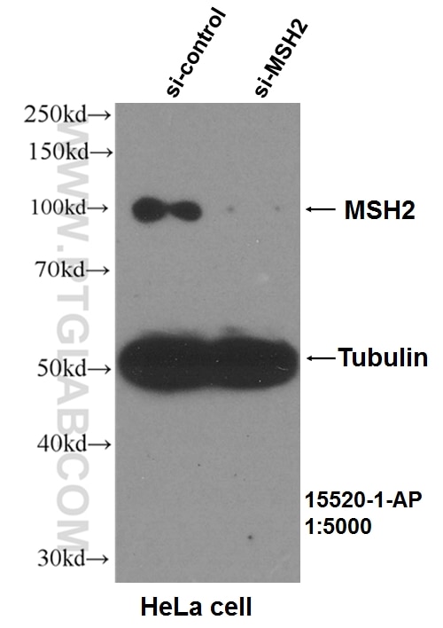
- Validé par KD/KO
Anticorps Polyclonal de lapin anti-MSH2
MSH2 Polyclonal Antibody for WB, IP, IF, IHC, ELISA, FC (Intra)
Hôte / Isotype
Lapin / IgG
Réactivité testée
Humain et plus (1)
Applications
WB, IHC, IF/ICC, FC (Intra), IP, ELISA
Conjugaison
Non conjugué
N° de cat : 15520-1-AP
Synonymes
Galerie de données de validation
Applications testées
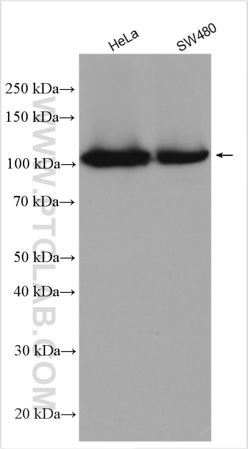
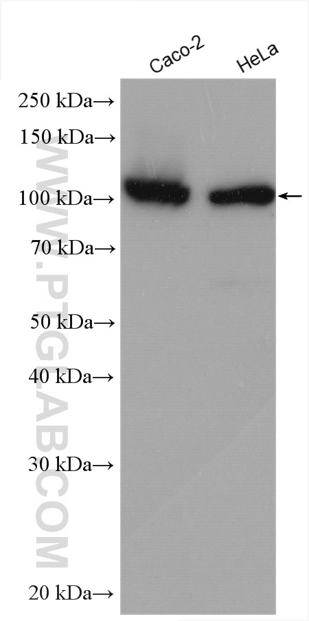
| Résultats positifs en WB | cellules HeLa, cellules Caco-2, cellules SW480 |
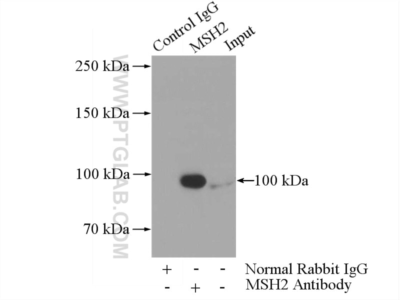
| Résultats positifs en IP | cellules HeLa |
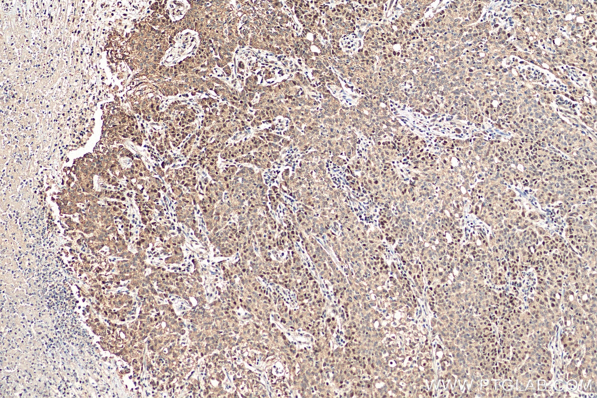
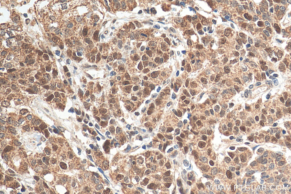
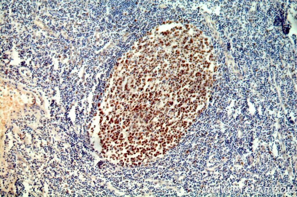
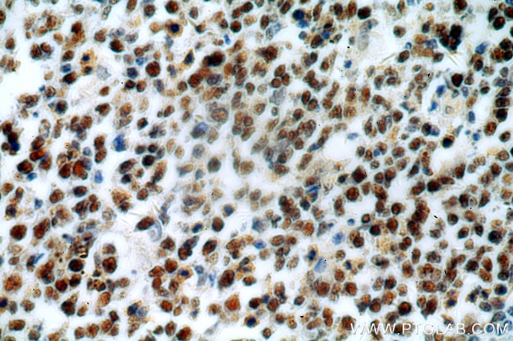
| Résultats positifs en IHC | tissu de cancer de l'estomac humain, tissu amygdalien humain il est suggéré de démasquer l'antigène avec un tampon de TE buffer pH 9.0; (*) À défaut, 'le démasquage de l'antigène peut être 'effectué avec un tampon citrate pH 6,0. |
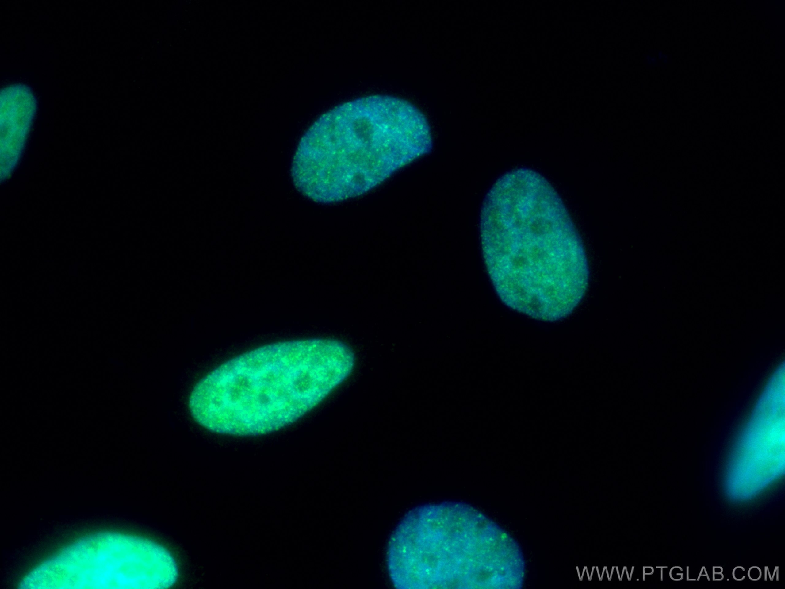
| Résultats positifs en IF/ICC | cellules HeLa, |
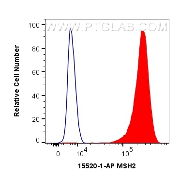
| Résultats positifs en FC (Intra) | cellules HeLa |
| Résultats positifs en cytométrie | cellules HeLa |
Dilution recommandée
| Application | Dilution |
|---|---|
| Western Blot (WB) | WB : 1:500-1:2000 |
| Immunoprécipitation (IP) | IP : 0.5-4.0 ug for 1.0-3.0 mg of total protein lysate |
| Immunohistochimie (IHC) | IHC : 1:50-1:500 |
| Immunofluorescence (IF)/ICC | IF/ICC : 1:50-1:500 |
| Flow Cytometry (FC) (INTRA) | FC (INTRA) : 0.40 ug per 10^6 cells in a 100 µl suspension |
| Flow Cytometry (FC) | FC : 0.40 ug per 10^6 cells in a 100 µl suspension |
| It is recommended that this reagent should be titrated in each testing system to obtain optimal results. | |
| Sample-dependent, check data in validation data gallery | |
Applications publiées
| KD/KO | See 4 publications below |
| WB | See 16 publications below |
| IHC | See 12 publications below |
| IF | See 3 publications below |
| IP | See 2 publications below |
Informations sur le produit
15520-1-AP cible MSH2 dans les applications de WB, IHC, IF/ICC, FC (Intra), IP, ELISA et montre une réactivité avec des échantillons Humain
| Réactivité | Humain |
| Réactivité citée | Humain, souris |
| Hôte / Isotype | Lapin / IgG |
| Clonalité | Polyclonal |
| Type | Anticorps |
| Immunogène | MSH2 Protéine recombinante Ag7835 |
| Nom complet | mutS homolog 2, colon cancer, nonpolyposis type 1 (E. coli) |
| Masse moléculaire calculée | 105 kDa |
| Poids moléculaire observé | 100 kDa-105 kDa |
| Numéro d’acquisition GenBank | BC021566 |
| Symbole du gène | MSH2 |
| Identification du gène (NCBI) | 4436 |
| Conjugaison | Non conjugué |
| Forme | Liquide |
| Méthode de purification | Purification par affinité contre l'antigène |
| Tampon de stockage | PBS avec azoture de sodium à 0,02 % et glycérol à 50 % pH 7,3 |
| Conditions de stockage | Stocker à -20°C. Stable pendant un an après l'expédition. L'aliquotage n'est pas nécessaire pour le stockage à -20oC Les 20ul contiennent 0,1% de BSA. |
Informations générales
MSH2, named for its homologous to the E.coli MutS gene, takes part in DNA mismatch repair(MMR) to maintain the stability of DNA. MSH2 contains a helix-turn-helix domain, which responds for binding to DNA. Also, MSH2 has a direct role in mutation avoidance and micro-satellite stability in cell.
Protocole
| Product Specific Protocols | |
|---|---|
| WB protocol for MSH2 antibody 15520-1-AP | Download protocol |
| IHC protocol for MSH2 antibody 15520-1-AP | Download protocol |
| IF protocol for MSH2 antibody 15520-1-AP | Download protocol |
| IP protocol for MSH2 antibody 15520-1-AP | Download protocol |
| FC protocol for MSH2 antibody 15520-1-AP | Download protocol |
| Standard Protocols | |
|---|---|
| Click here to view our Standard Protocols |
Publications
| Species | Application | Title |
|---|---|---|
Leukemia Targeting DNA polymerase β elicits synthetic lethality with mismatch repair deficiency in acute lymphoblastic leukemia
| ||
J Immunother Cancer KALRN mutations promote antitumor immunity and immunotherapy response in cancer. | ||
Hum Mol Genet Lack of association of somatic CAG repeat expansion with striatal neurodegeneration in HD knock-in animal models.
| ||
Cancer Prev Res (Phila) Estrogen stimulates the expression of mismatch repair gene hMLH1 in colonic epithelial cells. | ||
Front Oncol Molecular Classification in Patients With Endometrial Cancer After Fertility-Preserving Treatment: Application of ProMisE Classifier and Combination of Prognostic Evidence. | ||
J Biol Chem Ectopic expression of human MutS homologue 2 on renal carcinoma cells is induced by oxidative stress with interleukin-18 promotion via p38 mitogen-activated protein kinase (MAPK) and c-Jun N-terminal kinase (JNK) signaling pathways. |
