- Phare
- Validé par KD/KO
Anticorps Polyclonal de lapin anti-MGAT3
MGAT3 Polyclonal Antibody for WB, IHC, ELISA
Hôte / Isotype
Lapin / IgG
Réactivité testée
Humain, rat, souris
Applications
WB, IF, IHC, ELISA
Conjugaison
Non conjugué
N° de cat : 17869-1-AP
Synonymes
Galerie de données de validation
Applications testées
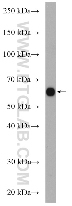
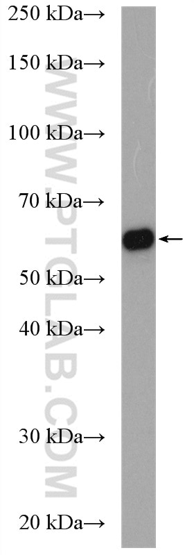
| Résultats positifs en WB | cellules COLO 320, cellules Caco-2 |
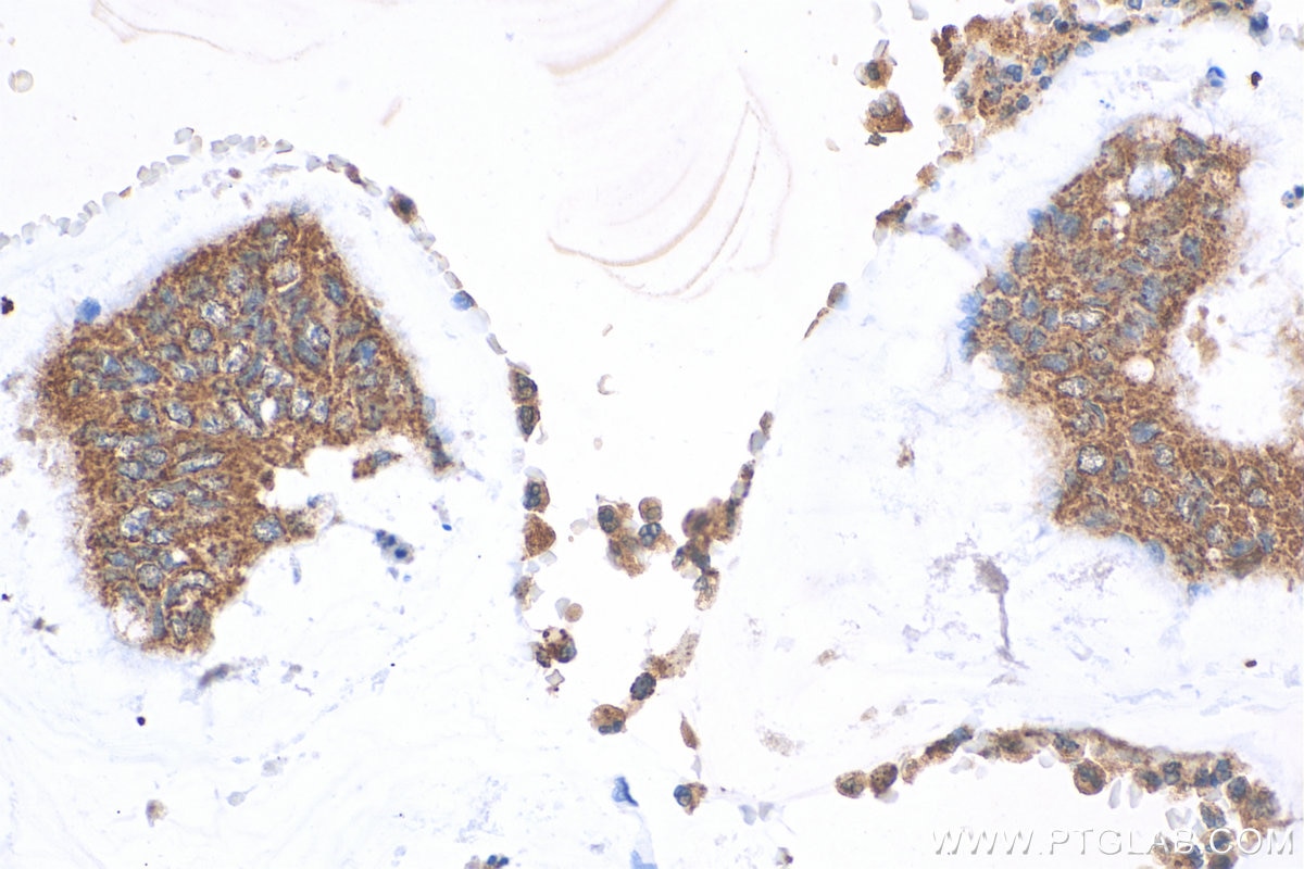
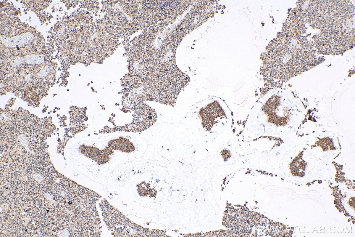
| Résultats positifs en IHC | tissu de cancer du côlon humain, il est suggéré de démasquer l'antigène avec un tampon de TE buffer pH 9.0; (*) À défaut, 'le démasquage de l'antigène peut être 'effectué avec un tampon citrate pH 6,0. |
Dilution recommandée
| Application | Dilution |
|---|---|
| Western Blot (WB) | WB : 1:500-1:1000 |
| Immunohistochimie (IHC) | IHC : 1:50-1:500 |
| It is recommended that this reagent should be titrated in each testing system to obtain optimal results. | |
| Sample-dependent, check data in validation data gallery | |
Applications publiées
| KD/KO | See 1 publications below |
| WB | See 2 publications below |
| IHC | See 2 publications below |
| IF | See 1 publications below |
Informations sur le produit
17869-1-AP cible MGAT3 dans les applications de WB, IF, IHC, ELISA et montre une réactivité avec des échantillons Humain, rat, souris
| Réactivité | Humain, rat, souris |
| Réactivité citée | Humain, souris |
| Hôte / Isotype | Lapin / IgG |
| Clonalité | Polyclonal |
| Type | Anticorps |
| Immunogène | MGAT3 Protéine recombinante Ag12352 |
| Nom complet | mannosyl (beta-1,4-)-glycoprotein beta-1,4-N-acetylglucosaminyltransferase |
| Masse moléculaire calculée | 533 aa, 61 kDa |
| Poids moléculaire observé | 61 kDa |
| Numéro d’acquisition GenBank | BC075026 |
| Symbole du gène | MGAT3 |
| Identification du gène (NCBI) | 4248 |
| Conjugaison | Non conjugué |
| Forme | Liquide |
| Méthode de purification | Purification par affinité contre l'antigène |
| Tampon de stockage | PBS avec azoture de sodium à 0,02 % et glycérol à 50 % pH 7,3 |
| Conditions de stockage | Stocker à -20°C. Stable pendant un an après l'expédition. L'aliquotage n'est pas nécessaire pour le stockage à -20oC Les 20ul contiennent 0,1% de BSA. |
Informations générales
In the small intestine, ~75% of triacylglycerol (TG) synthesis occurs via the monoacylglycerol acyltransferase (MGAT) pathway.Three MGAT isoforms (MGAT1, MGAT2 and MGAT3) have been identifed based on their extensive homology to DGAT2. MGAT1 is primarily expressed in the adipose tissue, stomach and kidney but not in the small intestine. MGAT2 and MGAT3 are most highly expressed in the small intestine, suggesting that these two MGAT forms are responsible for the resynthesis of dietary TG that is packaged into chylomicrons.
Protocole
| Product Specific Protocols | |
|---|---|
| WB protocol for MGAT3 antibody 17869-1-AP | Download protocol |
| IHC protocol for MGAT3 antibody 17869-1-AP | Download protocol |
| Standard Protocols | |
|---|---|
| Click here to view our Standard Protocols |
Publications
| Species | Application | Title |
|---|---|---|
Theranostics MGAT3-mediated glycosylation of tetraspanin CD82 at asparagine 157 suppresses ovarian cancer metastasis by inhibiting the integrin signaling pathway.
| ||
Neuropharmacology DHEC mesylate attenuates pathologies and aberrant bisecting N-glycosylation in Alzheimer's disease models | ||
Pharmacol Res Targeting aberrant glycosylation to modulate microglial response and improve cognition in models of Alzheimer's disease |


