- Phare
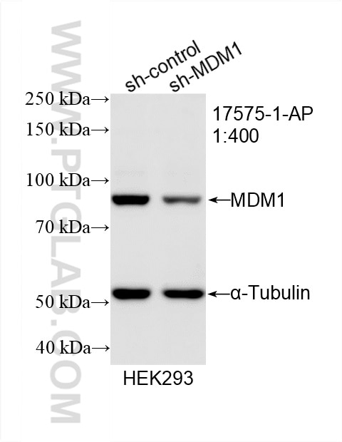
- Validé par KD/KO
Anticorps Polyclonal de lapin anti-MDM1
MDM1 Polyclonal Antibody for WB, IHC, IF/ICC, IF-P, ELISA
Hôte / Isotype
Lapin / IgG
Réactivité testée
Humain, rat, souris
Applications
WB, IHC, IF/ICC, IF-P, IP, ELISA
Conjugaison
Non conjugué
N° de cat : 17575-1-AP
Synonymes
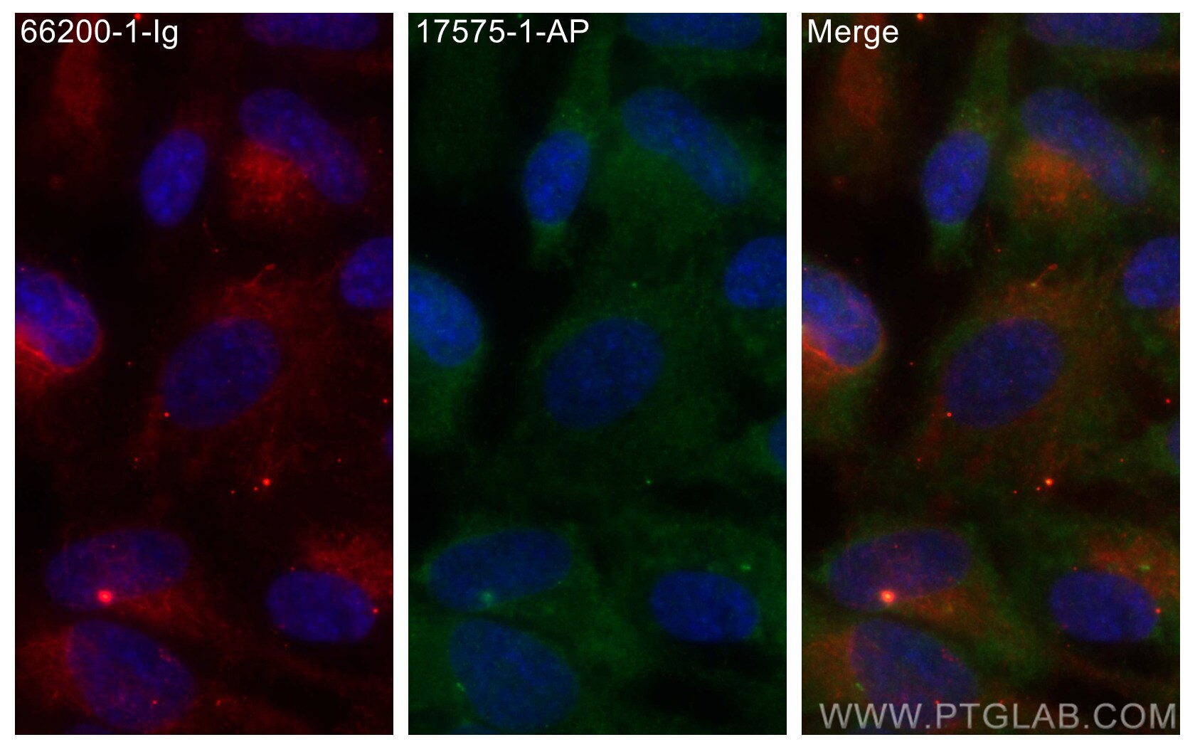
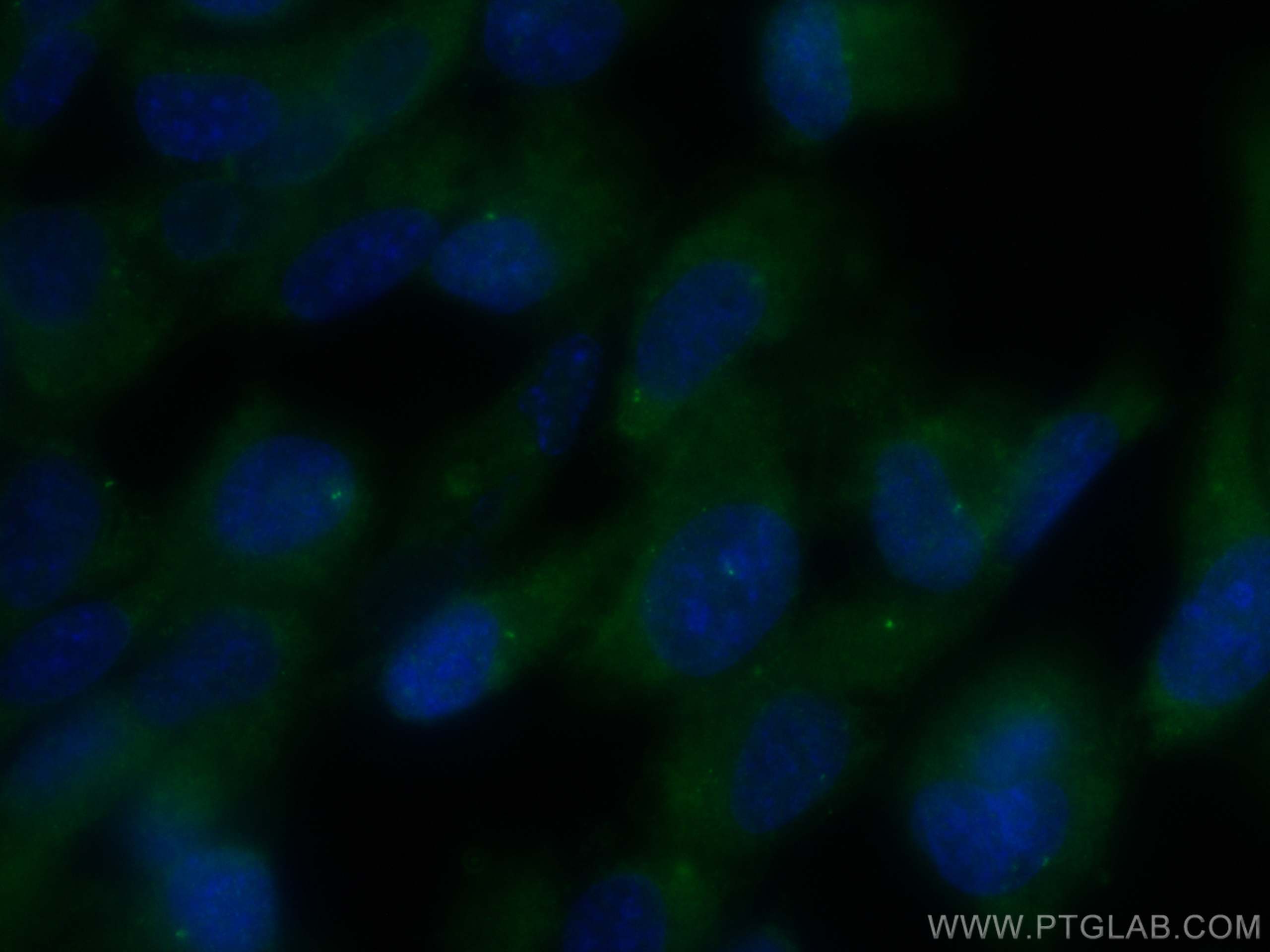
Galerie de données de validation
Applications testées
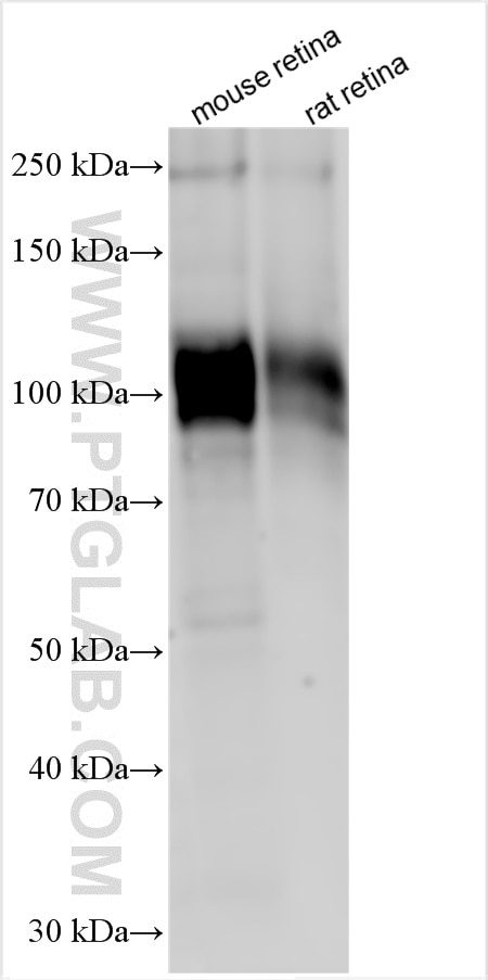
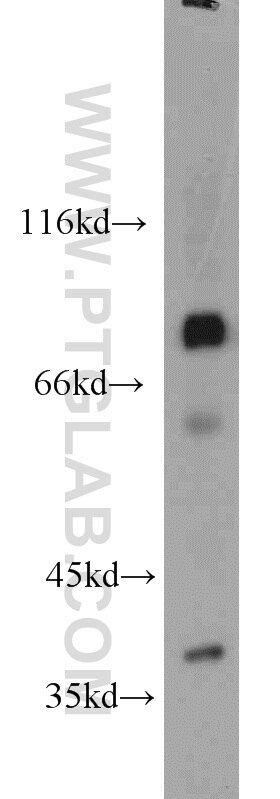
| Résultats positifs en WB | tissu rétinien de souris, cellules HEK-293, cellules Raji, tissu rétinien de rat |
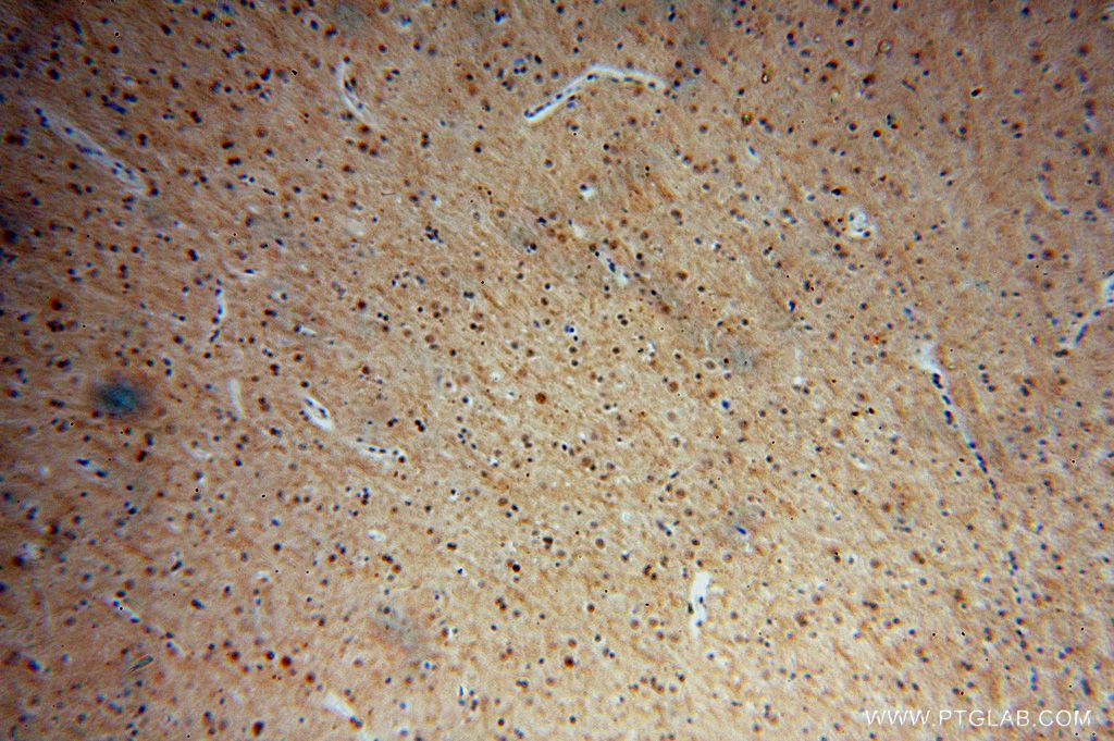
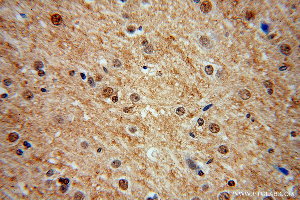
| Résultats positifs en IHC | tissu cérébral humain il est suggéré de démasquer l'antigène avec un tampon de TE buffer pH 9.0; (*) À défaut, 'le démasquage de l'antigène peut être 'effectué avec un tampon citrate pH 6,0. |
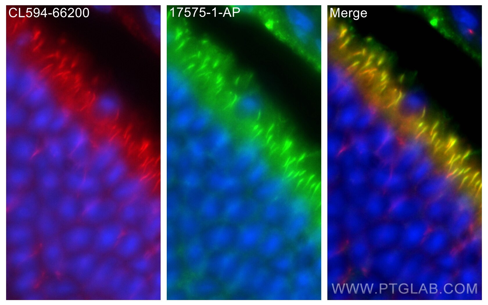
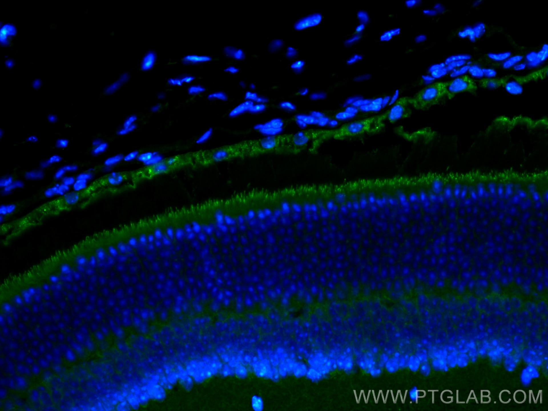
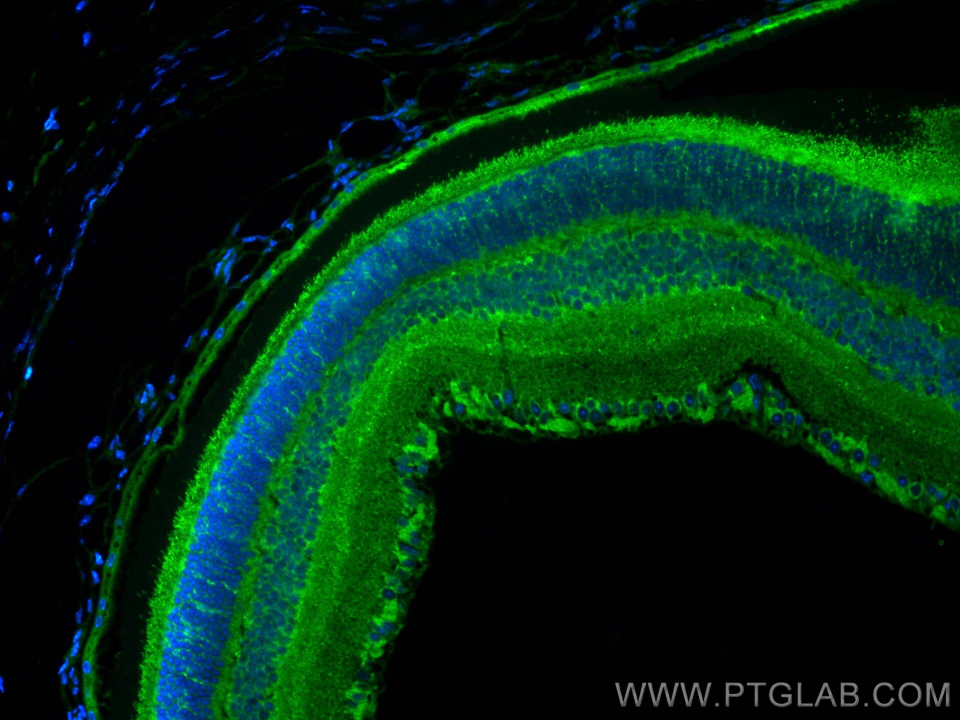
| Résultats positifs en IF-P | tissu oculaire de souris, |
| Résultats positifs en IF/ICC | cellules hTERT-RPE1, |
Dilution recommandée
| Application | Dilution |
|---|---|
| Western Blot (WB) | WB : 1:500-1:2000 |
| Immunohistochimie (IHC) | IHC : 1:20-1:200 |
| Immunofluorescence (IF)-P | IF-P : 1:50-1:500 |
| Immunofluorescence (IF)/ICC | IF/ICC : 1:200-1:800 |
| It is recommended that this reagent should be titrated in each testing system to obtain optimal results. | |
| Sample-dependent, check data in validation data gallery | |
Applications publiées
| KD/KO | See 1 publications below |
| WB | See 3 publications below |
| IP | See 1 publications below |
Informations sur le produit
17575-1-AP cible MDM1 dans les applications de WB, IHC, IF/ICC, IF-P, IP, ELISA et montre une réactivité avec des échantillons Humain, rat, souris
| Réactivité | Humain, rat, souris |
| Réactivité citée | Humain, souris |
| Hôte / Isotype | Lapin / IgG |
| Clonalité | Polyclonal |
| Type | Anticorps |
| Immunogène | MDM1 Protéine recombinante Ag10514 |
| Nom complet | Mdm1 nuclear protein homolog (mouse) |
| Masse moléculaire calculée | 81 kDa |
| Poids moléculaire observé | 81 kDa |
| Numéro d’acquisition GenBank | BC028355 |
| Symbole du gène | MDM1 |
| Identification du gène (NCBI) | 56890 |
| Conjugaison | Non conjugué |
| Forme | Liquide |
| Méthode de purification | Purification par affinité contre l'antigène |
| Tampon de stockage | PBS with 0.02% sodium azide and 50% glycerol |
| Conditions de stockage | Stocker à -20°C. Stable pendant un an après l'expédition. L'aliquotage n'est pas nécessaire pour le stockage à -20oC Les 20ul contiennent 0,1% de BSA. |
Informations générales
Mdm1 was upregulated during ciliogenesis in multiciliated tracheal epithelial cells and localized to centrioles and cilia when transfected into NIH/3T3 cells. Mdm1 is a microtubule-binding protein that inhibits centrosome duplication when overexpressed. Proteomic analysis revealed that Mdm1 was also detected in the mouse photoreceptor sensory cilium complex and the outer segment (OS) of photoreceptor cells in the retina, suggesting that Mdm1 might be involved in the regulation of the functions and structure of the centrosome, as well as cilia. (PMID: 36171205)
Protocole
| Product Specific Protocols | |
|---|---|
| WB protocol for MDM1 antibody 17575-1-AP | Download protocol |
| IHC protocol for MDM1 antibody 17575-1-AP | Download protocol |
| IF protocol for MDM1 antibody 17575-1-AP | Download protocol |
| Standard Protocols | |
|---|---|
| Click here to view our Standard Protocols |
Publications
| Species | Application | Title |
|---|---|---|
Cancer Biol Med MDM1 overexpression promotes p53 expression and cell apoptosis to enhance therapeutic sensitivity to chemoradiotherapy in patients with colorectal cancer | ||
Adv Sci (Weinh) m6A Reader hnRNPA2B1 Modulates Late Pachytene Progression in Male Meiosis Through Post-Transcriptional Control | ||
Cell Death Dis Mdm1 ablation results in retinal degeneration by specific intraflagellar transport defects of photoreceptor cells
|
