- Phare
- Validé par KD/KO
Anticorps Polyclonal de lapin anti-P3H2
P3H2 Polyclonal Antibody for WB, IP, IHC, ELISA
Hôte / Isotype
Lapin / IgG
Réactivité testée
Humain, souris
Applications
WB, IP, IHC, ELISA
Conjugaison
Non conjugué
N° de cat : 15723-1-AP
Synonymes
Galerie de données de validation
Applications testées
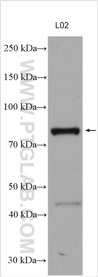
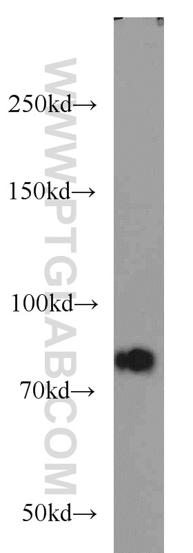
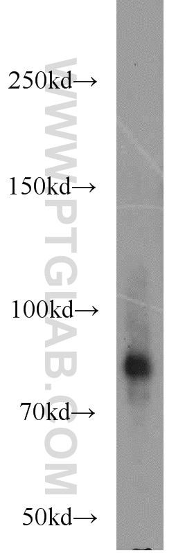
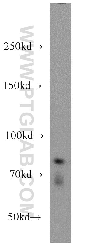
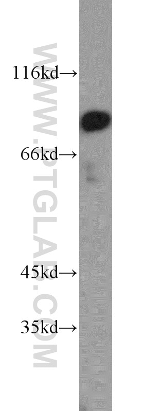
| Résultats positifs en WB | cellules L02, cellules HEK-293, tissu placentaire de souris, tissu placentaire humain, tissu rénal de souris |
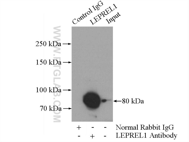
| Résultats positifs en IP | tissu rénal de souris |
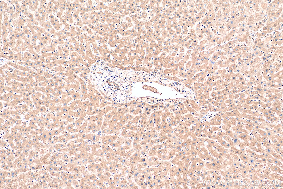
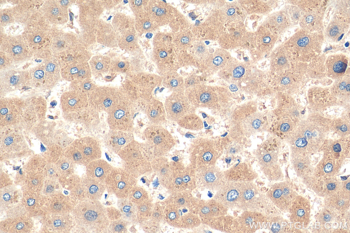
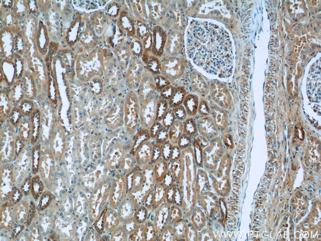
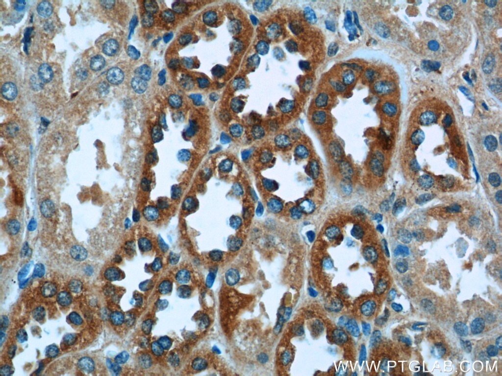
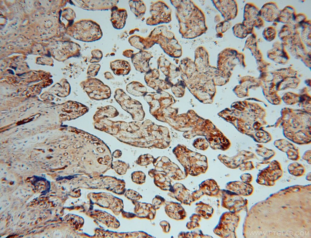
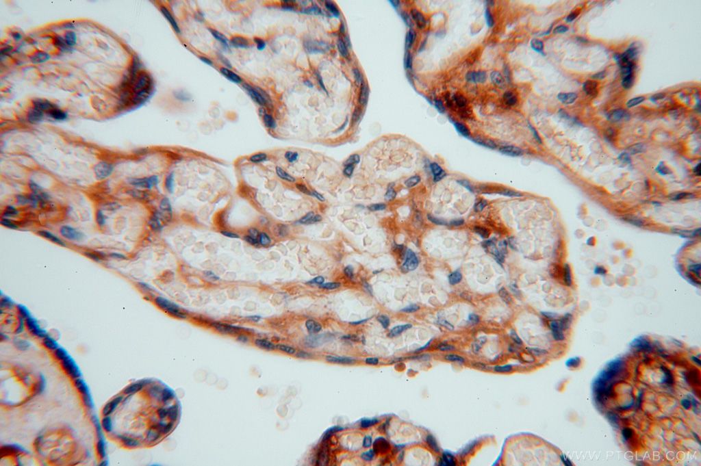
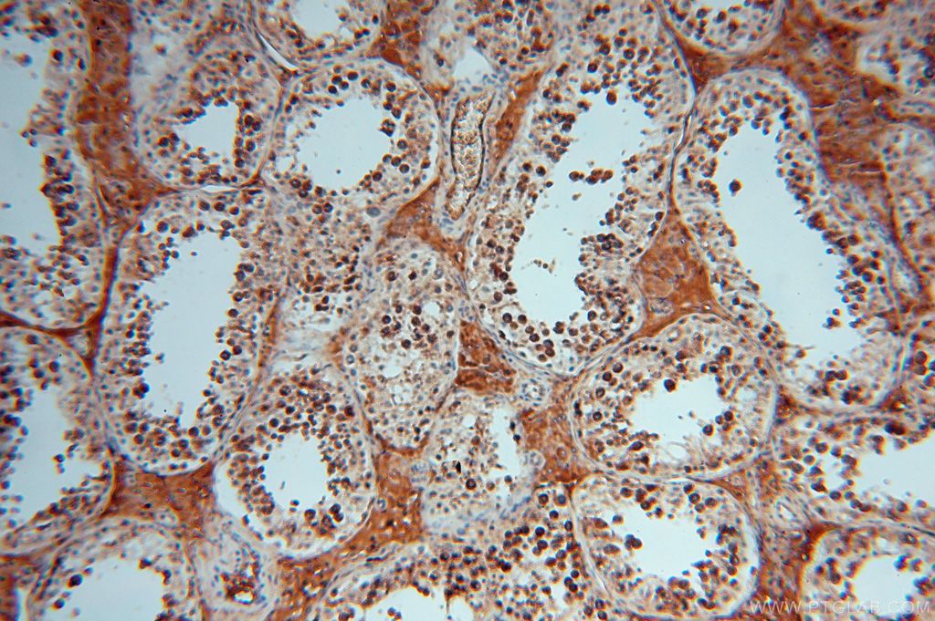
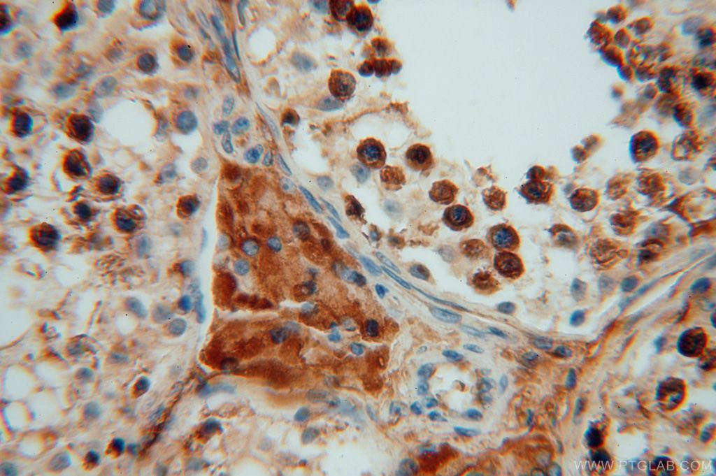
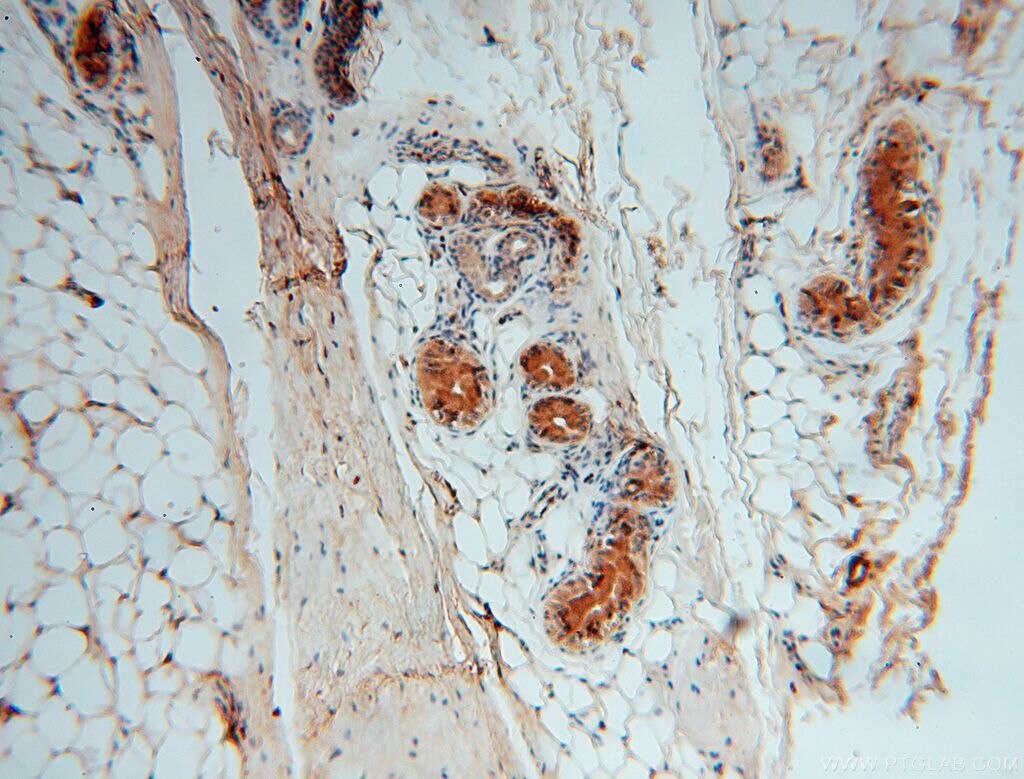
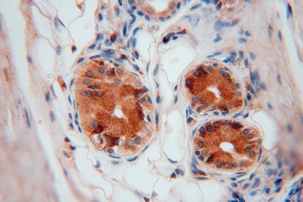
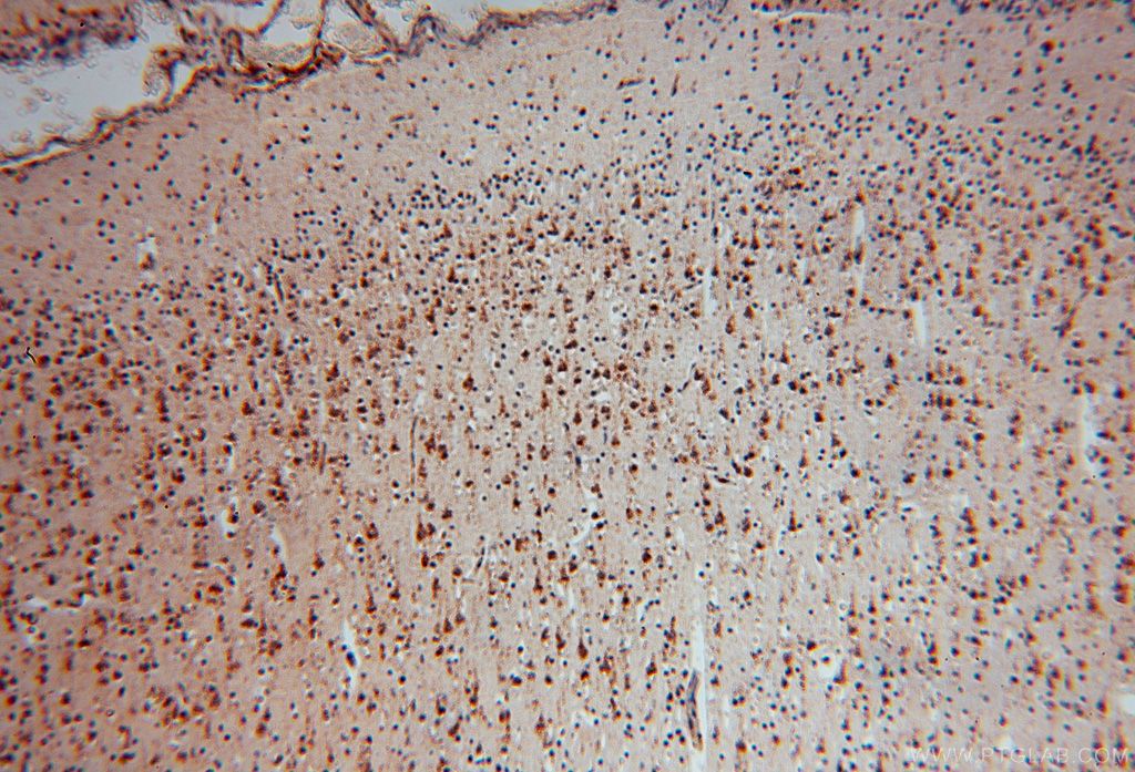
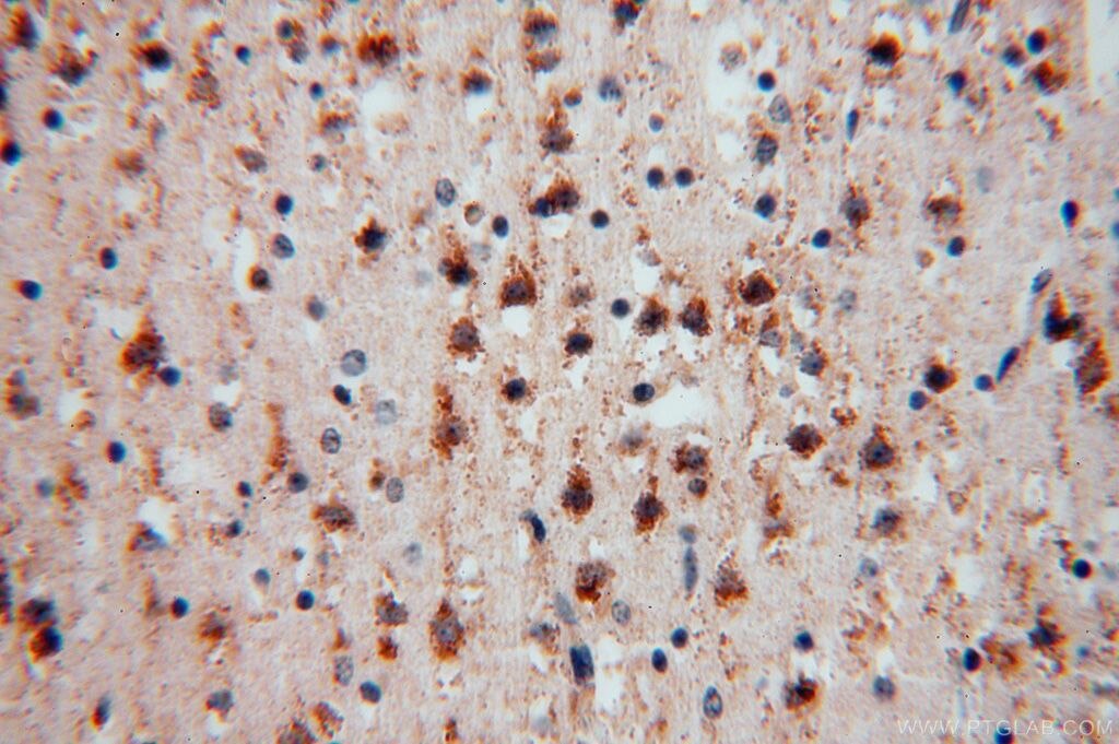
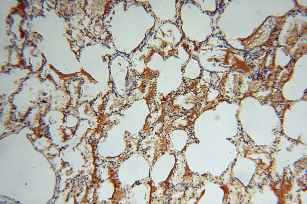
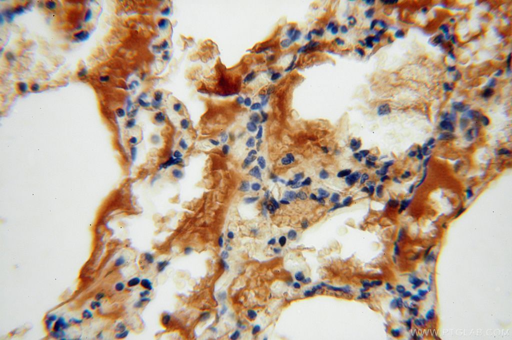
| Résultats positifs en IHC | tissu hépatique humain, tissu cérébral humain, tissu cutané humain, tissu placentaire humain, tissu pulmonaire humain, tissu rénal humain, tissu testiculaire humain il est suggéré de démasquer l'antigène avec un tampon de TE buffer pH 9.0; (*) À défaut, 'le démasquage de l'antigène peut être 'effectué avec un tampon citrate pH 6,0. |
Dilution recommandée
| Application | Dilution |
|---|---|
| Western Blot (WB) | WB : 1:1000-1:4000 |
| Immunoprécipitation (IP) | IP : 0.5-4.0 ug for 1.0-3.0 mg of total protein lysate |
| Immunohistochimie (IHC) | IHC : 1:50-1:500 |
| It is recommended that this reagent should be titrated in each testing system to obtain optimal results. | |
| Sample-dependent, check data in validation data gallery | |
Applications publiées
| KD/KO | See 1 publications below |
| WB | See 3 publications below |
Informations sur le produit
15723-1-AP cible P3H2 dans les applications de WB, IP, IHC, ELISA et montre une réactivité avec des échantillons Humain, souris
| Réactivité | Humain, souris |
| Réactivité citée | Humain, souris |
| Hôte / Isotype | Lapin / IgG |
| Clonalité | Polyclonal |
| Type | Anticorps |
| Immunogène | P3H2 Protéine recombinante Ag8444 |
| Nom complet | leprecan-like 1 |
| Masse moléculaire calculée | 708aa,81 kDa; 527aa,60 kDa |
| Poids moléculaire observé | 80 kDa |
| Numéro d’acquisition GenBank | BC005029 |
| Symbole du gène | LEPREL1 |
| Identification du gène (NCBI) | 55214 |
| Conjugaison | Non conjugué |
| Forme | Liquide |
| Méthode de purification | Purification par affinité contre l'antigène |
| Tampon de stockage | PBS avec azoture de sodium à 0,02 % et glycérol à 50 % pH 7,3 |
| Conditions de stockage | Stocker à -20°C. Stable pendant un an après l'expédition. L'aliquotage n'est pas nécessaire pour le stockage à -20oC Les 20ul contiennent 0,1% de BSA. |
Informations générales
P3H2 (prolyl 3-hydroxylase 2, also known as LEPREL1) is a member of the leprecan family of proteins, which also include P3H1, P3H3, CRTAP and SC56. Collagen prolyl hydroxylases are required for proper collagen biosynthesis, folding, and assembly. P3H2 shows prolyl 3-hydroxylase activity catalyzing the post-translational formation of 3-hydroxyproline in -Xaa-Pro-Gly-sequences in collagens, especially types II, IV and V. Mutations in this gene are associated with nonsyndromic severe myopia with cataract and vitreoretinal degeneration, and downregulation of this gene may play a role in breast cancer.
Protocole
| Product Specific Protocols | |
|---|---|
| WB protocol for P3H2 antibody 15723-1-AP | Download protocol |
| IHC protocol for P3H2 antibody 15723-1-AP | Download protocol |
| IP protocol for P3H2 antibody 15723-1-AP | Download protocol |
| Standard Protocols | |
|---|---|
| Click here to view our Standard Protocols |
Publications
| Species | Application | Title |
|---|---|---|
J Biol Chem Post-translationally abnormal collagens of prolyl 3-hydroxylase-2 null mice offer a pathobiological mechanism for the high myopia linked to human LEPREL1 mutations* | ||
Sci Rep Transcriptome analysis of a dog model of congestive heart failure shows that collagen-related 2-oxoglutarate-dependent dioxygenases contribute to heart failure | ||
J Biol Chem Post-translational modification of type IV collagen with 3-hydroxyproline affects its interactions with glycoprotein VI and nidogens 1 and 2.
|
Avis
The reviews below have been submitted by verified Proteintech customers who received an incentive forproviding their feedback.
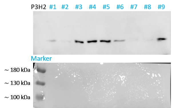
FH Shinford (Verified Customer) (10-26-2023) | We conducted a screening of basal expression levels in multiple human tumor cell lines, attempting antibody concentrations ranging from 1:500 to 2000, all of which exhibited clear bands, indicating stable working conditions. However, the expression depends on the cancer cell line itself.
 |
