- Phare
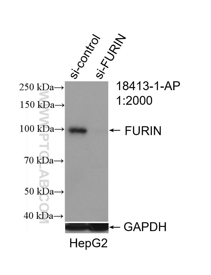
- Validé par KD/KO
Anticorps Polyclonal de lapin anti-FURIN
FURIN Polyclonal Antibody for WB, IP, IHC, ELISA
Hôte / Isotype
Lapin / IgG
Réactivité testée
Humain et plus (3)
Applications
WB, IP, IF, IHC, ELISA
Conjugaison
Non conjugué
N° de cat : 18413-1-AP
Synonymes
Galerie de données de validation
Applications testées
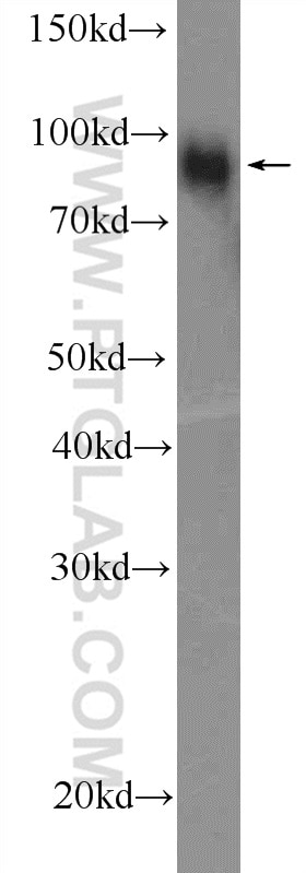
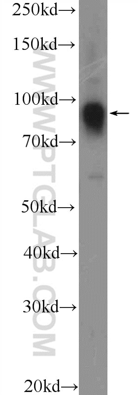
| Résultats positifs en WB | cellules HepG2, cellules COLO 320, cellules HeLa |
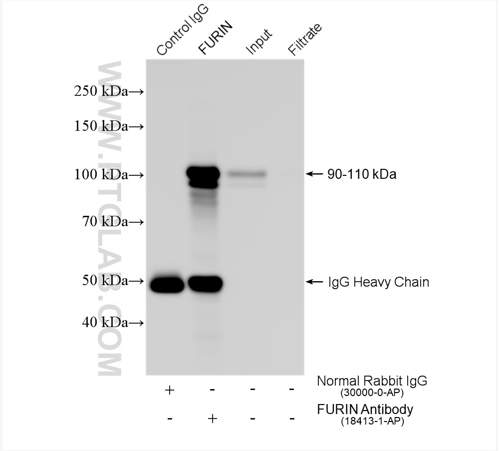
| Résultats positifs en IP | cellules HeLa, |
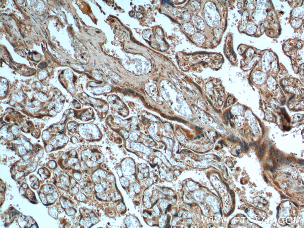
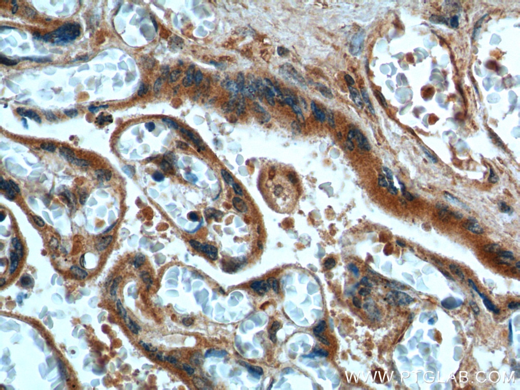
| Résultats positifs en IHC | tissu placentaire humain il est suggéré de démasquer l'antigène avec un tampon de TE buffer pH 9.0; (*) À défaut, 'le démasquage de l'antigène peut être 'effectué avec un tampon citrate pH 6,0. |
Dilution recommandée
| Application | Dilution |
|---|---|
| Western Blot (WB) | WB : 1:1000-1:4000 |
| Immunoprécipitation (IP) | IP : 0.5-4.0 ug for 1.0-3.0 mg of total protein lysate |
| Immunohistochimie (IHC) | IHC : 1:20-1:200 |
| It is recommended that this reagent should be titrated in each testing system to obtain optimal results. | |
| Sample-dependent, check data in validation data gallery | |
Applications publiées
| KD/KO | See 4 publications below |
| WB | See 16 publications below |
| IHC | See 3 publications below |
| IF | See 1 publications below |
Informations sur le produit
18413-1-AP cible FURIN dans les applications de WB, IP, IF, IHC, ELISA et montre une réactivité avec des échantillons Humain
| Réactivité | Humain |
| Réactivité citée | rat, Humain, poulet, souris |
| Hôte / Isotype | Lapin / IgG |
| Clonalité | Polyclonal |
| Type | Anticorps |
| Immunogène | FURIN Protéine recombinante Ag13261 |
| Nom complet | furin (paired basic amino acid cleaving enzyme) |
| Masse moléculaire calculée | 87 kDa |
| Poids moléculaire observé | 90-110 kDa |
| Numéro d’acquisition GenBank | BC012181 |
| Symbole du gène | FURIN |
| Identification du gène (NCBI) | 5045 |
| Conjugaison | Non conjugué |
| Forme | Liquide |
| Méthode de purification | Purification par affinité contre l'antigène |
| Tampon de stockage | PBS avec azoture de sodium à 0,02 % et glycérol à 50 % pH 7,3 |
| Conditions de stockage | Stocker à -20°C. Stable pendant un an après l'expédition. L'aliquotage n'est pas nécessaire pour le stockage à -20oC Les 20ul contiennent 0,1% de BSA. |
Informations générales
FURIN, also named FUR, PACE, PCSK3, SPC1, and Kex2p, is likely to represent the ubiquitous endoprotease activity within constitutive secretory pathways and is capable of cleavage at the RX(K/R)R consensus motif. Furin is synthesized as an inactive zymogen that may minimize the occurrence of premature enzymatic activity that would lead to alternative protein activation or degradation. The molecular weight of mature furin is 75-90 kDa and that of pre-pro furin is 95 kDa. With glycosylation and other post-translational modifications, the molecular weight of pre-pro furin will be migrated to 100-110 kDa. (PMID: 1644796, 1885622, 31336100, 35322530).
Protocole
| Product Specific Protocols | |
|---|---|
| WB protocol for FURIN antibody 18413-1-AP | Download protocol |
| IHC protocol for FURIN antibody 18413-1-AP | Download protocol |
| IP protocol for FURIN antibody 18413-1-AP | Download protocol |
| Standard Protocols | |
|---|---|
| Click here to view our Standard Protocols |
Publications
| Species | Application | Title |
|---|---|---|
Cell Rep Deep spatial proteomics reveals region-specific features of severe COVID-19-related pulmonary injury | ||
Oncogene Lipoic acid-induced oxidative stress abrogates IGF-1R maturation by inhibiting the CREB/furin axis in breast cancer cell lines. | ||
PLoS Pathog Antibody-mediated spike activation promotes cell-cell transmission of SARS-CoV-2
| ||
Oncogenesis Methyl-CpG-binding protein 2 drives the Furin/TGF-β1/Smad axis to promote epithelial-mesenchymal transition in pancreatic cancer cells. | ||
Br J Cancer Lipoic acid decreases breast cancer cell proliferation by inhibiting IGF-1R via furin downregulation.
| ||
J Virol Interplay between Furin and Sialoglycans in Modulating Adeno-Associated Viral Cell Entry
|
Avis
The reviews below have been submitted by verified Proteintech customers who received an incentive forproviding their feedback.
FH Wojciech (Verified Customer) (05-31-2024) | WB at 1:1000. Two (80 and 104 kDa) bands detected.
|
FH Wojciech (Verified Customer) (05-21-2024) | Used in WB and got 2 bands at 81 and 104 kDa at 1:1000 dilution.
|
FH Boyan (Verified Customer) (03-11-2019) | For WB, the expected band was strong; for IF, PFA fixation, it could label the cell surface-like localisation and the Golgi localisation of Furin.
|
FH Alex (Verified Customer) (10-23-2018) | Cells were fixed with 4% PFA, permeabilized with saponin, and stained with anti-furin.
|
