- Phare
- Validé par KD/KO
Anticorps Polyclonal de lapin anti-EXOSC10
EXOSC10 Polyclonal Antibody for WB, IP, IF, IHC, ELISA
Hôte / Isotype
Lapin / IgG
Réactivité testée
Humain, souris
Applications
WB, IHC, IF/ICC, IP, RIP, ELISA
Conjugaison
Non conjugué
N° de cat : 11178-1-AP
Synonymes
Galerie de données de validation
Applications testées
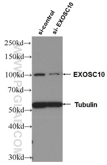
| Résultats positifs en WB | cellules HeLa, |
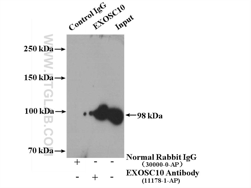
| Résultats positifs en IP | cellules MCF-7, |
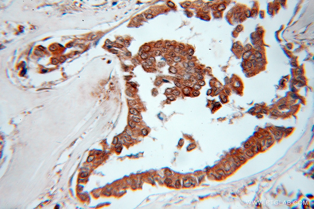
| Résultats positifs en IHC | tissu de cancer du sein humain il est suggéré de démasquer l'antigène avec un tampon de TE buffer pH 9.0; (*) À défaut, 'le démasquage de l'antigène peut être 'effectué avec un tampon citrate pH 6,0. |
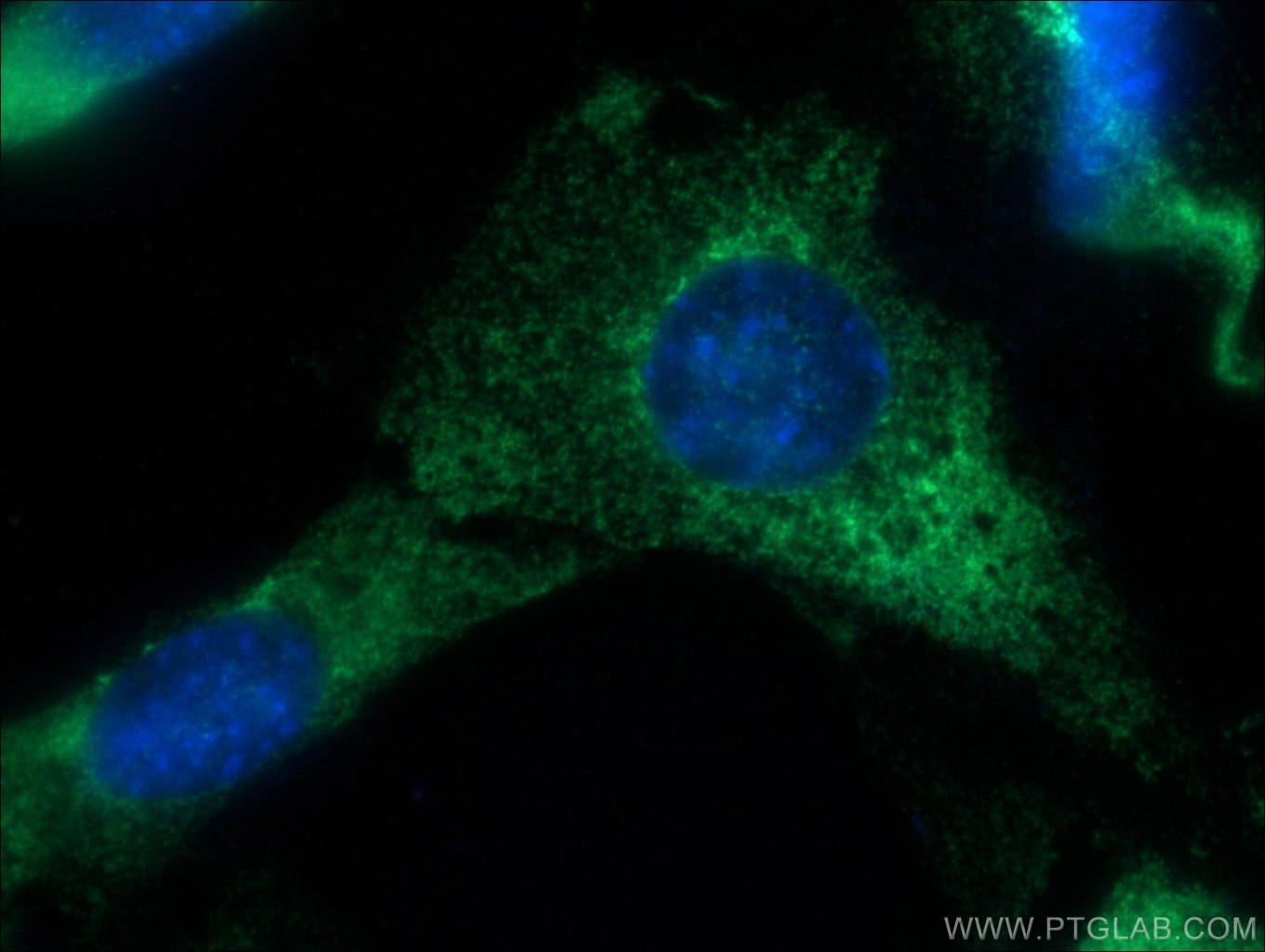
| Résultats positifs en IF/ICC | cellules NIH/3T3 |
Dilution recommandée
| Application | Dilution |
|---|---|
| Western Blot (WB) | WB : 1:500-1:2000 |
| Immunoprécipitation (IP) | IP : 0.5-4.0 ug for 1.0-3.0 mg of total protein lysate |
| Immunohistochimie (IHC) | IHC : 1:20-1:200 |
| Immunofluorescence (IF)/ICC | IF/ICC : 1:20-1:200 |
| It is recommended that this reagent should be titrated in each testing system to obtain optimal results. | |
| Sample-dependent, check data in validation data gallery | |
Applications publiées
| KD/KO | See 2 publications below |
| WB | See 4 publications below |
| IF | See 1 publications below |
| IP | See 1 publications below |
| RIP | See 1 publications below |
Informations sur le produit
11178-1-AP cible EXOSC10 dans les applications de WB, IHC, IF/ICC, IP, RIP, ELISA et montre une réactivité avec des échantillons Humain, souris
| Réactivité | Humain, souris |
| Réactivité citée | Humain, souris |
| Hôte / Isotype | Lapin / IgG |
| Clonalité | Polyclonal |
| Type | Anticorps |
| Immunogène | EXOSC10 Protéine recombinante Ag1666 |
| Nom complet | exosome component 10 |
| Masse moléculaire calculée | 98 kDa |
| Poids moléculaire observé | 100 kDa |
| Numéro d’acquisition GenBank | BC039901 |
| Symbole du gène | EXOSC10 |
| Identification du gène (NCBI) | 5394 |
| Conjugaison | Non conjugué |
| Forme | Liquide |
| Méthode de purification | Purification par affinité contre l'antigène |
| Tampon de stockage | PBS avec azoture de sodium à 0,02 % et glycérol à 50 % pH 7,3 |
| Conditions de stockage | Stocker à -20°C. Stable pendant un an après l'expédition. L'aliquotage n'est pas nécessaire pour le stockage à -20oC Les 20ul contiennent 0,1% de BSA. |
Informations générales
About 50% of patients with polymyositis/scleroderma (PM-Scl) overlap syndrome are reported to have autoantibodies to a neuclear
ucleolar particle termed PM-Scl. Exosome component 10 (EXOSC10), also named autoantigen PM/Scl 2, is the 100 kDa antigen component of PM-Scl and is recognized by most sera of PM-Scl paitents. EXOSC10 is strongly enriched in the nucleolus and a small amount has been found in cytoplasm supporting the existence of a nucleolar RNA exosome complex form. As a putative catalytic component of the RNA exosome complex which has 3'->5' exoribonuclease activity, EXOSC10 participates in a multitude of cellular RNA processing and degradation events.
Protocole
| Product Specific Protocols | |
|---|---|
| WB protocol for EXOSC10 antibody 11178-1-AP | Download protocol |
| IHC protocol for EXOSC10 antibody 11178-1-AP | Download protocol |
| IF protocol for EXOSC10 antibody 11178-1-AP | Download protocol |
| IP protocol for EXOSC10 antibody 11178-1-AP | Download protocol |
| Standard Protocols | |
|---|---|
| Click here to view our Standard Protocols |
Publications
| Species | Application | Title |
|---|---|---|
Nat Commun TASOR epigenetic repressor cooperates with a CNOT1 RNA degradation pathway to repress HIV. | ||
Development Post-transcription regulation by the exosome complex is required for cell survival and forebrain development by repressing P53 signaling.
| ||
Life Sci Alliance Low expression of EXOSC2 protects against clinical COVID-19 and impedes SARS-CoV-2 replication | ||
Cancer Res NAT10 drives cisplatin chemoresistance by enhancing ac4C-associated DNA repair in bladder cancer |
Avis
The reviews below have been submitted by verified Proteintech customers who received an incentive forproviding their feedback.
FH Roy (Verified Customer) (06-12-2024) | Did detect EXOSC10 by WB (1/1000 - overnight incubation at 4°C) in HeLa cells.
|
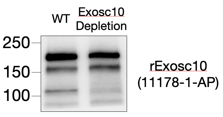
FH Tsimafei (Verified Customer) (12-17-2023) | Antibody detect Exosc10 (around 100 kDa on the image). However non-specific bands are observed in higher molecular weight area
 |
