Anticorps Polyclonal de lapin anti-DR1
DR1 Polyclonal Antibody for WB, IHC, ELISA
Hôte / Isotype
Lapin / IgG
Réactivité testée
Humain, rat, souris
Applications
WB, IHC, ELISA
Conjugaison
Non conjugué
N° de cat : 10406-1-AP
Synonymes
Galerie de données de validation
Applications testées
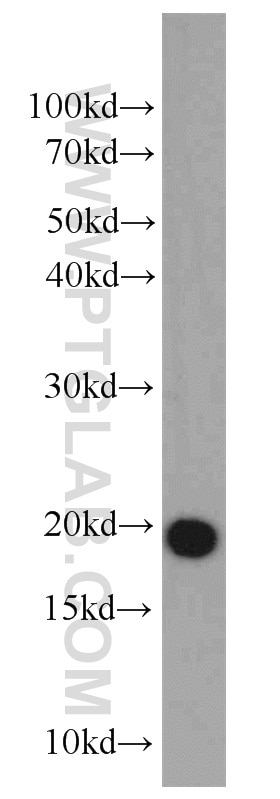
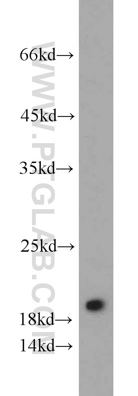
| Résultats positifs en WB | tissu testiculaire de souris, cellules PC-3 |
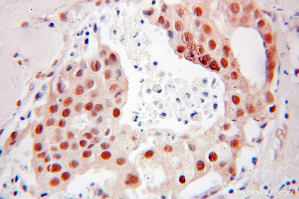
| Résultats positifs en IHC | tissu de cancer du sein humain il est suggéré de démasquer l'antigène avec un tampon de TE buffer pH 9.0; (*) À défaut, 'le démasquage de l'antigène peut être 'effectué avec un tampon citrate pH 6,0. |
Dilution recommandée
| Application | Dilution |
|---|---|
| Western Blot (WB) | WB : 1:500-1:2000 |
| Immunohistochimie (IHC) | IHC : 1:10-1:100 |
| It is recommended that this reagent should be titrated in each testing system to obtain optimal results. | |
| Sample-dependent, check data in validation data gallery | |
Applications publiées
| WB | See 2 publications below |
Informations sur le produit
10406-1-AP cible DR1 dans les applications de WB, IHC, ELISA et montre une réactivité avec des échantillons Humain, rat, souris
| Réactivité | Humain, rat, souris |
| Réactivité citée | Humain |
| Hôte / Isotype | Lapin / IgG |
| Clonalité | Polyclonal |
| Type | Anticorps |
| Immunogène | DR1 Protéine recombinante Ag0664 |
| Nom complet | down-regulator of transcription 1, TBP-binding (negative cofactor 2) |
| Masse moléculaire calculée | 19 kDa |
| Poids moléculaire observé | 19 kDa |
| Numéro d’acquisition GenBank | BC002809 |
| Symbole du gène | DR1 |
| Identification du gène (NCBI) | 1810 |
| Conjugaison | Non conjugué |
| Forme | Liquide |
| Méthode de purification | Purification par affinité contre l'antigène |
| Tampon de stockage | PBS avec azoture de sodium à 0,02 % et glycérol à 50 % pH 7,3 |
| Conditions de stockage | Stocker à -20°C. Stable pendant un an après l'expédition. L'aliquotage n'est pas nécessaire pour le stockage à -20oC Les 20ul contiennent 0,1% de BSA. |
Informations générales
Down-regulator of transcription 1, TATA box-binding protein (TBP)-binding (negative cofactor 2) is a TBP associated phosphoprotein that represses both basal and activated levels of TBP transcription. DR1 is phosphorylated in vivo and this phosphorylation affects its interaction with TBP. DR1 contains a histone fold motif at the amino terminus, a TBP-binding domain, and a glutamine- and alanine-rich region. The binding of DR1 repressor complexes to TBP-promoter complexes may establish a mechanism in which an altered DNA conformation, together with the formation of higher order complexes, inhibits the assembly of the preinitiation complex and controls the rate of RNA polymerase II transcription.
Protocole
| Product Specific Protocols | |
|---|---|
| WB protocol for DR1 antibody 10406-1-AP | Download protocol |
| IHC protocol for DR1 antibody 10406-1-AP | Download protocol |
| Standard Protocols | |
|---|---|
| Click here to view our Standard Protocols |
Publications
| Species | Application | Title |
|---|---|---|
Mol Cell Biol The double-histone-acetyltransferase complex ATAC is essential for mammalian development. | ||
Exp Cell Res Dopamine D1 receptor activation ameliorates ox-LDL-induced endothelial cell senescence via CREB/Nrf2 pathway |


