- Phare
- Validé par KD/KO
Anticorps Polyclonal de lapin anti-DOCK8
DOCK8 Polyclonal Antibody for WB, IHC, ELISA
Hôte / Isotype
Lapin / IgG
Réactivité testée
Humain, rat, souris
Applications
WB, IHC, IF, IP, ELISA
Conjugaison
Non conjugué
N° de cat : 11622-1-AP
Synonymes
Galerie de données de validation
Applications testées
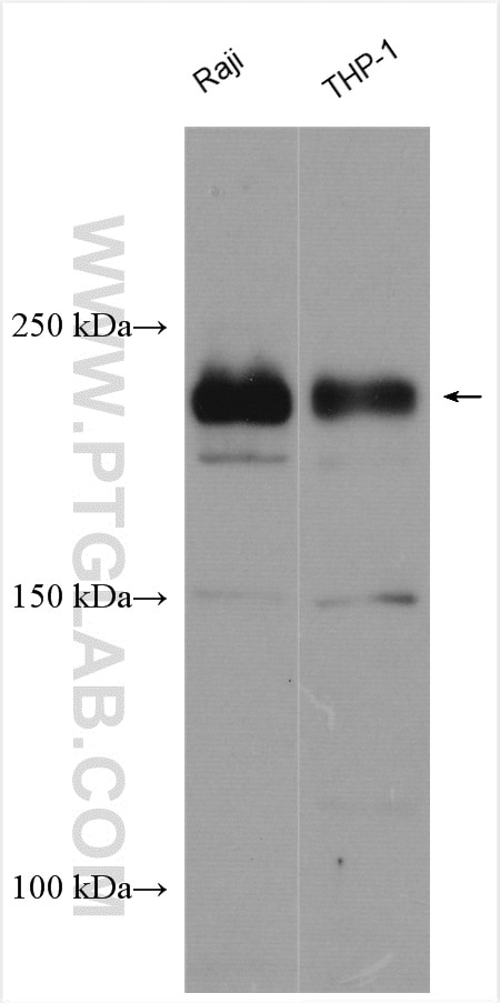
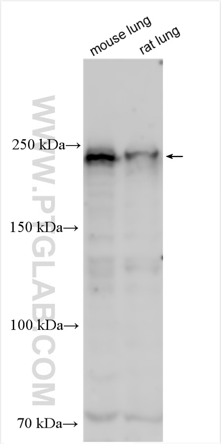
| Résultats positifs en WB | cellules Raji, cellules THP-1, tissu pulmonaire de rat |
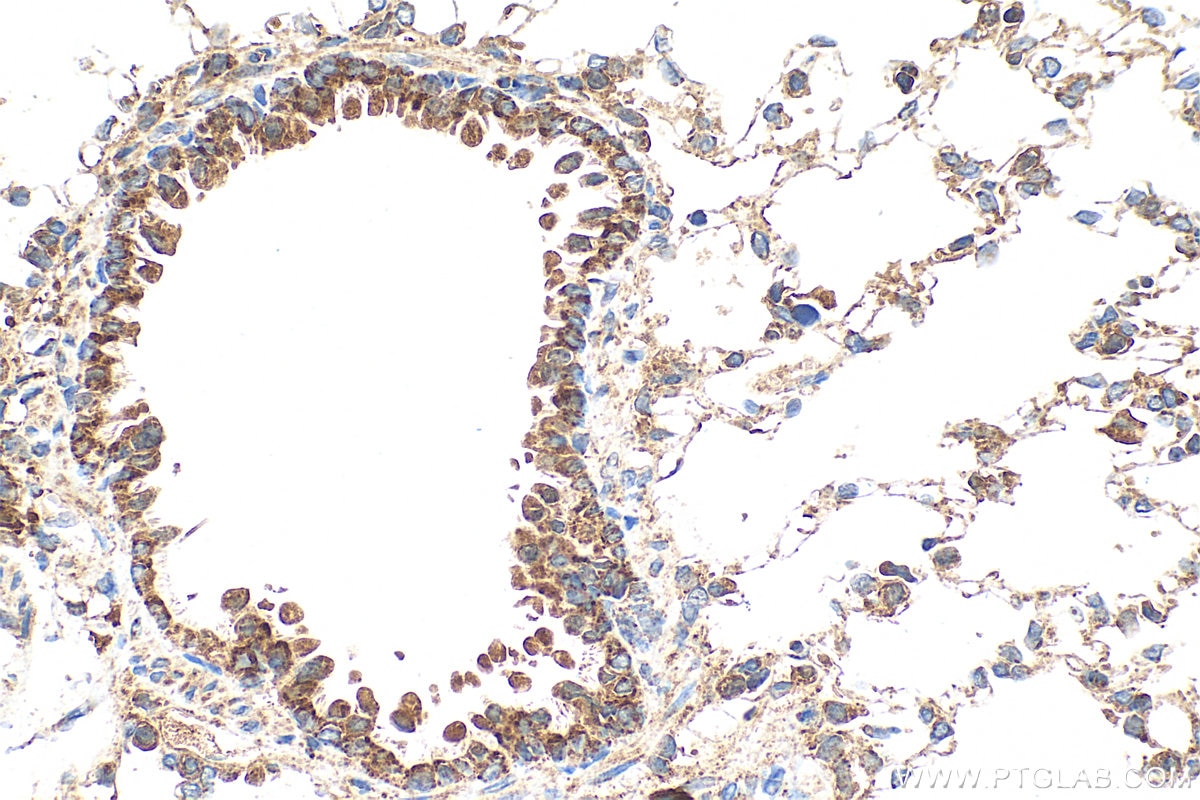
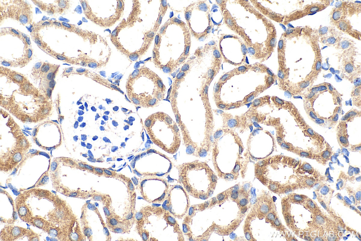
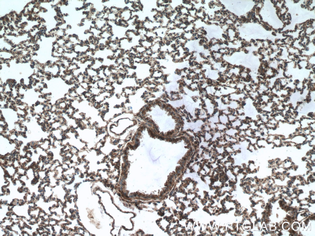
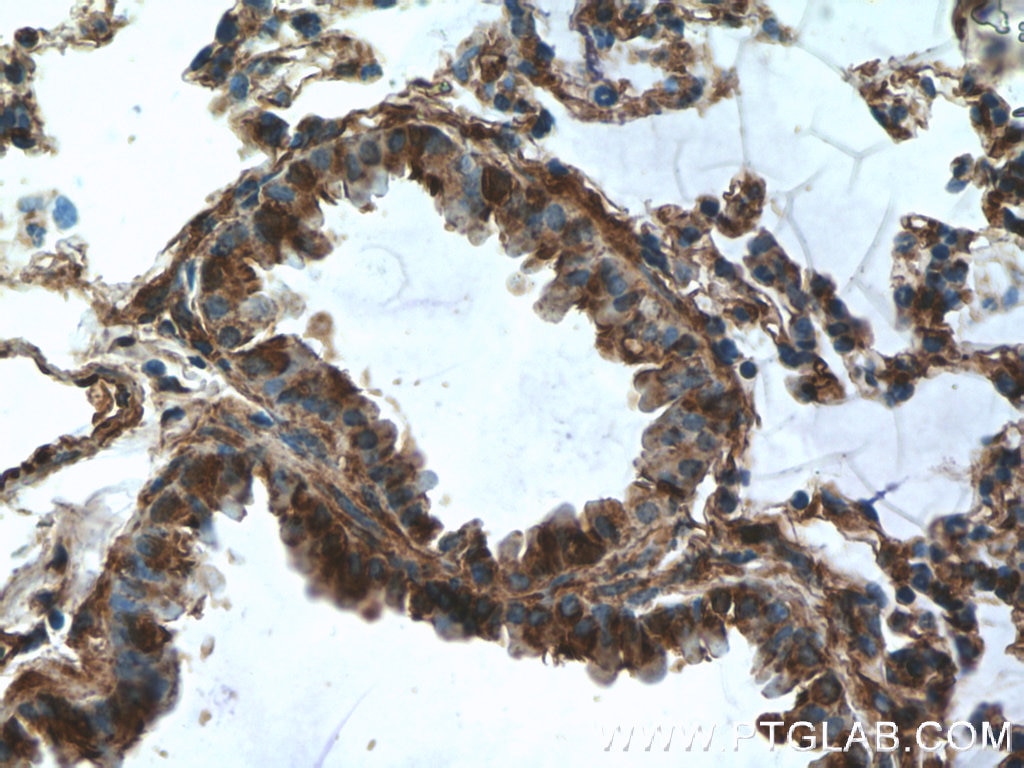
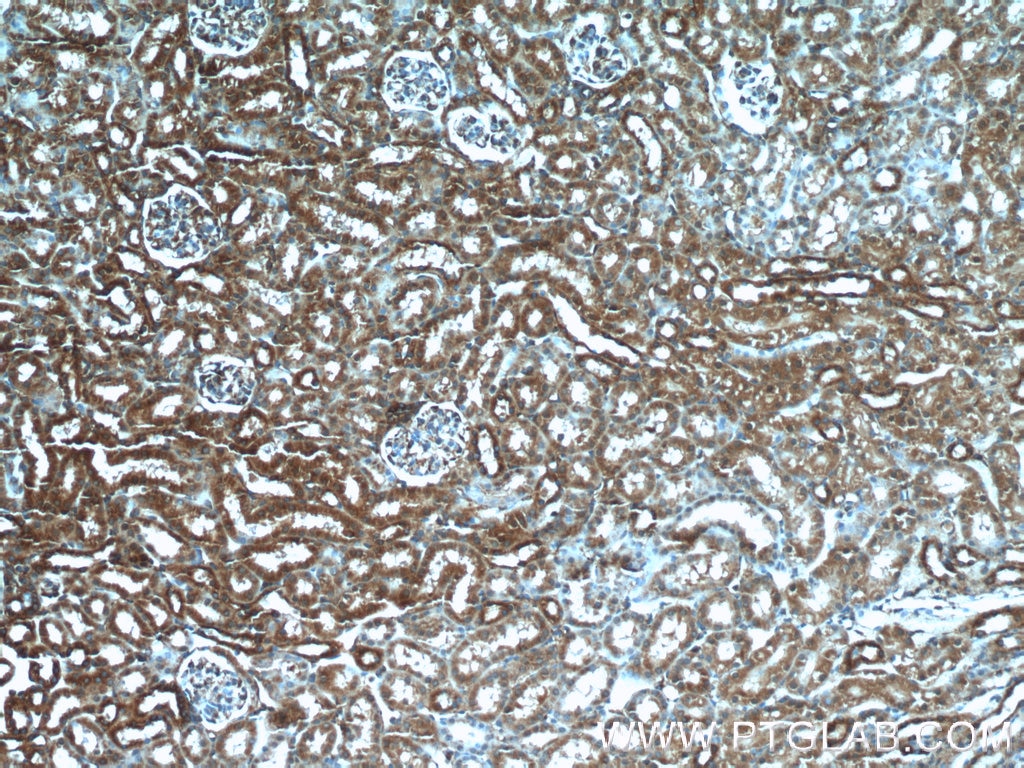
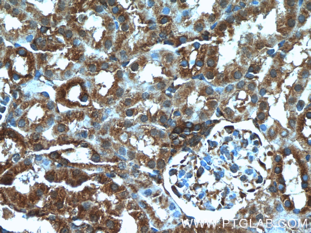
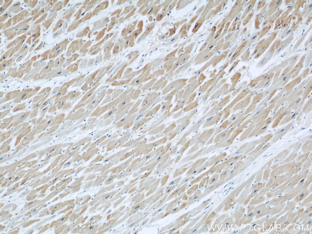
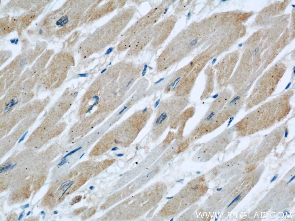
| Résultats positifs en IHC | tissu pulmonaire de rat, tissu cardiaque humain, tissu pulmonaire de souris, tissu rénal de souris il est suggéré de démasquer l'antigène avec un tampon de TE buffer pH 9.0; (*) À défaut, 'le démasquage de l'antigène peut être 'effectué avec un tampon citrate pH 6,0. |
Dilution recommandée
| Application | Dilution |
|---|---|
| Western Blot (WB) | WB : 1:500-1:1000 |
| Immunohistochimie (IHC) | IHC : 1:50-1:500 |
| It is recommended that this reagent should be titrated in each testing system to obtain optimal results. | |
| Sample-dependent, check data in validation data gallery | |
Applications publiées
| KD/KO | See 4 publications below |
| WB | See 8 publications below |
| IF | See 2 publications below |
| IP | See 2 publications below |
Informations sur le produit
11622-1-AP cible DOCK8 dans les applications de WB, IHC, IF, IP, ELISA et montre une réactivité avec des échantillons Humain, rat, souris
| Réactivité | Humain, rat, souris |
| Réactivité citée | Humain, souris |
| Hôte / Isotype | Lapin / IgG |
| Clonalité | Polyclonal |
| Type | Anticorps |
| Immunogène | DOCK8 Protéine recombinante Ag2040 |
| Nom complet | dedicator of cytokinesis 8 |
| Masse moléculaire calculée | 2031 aa, 231 kDa |
| Poids moléculaire observé | 230-239 kDa |
| Numéro d’acquisition GenBank | BC019102 |
| Symbole du gène | DOCK8 |
| Identification du gène (NCBI) | 81704 |
| Conjugaison | Non conjugué |
| Forme | Liquide |
| Méthode de purification | Purification par affinité contre l'antigène |
| Tampon de stockage | PBS with 0.02% sodium azide and 50% glycerol |
| Conditions de stockage | Stocker à -20°C. Stable pendant un an après l'expédition. L'aliquotage n'est pas nécessaire pour le stockage à -20oC Les 20ul contiennent 0,1% de BSA. |
Informations générales
Background
Dedicator of Cytokinesis 8 (DOCK8) is a protein that regulates the actin cytoskeleton, with particular importance in immune cells and a key role in innate and adaptive immune responses.
What is the molecular weight of DOCK8?
231 kDa. DOCK8 is a protein composed of 2031 amino acids and is a guanine nucleotide exchange factor (GEF).
What is the function of DOCK8?
DOCK8 is a member of the DOCK family of proteins, which have a unique DRH2 domain enabling them to act as GEFs and so controlling a range of cellular processes in various signaling pathways (PMID: 12432077). The specific target of DOCK8 is Cell division control protein 42 homolog (Cdc42), a small GTPase that is involved in regulation of the cell cycle and forms a complex. DOCK8 also acts as a scaffold molecule in this complex that initiates actin polymerization via the Wiskott-Aldrich Syndrome protein (WASp) (PubMed: 28028151, PubMed: 22461490).
What diseases are associated with DOCK8?
The role of DOCK8 in immunity was first identified with the study of DOCK8-deficient patients who presented with combined immunodeficiency (PMID: 19776401; PMID: 20004785). The subsequent study of DOCK8 in immune cells such as T cells, natural killer (NK) cells, and B cells has revealed how it regulates their normal function. This includes the regulation of immune synapse formation, immune cell trafficking, regulation of dendritic cell polarization, and cytokine production (PMID: 28366940).
DOCK8 deficiency is caused by a number of different mutations in the gene. It leads to the autosomal recessive form of the immunodeficiency disease Hyper-IgE syndrome, or Job's syndrome. The symptoms of DOCK8 deficiency include eczema, high levels of serum IgE, hypereosinophilia, and recurrent respiratory and skin infections as a result of impaired immune cell function.
Protocole
| Product Specific Protocols | |
|---|---|
| WB protocol for DOCK8 antibody 11622-1-AP | Download protocol |
| IHC protocol for DOCK8 antibody 11622-1-AP | Download protocol |
| Standard Protocols | |
|---|---|
| Click here to view our Standard Protocols |
Publications
| Species | Application | Title |
|---|---|---|
Nat Immunol Heme drives hemolysis-induced susceptibility to infection via disruption of phagocyte functions.
| ||
Cell Death Differ CCL2 regulation of MST1-mTOR-STAT1 signaling axis controls BCR signaling and B-cell differentiation. | ||
Diabetes Dedicator of Cytokinesis 5 Regulates Keratinocyte Function and Promotes Diabetic Wound Healing. | ||
Oncotarget CD147 promotes Src-dependent activation of Rac1 signaling through STAT3/DOCK8 during the motility of hepatocellular carcinoma cells.
| ||
iScience DOCK8-expressing T follicular helper cells newly generated beyond self-organized criticality cause systemic lupus erythematosus.
| ||
J Alzheimers Dis A novel finding relates to the involvement of ATF3/DOCK8 in Alzheimer's disease pathogenesis |
