- Phare
- Validé par KD/KO
Anticorps Polyclonal de lapin anti-DIAPH3
DIAPH3 Polyclonal Antibody for WB, IP, IF, IHC, ELISA
Hôte / Isotype
Lapin / IgG
Réactivité testée
Humain, rat, souris
Applications
WB, IHC, IF/ICC, IP, ELISA
Conjugaison
Non conjugué
N° de cat : 14342-1-AP
Synonymes
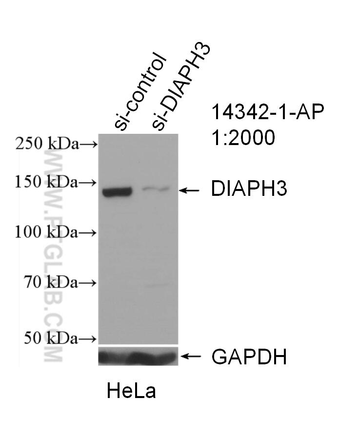
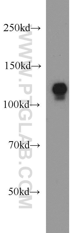
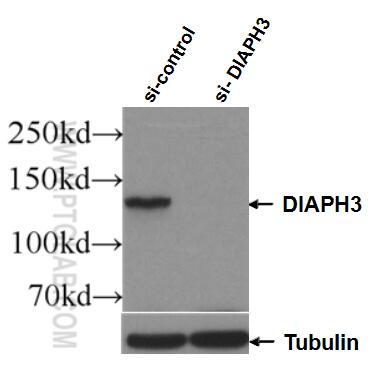
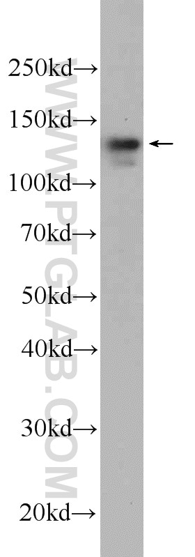
Galerie de données de validation
Applications testées
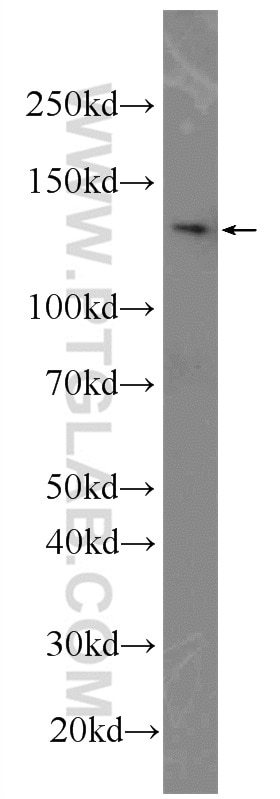
| Résultats positifs en WB | cellules HeLa, cellules DU 145, cellules HEK-293 |
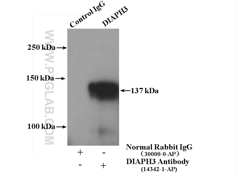
| Résultats positifs en IP | cellules HeLa |
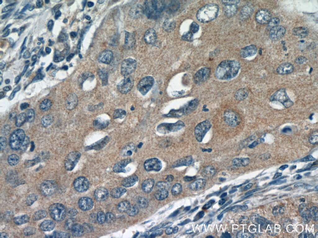
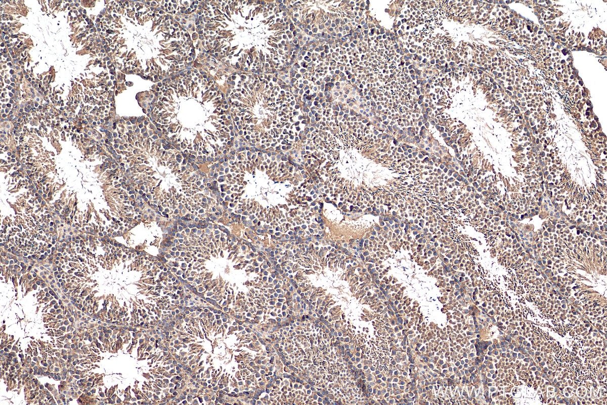
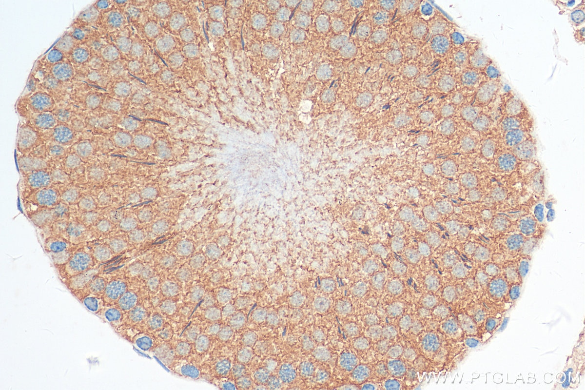
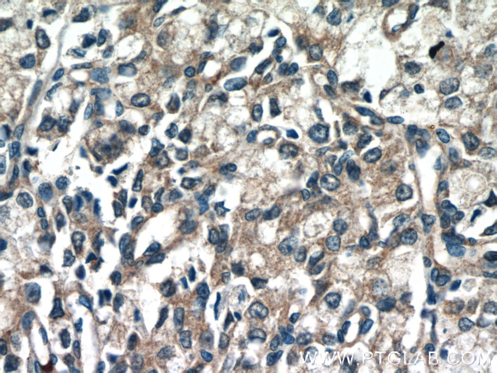
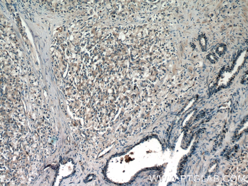
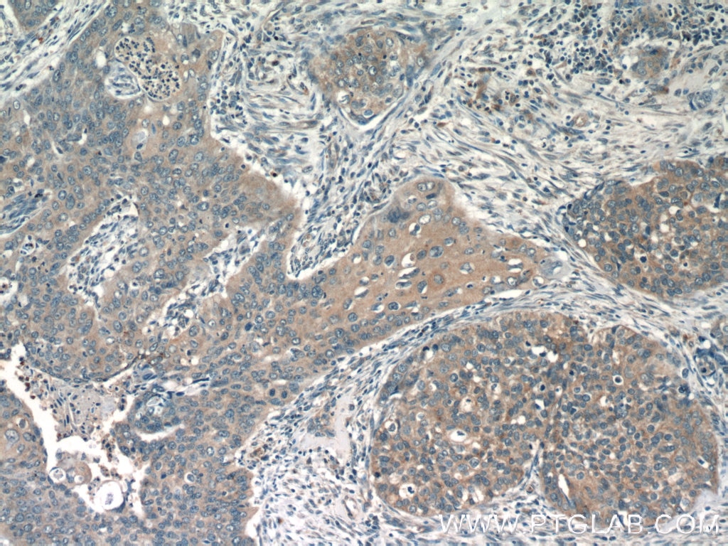
| Résultats positifs en IHC | tissu testiculaire de souris, tissu de cancer de la prostate humain, tissu de cancer du col de l'utérus humain, tissu testiculaire de rat il est suggéré de démasquer l'antigène avec un tampon de TE buffer pH 9.0; (*) À défaut, 'le démasquage de l'antigène peut être 'effectué avec un tampon citrate pH 6,0. |
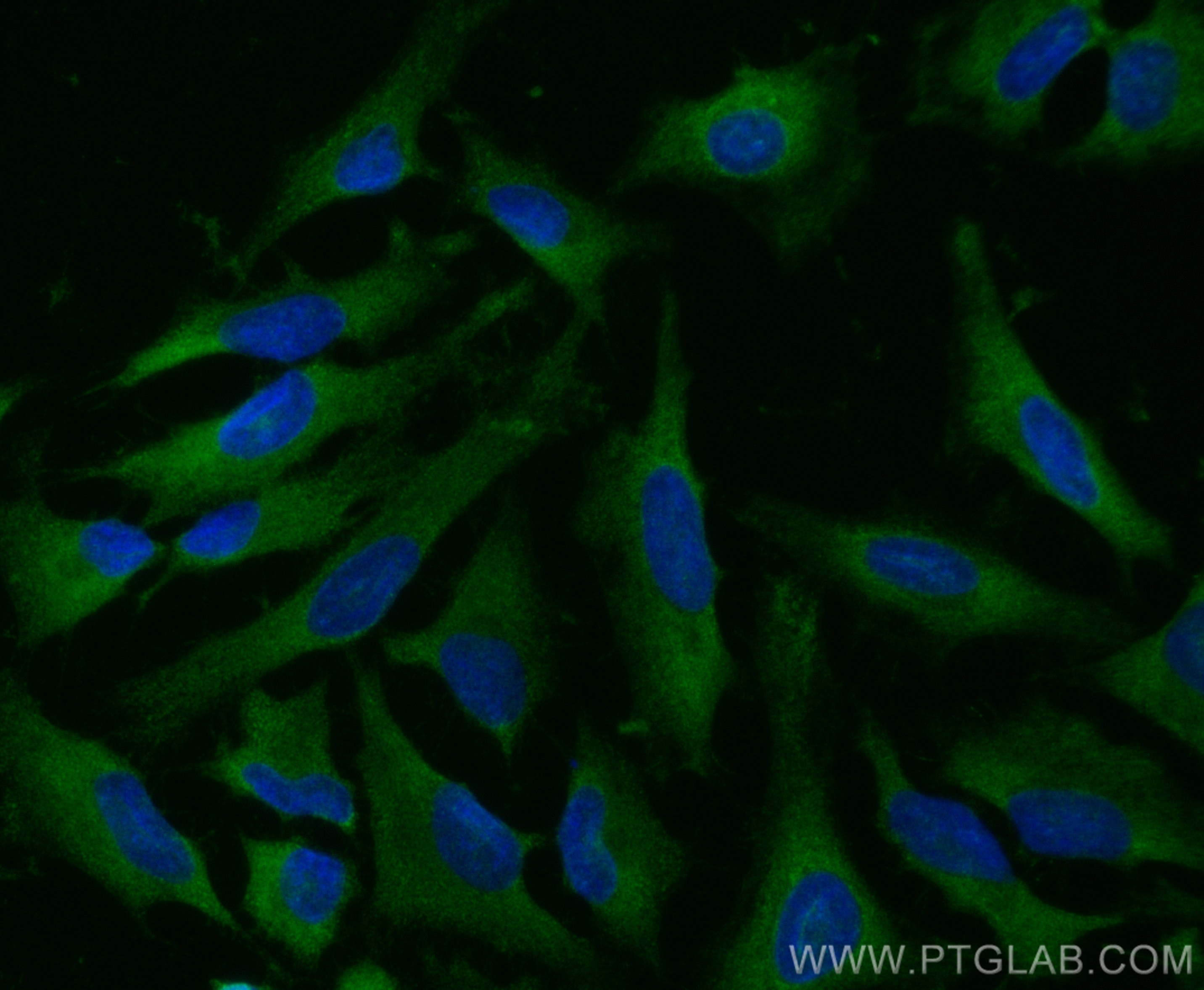
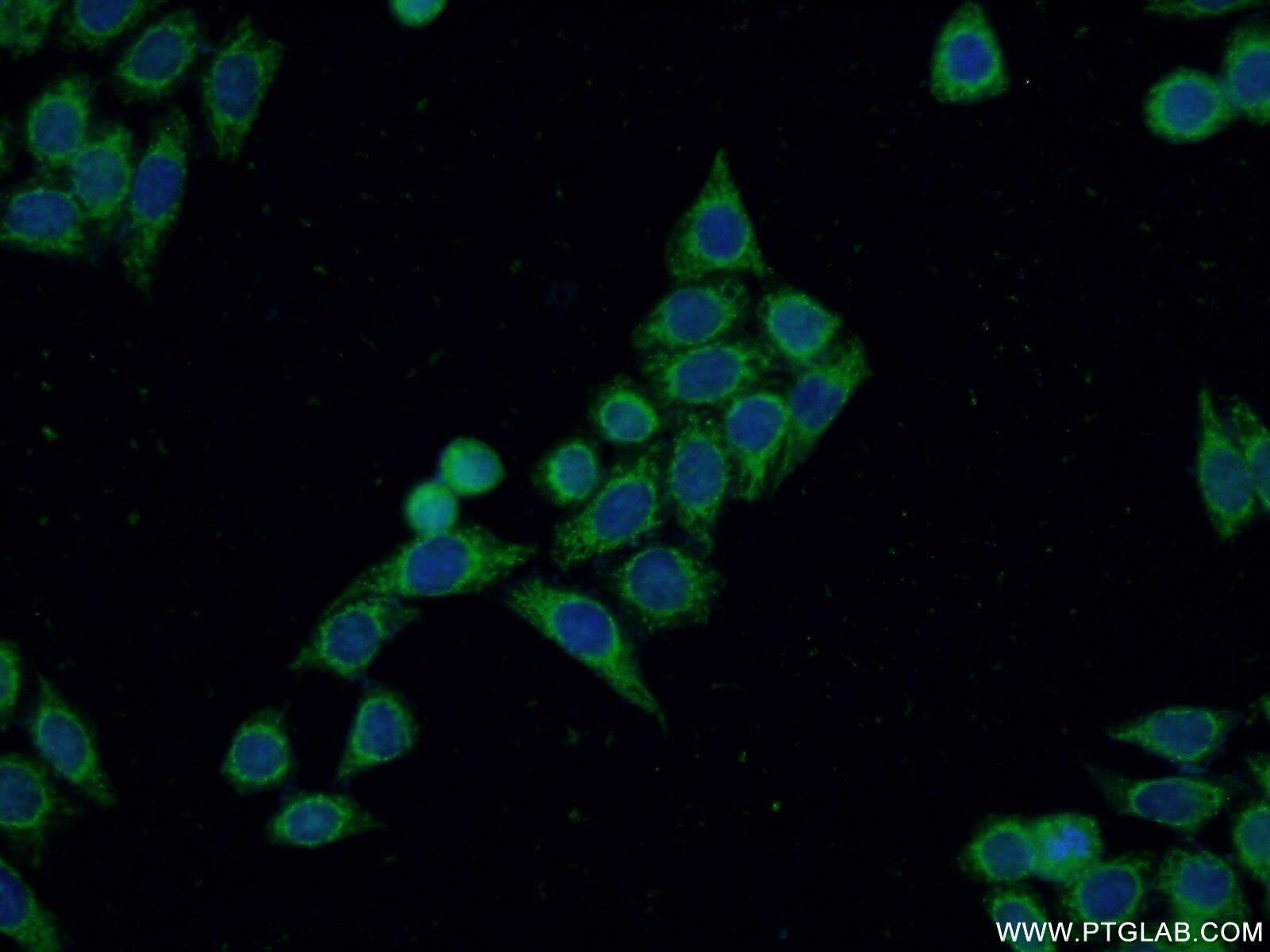
| Résultats positifs en IF/ICC | cellules HeLa, |
Dilution recommandée
| Application | Dilution |
|---|---|
| Western Blot (WB) | WB : 1:500-1:2000 |
| Immunoprécipitation (IP) | IP : 0.5-4.0 ug for 1.0-3.0 mg of total protein lysate |
| Immunohistochimie (IHC) | IHC : 1:50-1:500 |
| Immunofluorescence (IF)/ICC | IF/ICC : 1:200-1:800 |
| It is recommended that this reagent should be titrated in each testing system to obtain optimal results. | |
| Sample-dependent, check data in validation data gallery | |
Applications publiées
| KD/KO | See 7 publications below |
| WB | See 17 publications below |
| IHC | See 3 publications below |
| IF | See 6 publications below |
| IP | See 1 publications below |
Informations sur le produit
14342-1-AP cible DIAPH3 dans les applications de WB, IHC, IF/ICC, IP, ELISA et montre une réactivité avec des échantillons Humain, rat, souris
| Réactivité | Humain, rat, souris |
| Réactivité citée | Humain, souris |
| Hôte / Isotype | Lapin / IgG |
| Clonalité | Polyclonal |
| Type | Anticorps |
| Immunogène | DIAPH3 Protéine recombinante Ag5656 |
| Nom complet | diaphanous homolog 3 (Drosophila) |
| Masse moléculaire calculée | 137 kDa |
| Poids moléculaire observé | 125-137 kDa |
| Numéro d’acquisition GenBank | BC048963 |
| Symbole du gène | DIAPH3 |
| Identification du gène (NCBI) | 81624 |
| Conjugaison | Non conjugué |
| Forme | Liquide |
| Méthode de purification | Purification par affinité contre l'antigène |
| Tampon de stockage | PBS avec azoture de sodium à 0,02 % et glycérol à 50 % pH 7,3 |
| Conditions de stockage | Stocker à -20°C. Stable pendant un an après l'expédition. L'aliquotage n'est pas nécessaire pour le stockage à -20oC Les 20ul contiennent 0,1% de BSA. |
Informations générales
DIAPH3 is also refered as diaphanous-related formin-3 which binds to GTP-bound form of Rho and to profilin, acts in a Rho-dependent manner to recruit profilin to the membrane. It is required for cytokinesis, stress fiber formation, and transcriptional activation of the serum response factor. Seven isoforms of DIAPH3 exist due to the alternative splicing. Recently it has been reported that dimerization and actin assembly activity of DIAPH3 isoforms 1 and 7 are essential for induction of specific cell protrusions. It may propose that isoform-selective actin assembly by DIAPH3 exerts specific and differentially regulated functions during cell adhesion and motility.
Protocole
| Product Specific Protocols | |
|---|---|
| WB protocol for DIAPH3 antibody 14342-1-AP | Download protocol |
| IHC protocol for DIAPH3 antibody 14342-1-AP | Download protocol |
| IF protocol for DIAPH3 antibody 14342-1-AP | Download protocol |
| IP protocol for DIAPH3 antibody 14342-1-AP | Download protocol |
| Standard Protocols | |
|---|---|
| Click here to view our Standard Protocols |
Publications
| Species | Application | Title |
|---|---|---|
Blood Membrane skeleton modulates erythroid proteome remodeling and organelle clearance.
| ||
Sci Adv Recessive NOS1AP variants impair actin remodeling and cause glomerulopathy in humans and mice. | ||
Aging (Albany NY) The pan-cancer analysis identified DIAPH3 as a diagnostic biomarker of clinical cancer | ||
Cell Res Differing and isoform-specific roles for the formin DIAPH3 in plasma membrane blebbing and filopodia formation.
| ||
EMBO J TFAM loss induces nuclear actin assembly upon mDia2 malonylation to promote liver cancer metastasis. | ||
Elife Generation of stress fibers through myosin-driven reorganization of the actin cortex
|
