Phospho-Histone H3 (Ser10) Monoklonaler Antikörper
Phospho-Histone H3 (Ser10) Monoklonal Antikörper für IF/ICC
Wirt / Isotyp
Maus / IgG1
Getestete Reaktivität
Hausschwein, human, Maus, Ratte
Anwendung
IF/ICC
Konjugation
CoraLite® Plus 647 Fluorescent Dye
CloneNo.
4C7G2
Kat-Nr. : CL647-66863
Synonyme
Geprüfte Anwendungen
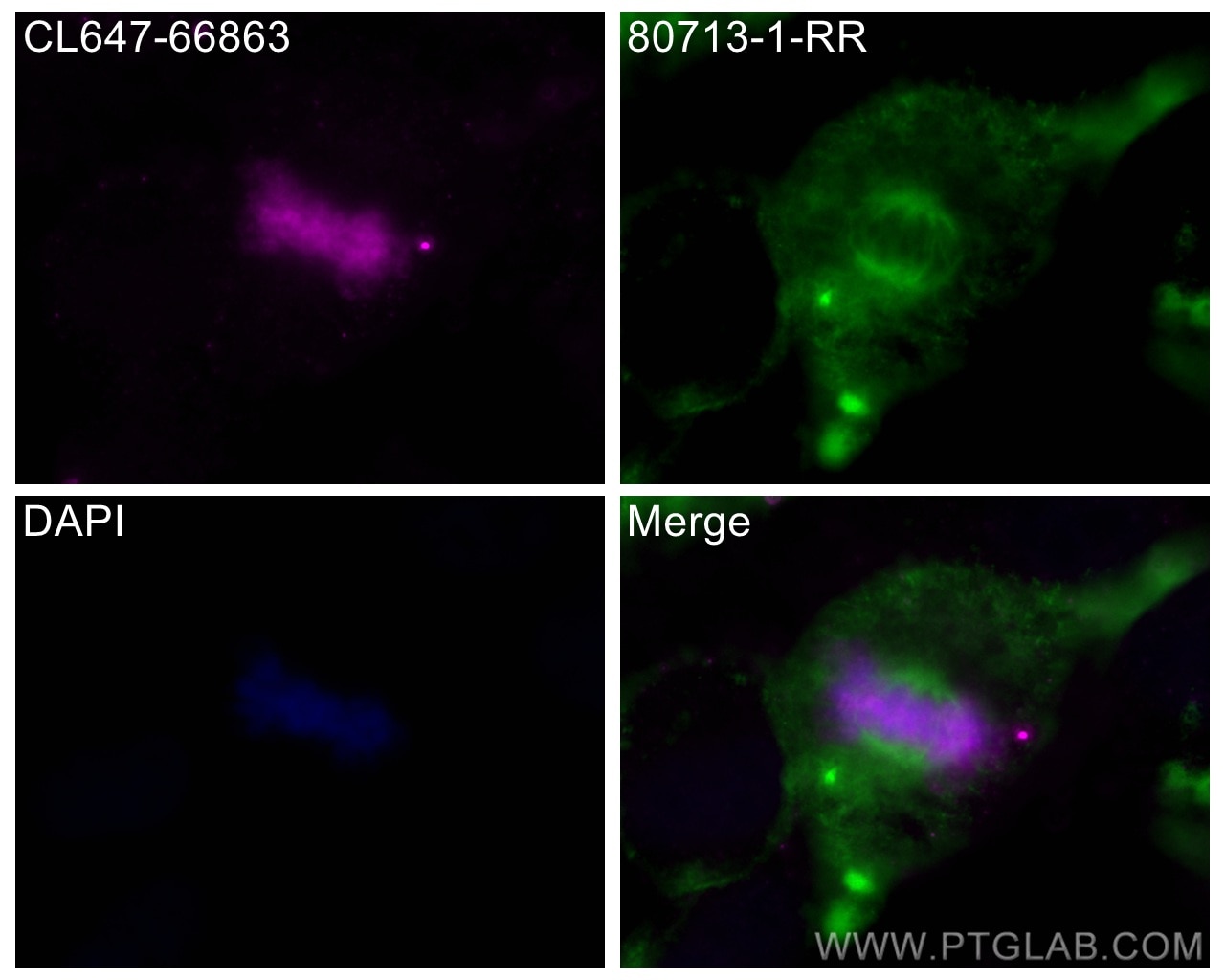
| Erfolgreiche Detektion in IF/ICC | HeLa-Zellen |
Empfohlene Verdünnung
| Anwendung | Verdünnung |
|---|---|
| Immunfluoreszenz (IF)/ICC | IF/ICC : 1:50-1:500 |
| It is recommended that this reagent should be titrated in each testing system to obtain optimal results. | |
| Sample-dependent, check data in validation data gallery | |
Produktinformation
CL647-66863 bindet in IF/ICC Phospho-Histone H3 (Ser10) und zeigt Reaktivität mit Hausschwein, human, Maus, Ratten
| Getestete Reaktivität | Hausschwein, human, Maus, Ratte |
| Wirt / Isotyp | Maus / IgG1 |
| Klonalität | Monoklonal |
| Typ | Antikörper |
| Immunogen | Peptid |
| Vollständiger Name | histone cluster 1, H3a |
| Berechnetes Molekulargewicht | 15 kDa |
| Beobachtetes Molekulargewicht | 15-17 kDa |
| GenBank-Zugangsnummer | NM_003529 |
| Gene symbol | HIST1H3A |
| Gene ID (NCBI) | 8350 |
| Konjugation | CoraLite® Plus 647 Fluorescent Dye |
| Excitation/Emission maxima wavelengths | 654 nm / 674 nm |
| Form | Liquid |
| Reinigungsmethode | Protein-G-Reinigung |
| Lagerungspuffer | PBS with 50% glycerol, 0.05% Proclin300, 0.5% BSA |
| Lagerungsbedingungen | Bei -20°C lagern. Vor Licht schützen. Nach dem Versand ein Jahr stabil. Aliquotieren ist bei -20oC Lagerung nicht notwendig. 20ul Größen enthalten 0,1% BSA. |
Hintergrundinformationen
Phospho-histone-H3 (PHH3) is a core histone protein, which in its phosphorylated state forms the principal constituents of eukaryotic chromatin, with histone H3 being phosphorylated at serine (Ser) 10 or Ser28 as well as its phosphorylation of Ser10 being strongly correlated with the late G2 to M-phase transition in mammalian mitotic cells. On the basis of previous research, a few cell line- and animal model-based researches have displayed an increase in phosphorylation of histone H3 at Ser10 (H3S10ph), the only histone marker that is involved in carcinogenesis and cellular transformation. Histone H3 phosphorylation on serine-10 is specific to mitosis and phosphorylated histone H3 (PHH3) proliferation markers (as counts defined per area or as indices defined per cell numbers) are increasingly being used to evaluate proliferation in various tumors.
Protokolle
| PRODUKTSPEZIFISCHE PROTOKOLLE | |
|---|---|
| IF protocol for CL Plus 647 Phospho-Histone H3 (Ser10) antibody CL647-66863 | Protokoll herunterladen |
| STANDARD-PROTOKOLLE | |
|---|---|
| Klicken Sie hier, um unsere Standardprotokolle anzuzeigen |


