GFP tag Monoklonaler Antikörper
GFP tag Monoklonal Antikörper für IF/ICC, FC (Intra)
Wirt / Isotyp
Maus / IgG2a
Getestete Reaktivität
rekombinanten Protein
Anwendung
IF/ICC, FC (Intra)
Konjugation
CoraLite® Plus 647 Fluorescent Dye
CloneNo.
1E10H7
Kat-Nr. : CL647-66002
Synonyme
Geprüfte Anwendungen
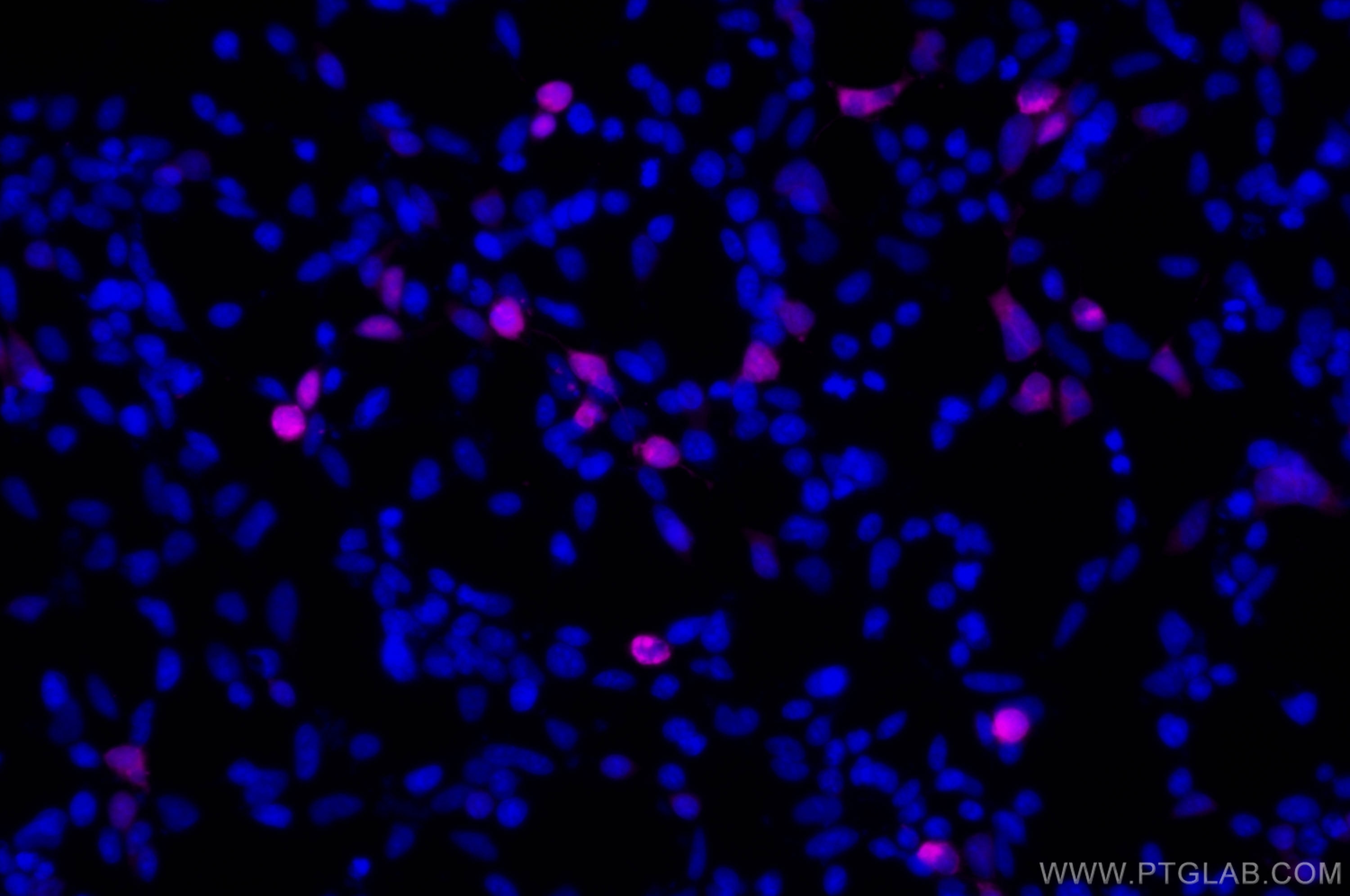
| Erfolgreiche Detektion in IF/ICC | Transfizierte HEK-293-Zellen |
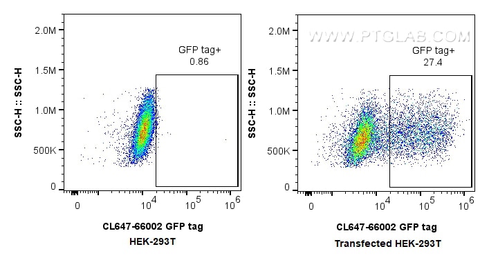
| Erfolgreiche Detektion in FC (Intra) | Transfected HEK-293T cells |
Empfohlene Verdünnung
| Anwendung | Verdünnung |
|---|---|
| Immunfluoreszenz (IF)/ICC | IF/ICC : 1:50-1:500 |
| Durchflusszytometrie (FC) (INTRA) | FC (INTRA) : 0.4 ug per 10^6 cells in a 100 µl suspension |
| It is recommended that this reagent should be titrated in each testing system to obtain optimal results. | |
| Sample-dependent, check data in validation data gallery | |
Produktinformation
CL647-66002 bindet in IF/ICC, FC (Intra) GFP tag und zeigt Reaktivität mit rekombinanten Protein
| Getestete Reaktivität | rekombinanten Protein |
| Wirt / Isotyp | Maus / IgG2a |
| Klonalität | Monoklonal |
| Typ | Antikörper |
| Immunogen | GFP tag fusion protein Ag2128 |
| Vollständiger Name | GFP tag |
| Berechnetes Molekulargewicht | 26 kDa |
| GenBank-Zugangsnummer | M62653 |
| Gene symbol | |
| Gene ID (NCBI) | |
| Konjugation | CoraLite® Plus 647 Fluorescent Dye |
| Excitation/Emission maxima wavelengths | 654 nm / 674 nm |
| Form | Liquid |
| Reinigungsmethode | Protein-A-Reinigung |
| Lagerungspuffer | PBS with 50% glycerol, 0.05% Proclin300, 0.5% BSA |
| Lagerungsbedingungen | Bei -20°C lagern. Vor Licht schützen. Nach dem Versand ein Jahr stabil. Aliquotieren ist bei -20oC Lagerung nicht notwendig. 20ul Größen enthalten 0,1% BSA. |
Hintergrundinformationen
Green Fluorescent Proteins (GFPs) encompass a diverse range of proteins carrying a green chromophore, originating from various species and forming different protein lineages.
Wildtype GFP consists of 238 amino acid residues (26.9 kDa). GFP was first identified in the jellyfish Aequorea victoria. It emits green light with a peak wavelength of 509 nm upon excitation by blue light at 395 nm.
When fused with other proteins, GFP serves as a versatile reporter protein e.g. for quantifying expression levels or facilitates visualization of subcellular localization through fluorescence microscopy.
This antibody is a mouse (IgG2a) monoclonal antibody against GFP reactive to GFP, eGFP, eYFP, YFP and CFP. This antibody is labeled with CoraLite Plus 647.
Protokolle
| PRODUKTSPEZIFISCHE PROTOKOLLE | |
|---|---|
| IF protocol for CL Plus 647 GFP tag antibody CL647-66002 | Protokoll herunterladen |
| STANDARD-PROTOKOLLE | |
|---|---|
| Klicken Sie hier, um unsere Standardprotokolle anzuzeigen |


