- Featured Product
- KD/KO Validated
DOCK8 Polyklonaler Antikörper
DOCK8 Polyklonal Antikörper für WB, IHC, ELISA
Wirt / Isotyp
Kaninchen / IgG
Getestete Reaktivität
human, Maus, Ratte
Anwendung
WB, IHC, IF, IP, ELISA
Konjugation
Unkonjugiert
Kat-Nr. : 11622-1-AP
Synonyme
Geprüfte Anwendungen
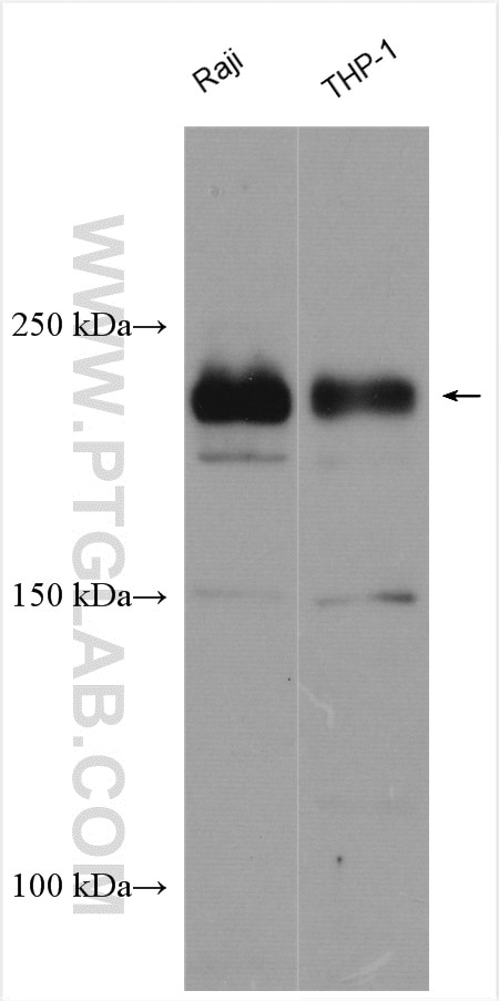
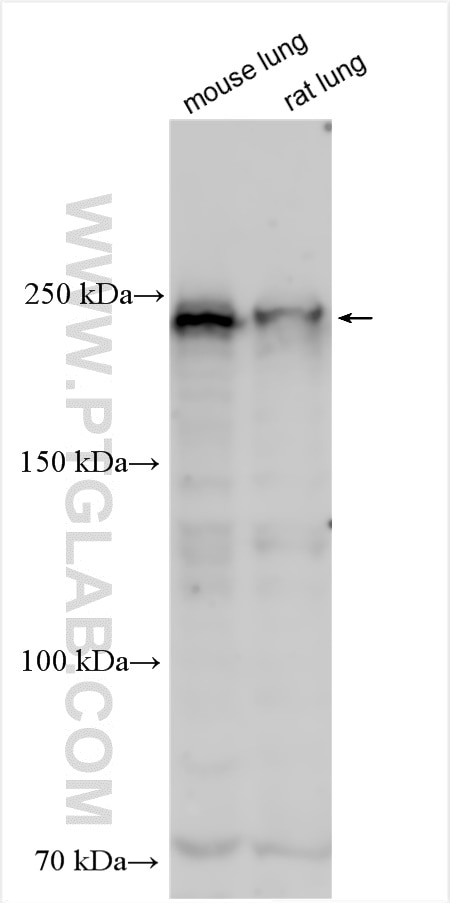
| Erfolgreiche Detektion in WB | Raji-Zellen, Rattenlungengewebe, THP-1-Zellen |
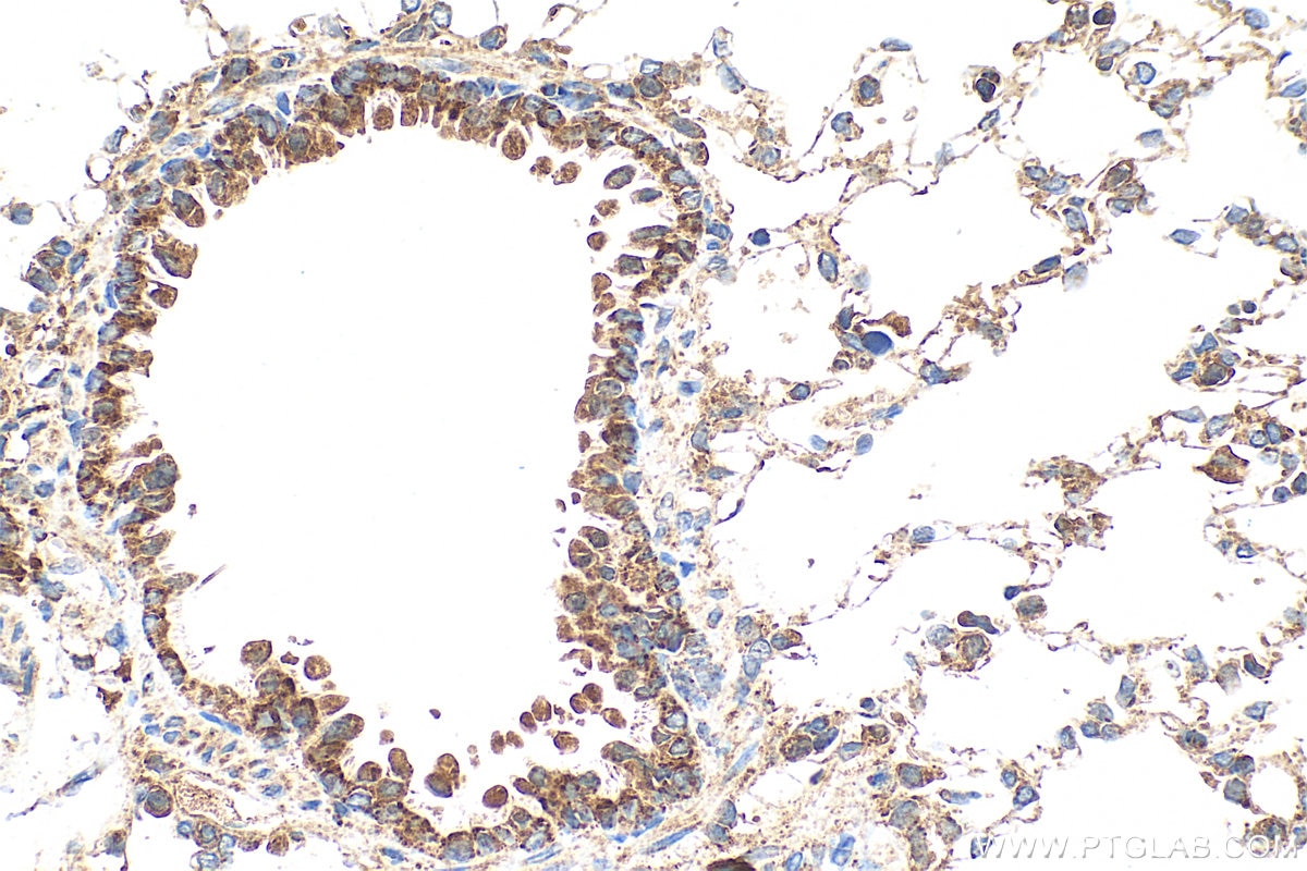
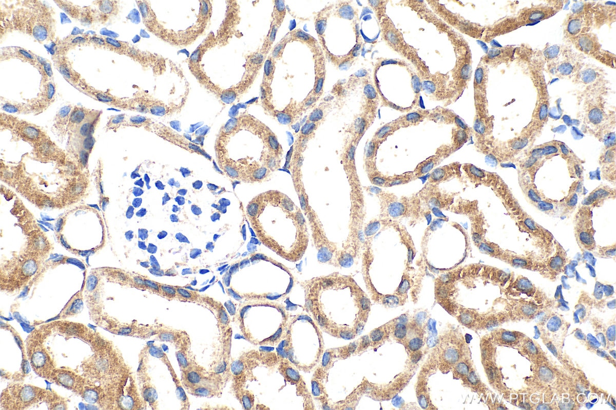
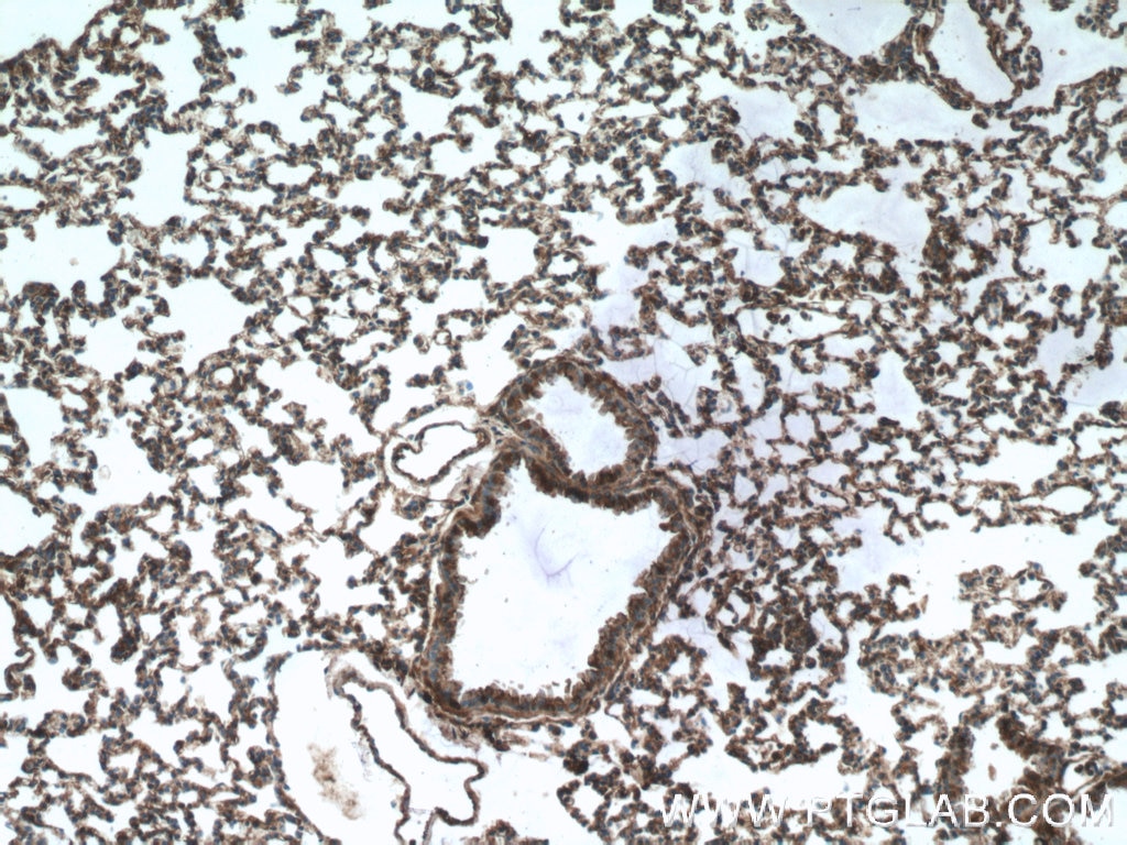
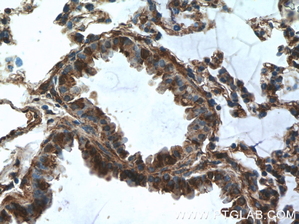
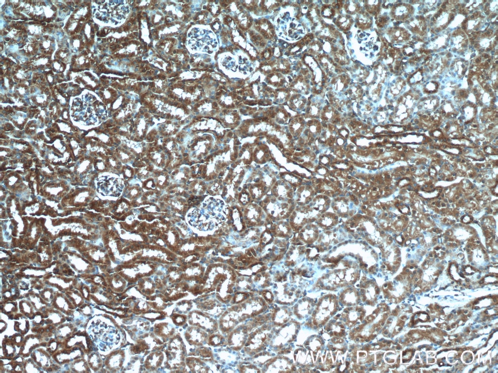
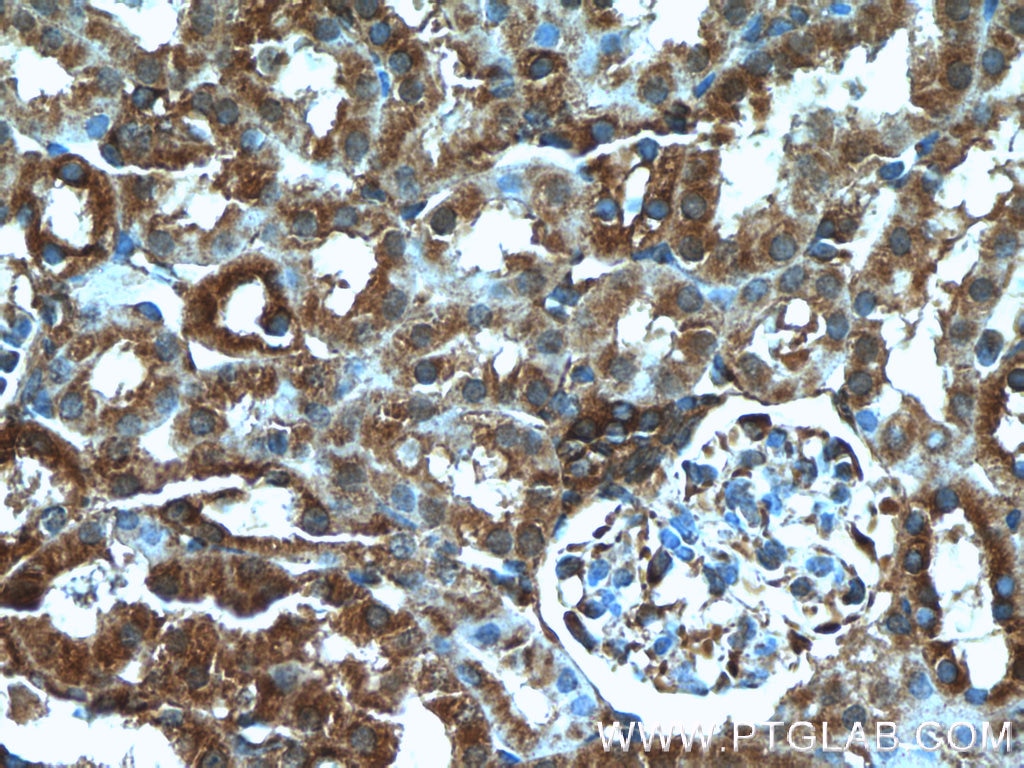
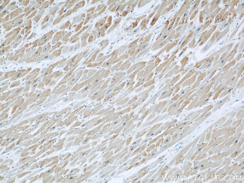
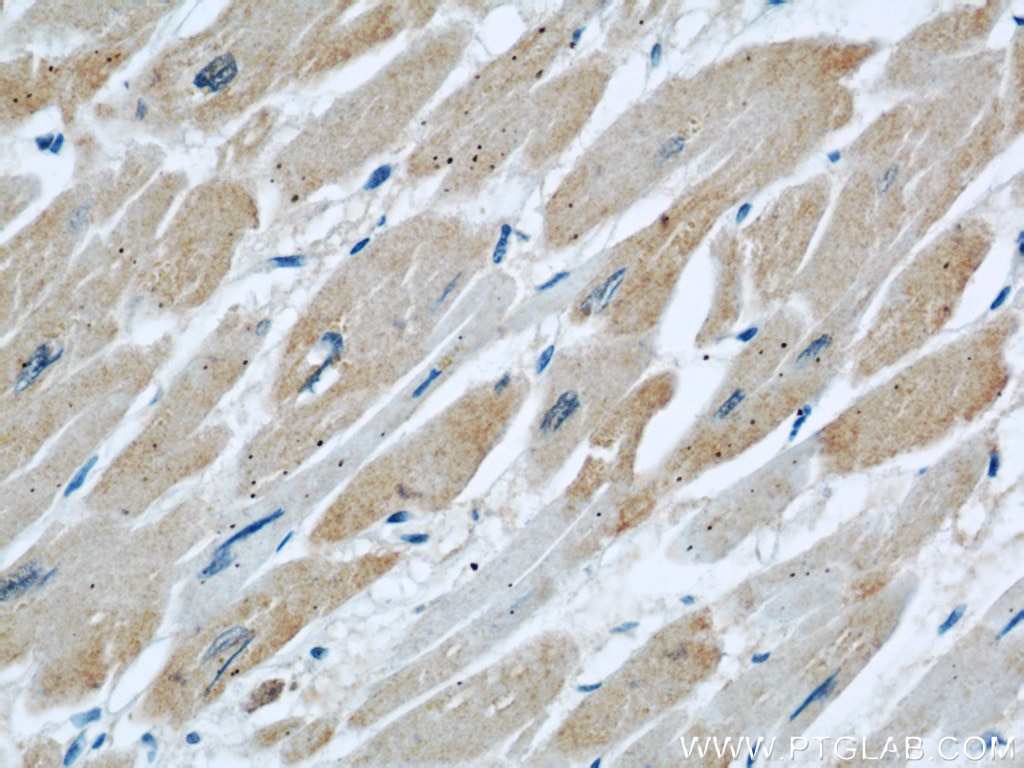
| Erfolgreiche Detektion in IHC | Rattenlungengewebe, humanes Herzgewebe, Mausnierengewebe, Mauslungengewebe Hinweis: Antigendemaskierung mit TE-Puffer pH 9,0 empfohlen. (*) Wahlweise kann die Antigendemaskierung auch mit Citratpuffer pH 6,0 erfolgen. |
Empfohlene Verdünnung
| Anwendung | Verdünnung |
|---|---|
| Western Blot (WB) | WB : 1:500-1:1000 |
| Immunhistochemie (IHC) | IHC : 1:50-1:500 |
| It is recommended that this reagent should be titrated in each testing system to obtain optimal results. | |
| Sample-dependent, check data in validation data gallery | |
Veröffentlichte Anwendungen
| KD/KO | See 4 publications below |
| WB | See 8 publications below |
| IF | See 2 publications below |
| IP | See 2 publications below |
Produktinformation
11622-1-AP bindet in WB, IHC, IF, IP, ELISA DOCK8 und zeigt Reaktivität mit human, Maus, Ratten
| Getestete Reaktivität | human, Maus, Ratte |
| In Publikationen genannte Reaktivität | human, Maus |
| Wirt / Isotyp | Kaninchen / IgG |
| Klonalität | Polyklonal |
| Typ | Antikörper |
| Immunogen | DOCK8 fusion protein Ag2040 |
| Vollständiger Name | dedicator of cytokinesis 8 |
| Berechnetes Molekulargewicht | 2031 aa, 231 kDa |
| Beobachtetes Molekulargewicht | 230-239 kDa |
| GenBank-Zugangsnummer | BC019102 |
| Gene symbol | DOCK8 |
| Gene ID (NCBI) | 81704 |
| Konjugation | Unkonjugiert |
| Form | Liquid |
| Reinigungsmethode | Antigen-Affinitätsreinigung |
| Lagerungspuffer | PBS with 0.02% sodium azide and 50% glycerol |
| Lagerungsbedingungen | Bei -20°C lagern. Nach dem Versand ein Jahr lang stabil Aliquotieren ist bei -20oC Lagerung nicht notwendig. 20ul Größen enthalten 0,1% BSA. |
Hintergrundinformationen
Background
Dedicator of Cytokinesis 8 (DOCK8) is a protein that regulates the actin cytoskeleton, with particular importance in immune cells and a key role in innate and adaptive immune responses.
What is the molecular weight of DOCK8?
231 kDa. DOCK8 is a protein composed of 2031 amino acids and is a guanine nucleotide exchange factor (GEF).
What is the function of DOCK8?
DOCK8 is a member of the DOCK family of proteins, which have a unique DRH2 domain enabling them to act as GEFs and so controlling a range of cellular processes in various signaling pathways (PMID: 12432077). The specific target of DOCK8 is Cell division control protein 42 homolog (Cdc42), a small GTPase that is involved in regulation of the cell cycle and forms a complex. DOCK8 also acts as a scaffold molecule in this complex that initiates actin polymerization via the Wiskott-Aldrich Syndrome protein (WASp) (PubMed: 28028151, PubMed: 22461490).
What diseases are associated with DOCK8?
The role of DOCK8 in immunity was first identified with the study of DOCK8-deficient patients who presented with combined immunodeficiency (PMID: 19776401; PMID: 20004785). The subsequent study of DOCK8 in immune cells such as T cells, natural killer (NK) cells, and B cells has revealed how it regulates their normal function. This includes the regulation of immune synapse formation, immune cell trafficking, regulation of dendritic cell polarization, and cytokine production (PMID: 28366940).
DOCK8 deficiency is caused by a number of different mutations in the gene. It leads to the autosomal recessive form of the immunodeficiency disease Hyper-IgE syndrome, or Job's syndrome. The symptoms of DOCK8 deficiency include eczema, high levels of serum IgE, hypereosinophilia, and recurrent respiratory and skin infections as a result of impaired immune cell function.
Protokolle
| PRODUKTSPEZIFISCHE PROTOKOLLE | |
|---|---|
| WB protocol for DOCK8 antibody 11622-1-AP | Protokoll herunterladen |
| IHC protocol for DOCK8 antibody 11622-1-AP | Protokoll herunterladenl |
| STANDARD-PROTOKOLLE | |
|---|---|
| Klicken Sie hier, um unsere Standardprotokolle anzuzeigen |
Publikationen
| Species | Application | Title |
|---|---|---|
Nat Immunol Heme drives hemolysis-induced susceptibility to infection via disruption of phagocyte functions.
| ||
Cell Death Differ CCL2 regulation of MST1-mTOR-STAT1 signaling axis controls BCR signaling and B-cell differentiation. | ||
Diabetes Dedicator of Cytokinesis 5 Regulates Keratinocyte Function and Promotes Diabetic Wound Healing. | ||
Oncotarget CD147 promotes Src-dependent activation of Rac1 signaling through STAT3/DOCK8 during the motility of hepatocellular carcinoma cells.
| ||
iScience DOCK8-expressing T follicular helper cells newly generated beyond self-organized criticality cause systemic lupus erythematosus.
| ||
J Alzheimers Dis A novel finding relates to the involvement of ATF3/DOCK8 in Alzheimer's disease pathogenesis |
