APC Polyklonaler Antikörper
APC Polyklonal Antikörper für IHC, ELISA
Wirt / Isotyp
Kaninchen / IgG
Getestete Reaktivität
human und mehr (1)
Anwendung
WB, IHC, ELISA
Konjugation
Unkonjugiert
Kat-Nr. : 19782-1-AP
Synonyme
Geprüfte Anwendungen
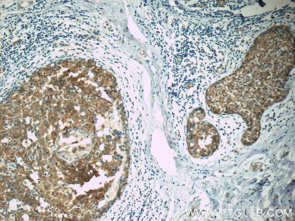
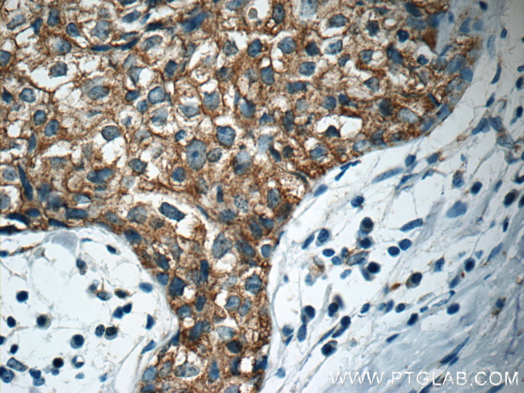
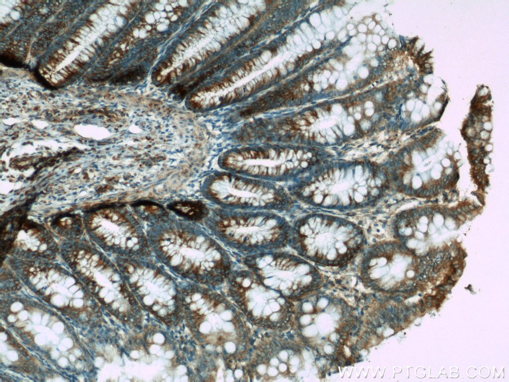
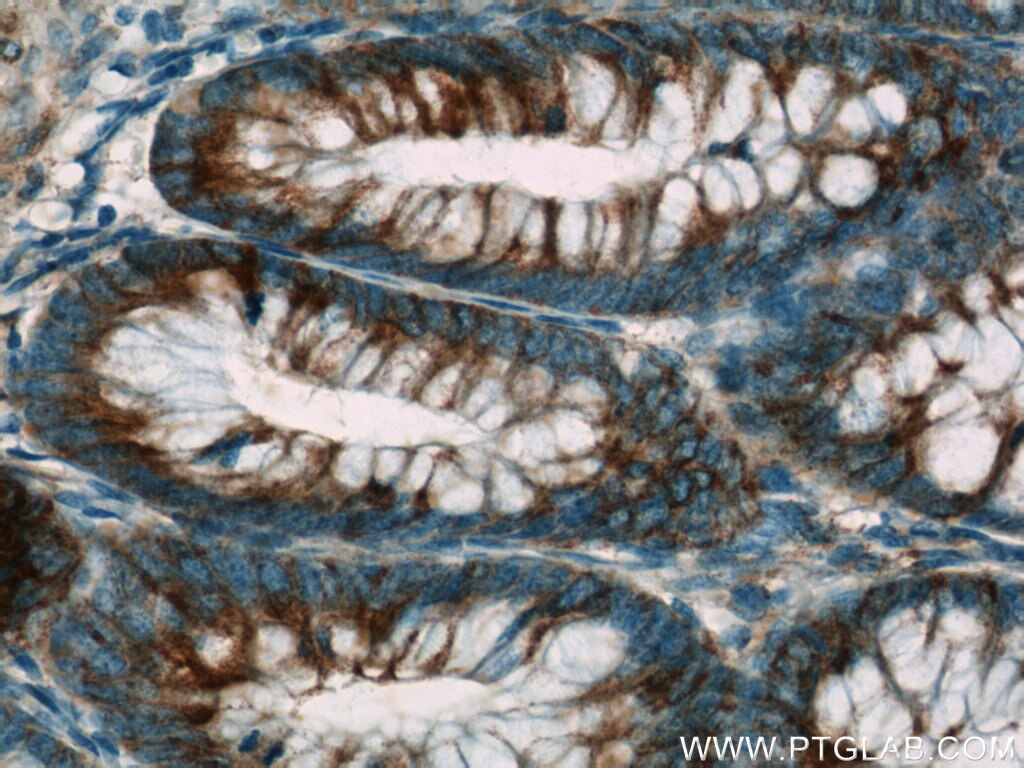
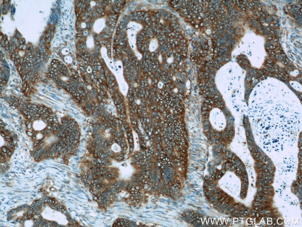
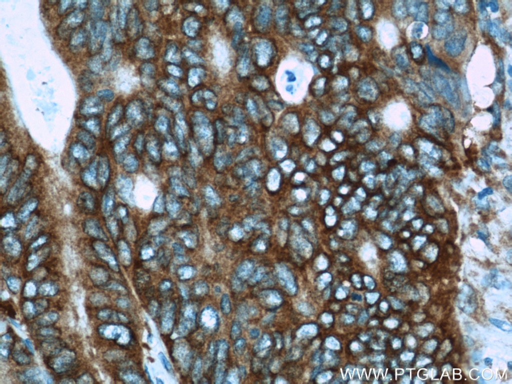
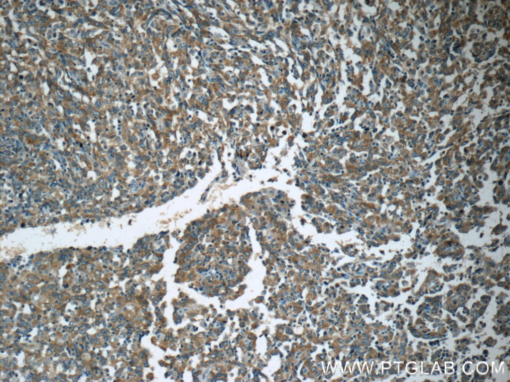
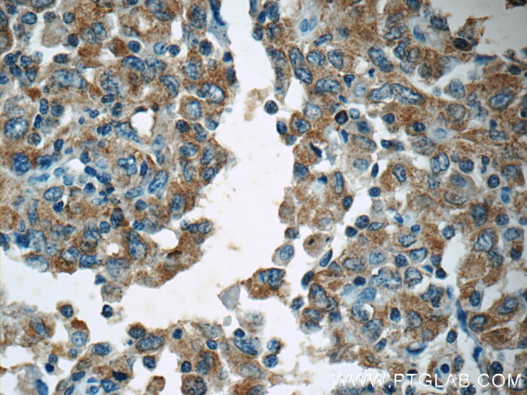
| Erfolgreiche Detektion in IHC | humanes Mammakarzinomgewebe, humanes Kolonkarzinomgewebe, humanes Kolongewebe, humanes Endometriumkarzinomgewebe Hinweis: Antigendemaskierung mit TE-Puffer pH 9,0 empfohlen. (*) Wahlweise kann die Antigendemaskierung auch mit Citratpuffer pH 6,0 erfolgen. |
Empfohlene Verdünnung
| Anwendung | Verdünnung |
|---|---|
| Immunhistochemie (IHC) | IHC : 1:20-1:200 |
| It is recommended that this reagent should be titrated in each testing system to obtain optimal results. | |
| Sample-dependent, check data in validation data gallery | |
Veröffentlichte Anwendungen
| WB | See 9 publications below |
Produktinformation
19782-1-AP bindet in WB, IHC, ELISA APC und zeigt Reaktivität mit human
| Getestete Reaktivität | human |
| In Publikationen genannte Reaktivität | human, Maus |
| Wirt / Isotyp | Kaninchen / IgG |
| Klonalität | Polyklonal |
| Typ | Antikörper |
| Immunogen | Peptid |
| Vollständiger Name | adenomatous polyposis coli |
| Berechnetes Molekulargewicht | 312 kDa |
| GenBank-Zugangsnummer | NM_000038 |
| Gene symbol | APC |
| Gene ID (NCBI) | 324 |
| Konjugation | Unkonjugiert |
| Form | Liquid |
| Reinigungsmethode | Antigen-Affinitätsreinigung |
| Lagerungspuffer | PBS with 0.02% sodium azide and 50% glycerol |
| Lagerungsbedingungen | Bei -20°C lagern. Nach dem Versand ein Jahr lang stabil Aliquotieren ist bei -20oC Lagerung nicht notwendig. 20ul Größen enthalten 0,1% BSA. |
Hintergrundinformationen
APC, also named as DP2.5, belongs to the adenomatous polyposis coli (APC) family. APC is a tumor suppressor that regulates cell division, helps ensure that the number of chromosomes in a cell is correct following cell division, and associates with other proteins involved in cell attachment and signaling. APC promotes rapid degradation of CTNNB1 and participates in Wnt signaling as a negative regulator. It plays a critical role in several cellular processes. APC regulates beta-catenin levels through Wnt-signaling and is involved in actin cytoskeletal integrity, cell-cell adhesion and cell migration. APC activity is correlated with its phosphorylation state. Defects in APC are a cause of familial adenomatous polyposis (FAP) which includes also Gardner syndrome (GS). Defects in APC are a cause of hereditary desmoid disease (HDD) which also known as familial infiltrative fibromatosis (FIF). Defects in APC are a cause of medulloblastoma (MDB) which is a malignant, invasive embryonal tumor of the cerebellum with a preferential manifestation in children. Defects in APC are a cause of mismatch repair cancer syndrome (MMRCS) which also known as Turcot syndrome or brain tumor-polyposis syndrome 1 (BTPS1).
Protokolle
| PRODUKTSPEZIFISCHE PROTOKOLLE | |
|---|---|
| IHC protocol for APC antibody 19782-1-AP | Protokoll herunterladenl |
| STANDARD-PROTOKOLLE | |
|---|---|
| Klicken Sie hier, um unsere Standardprotokolle anzuzeigen |
Publikationen
| Species | Application | Title |
|---|---|---|
Biomed Pharmacother Glycogen phosphorylase B promotes ovarian cancer progression via Wnt/β-catenin signaling and is regulated by miR-133a-3p. | ||
World J Gastroenterol MiR-205 mediated APC regulation contributes to pancreatic cancer cell proliferation. | ||
Cancer Med MiR-26b suppresses hepatocellular carcinoma development by negatively regulating ZNRD1 and Wnt/β-catenin signaling. | ||
J Bone Oncol BAIAP2L2 promotes the proliferation, migration and invasion of osteosarcoma associated with the Wnt/β-catenin pathway | ||
Evid Based Complement Alternat Med Intervention Mechanism of Hunag-Lian Jie-Du Decoction on Canonical Wnt/β-Catenin Signaling Pathway in Psoriasis Mouse Model. |
Rezensionen
The reviews below have been submitted by verified Proteintech customers who received an incentive for providing their feedback.
FH Brianna (Verified Customer) (12-13-2018) |
|
