- Phare
- Validé par KD/KO
Anticorps Polyclonal de lapin anti-mDia1
mDia1 Polyclonal Antibody for FC (Intra), IF
Hôte / Isotype
Lapin / IgG
Réactivité testée
Humain, rat, singe, souris
Applications
IF, FC (Intra)
Conjugaison
CoraLite® Plus 488 Fluorescent Dye
N° de cat : CL488-20624
Synonymes
Galerie de données de validation
Applications testées
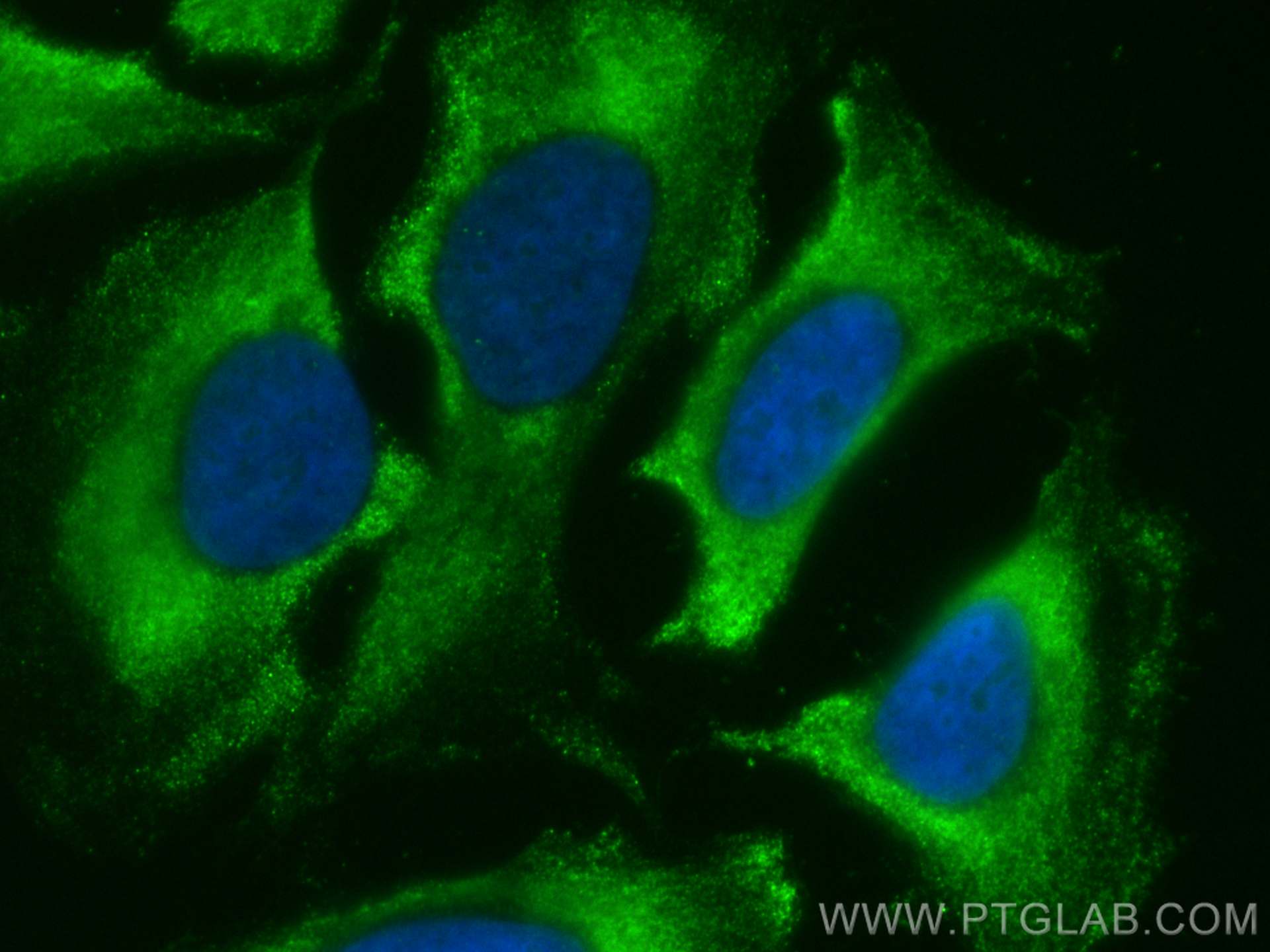
| Résultats positifs en IF | cellules HeLa, |
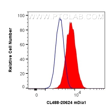
| Résultats positifs en cytométrie | cellules HeLa |
Dilution recommandée
| Application | Dilution |
|---|---|
| Immunofluorescence (IF) | IF : 1:50-1:500 |
| Flow Cytometry (FC) | FC : 0.80 ug per 10^6 cells in a 100 µl suspension |
| It is recommended that this reagent should be titrated in each testing system to obtain optimal results. | |
| Sample-dependent, check data in validation data gallery | |
Informations sur le produit
CL488-20624 cible mDia1 dans les applications de IF, FC (Intra) et montre une réactivité avec des échantillons Humain, rat, singe, souris
| Réactivité | Humain, rat, singe, souris |
| Hôte / Isotype | Lapin / IgG |
| Clonalité | Polyclonal |
| Type | Anticorps |
| Immunogène | mDia1 Protéine recombinante Ag14523 |
| Nom complet | diaphanous homolog 1 (Drosophila) |
| Masse moléculaire calculée | 1272 aa, 141 kDa |
| Poids moléculaire observé | 140-150 kDa, 70 kDa |
| Numéro d’acquisition GenBank | BC007411 |
| Symbole du gène | DIAPH1 |
| Identification du gène (NCBI) | 1729 |
| Conjugaison | CoraLite® Plus 488 Fluorescent Dye |
| Excitation/Emission maxima wavelengths | 493 nm / 522 nm |
| Forme | Liquide |
| Méthode de purification | Purification par affinité contre l'antigène |
| Tampon de stockage | PBS avec glycérol à 50 %, Proclin300 à 0,05 % et BSA à 0,5 %, pH 7,3. |
| Conditions de stockage | Stocker à -20 °C. Éviter toute exposition à la lumière. Stable pendant un an après l'expédition. L'aliquotage n'est pas nécessaire pour le stockage à -20oC Les 20ul contiennent 0,1% de BSA. |
Informations générales
mDia1, also known as DIAPH1 or Diap1, is a mammalian diaphanous-related formin which is implicated in multiple physical and pathological events including cytoskeletal dynamics, autosomal hearing loss, and myelodysplasia. Depending upon the cell type and position in the cell cycle, mDia1 has been shown to localize to the cell cortex, trafficking endosomes, cleavage furrow, mid-bodies, and centrosomes, the cytoplasmic microtubule-organizing center crucial for cell division. Mutation of mDia1 has been linked to microcephaly. This antibody recognizes the endogenous mDia1 mainly around 140-150 kDa, while sometimes an additional 70 kDa can also be observed which is proposed to be a fragment of 140-150 kDa molecules (26011179).
Protocole
| Product Specific Protocols | |
|---|---|
| IF protocol for CL Plus 488 mDia1 antibody CL488-20624 | Download protocol |
| FC protocol for CL Plus 488 mDia1 antibody CL488-20624 | Download protocol |
| Standard Protocols | |
|---|---|
| Click here to view our Standard Protocols |


