- Phare
- Validé par KD/KO
Anticorps Monoclonal anti-c-Fos
c-Fos Monoclonal Antibody for FC (Intra)
Hôte / Isotype
Mouse / IgG1
Réactivité testée
Humain, rat, souris
Applications
FC (Intra)
Conjugaison
CoraLite® Plus 488 Fluorescent Dye
CloneNo.
1G2C5
N° de cat : CL488-66590
Synonymes
Galerie de données de validation
Applications testées
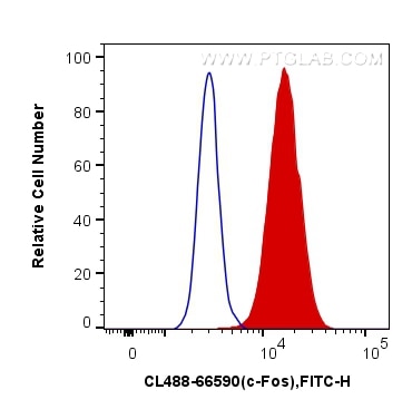
| Résultats positifs en cytométrie | cellules HeLa |
Dilution recommandée
| Application | Dilution |
|---|---|
| Flow Cytometry (FC) | FC : 0.40 ug per 10^6 cells in a 100 µl suspension |
| It is recommended that this reagent should be titrated in each testing system to obtain optimal results. | |
| Sample-dependent, check data in validation data gallery | |
Informations sur le produit
CL488-66590 cible c-Fos dans les applications de FC (Intra) et montre une réactivité avec des échantillons Humain, rat, souris
| Réactivité | Humain, rat, souris |
| Hôte / Isotype | Mouse / IgG1 |
| Clonalité | Monoclonal |
| Type | Anticorps |
| Immunogène | c-Fos Protéine recombinante Ag24340 |
| Nom complet | FOS |
| Masse moléculaire calculée | 41 kDa |
| Poids moléculaire observé | 55-60 kDa |
| Numéro d’acquisition GenBank | BC004490 |
| Symbole du gène | FOS |
| Identification du gène (NCBI) | 2353 |
| Conjugaison | CoraLite® Plus 488 Fluorescent Dye |
| Excitation/Emission maxima wavelengths | 493 nm / 522 nm |
| Forme | Liquide |
| Méthode de purification | Purification par protéine G |
| Tampon de stockage | PBS avec glycérol à 50 %, Proclin300 à 0,05 % et BSA à 0,5 %, pH 7,3. |
| Conditions de stockage | Stocker à -20 °C. Éviter toute exposition à la lumière. Stable pendant un an après l'expédition. L'aliquotage n'est pas nécessaire pour le stockage à -20oC Les 20ul contiennent 0,1% de BSA. |
Informations générales
c-FOS, also named as Proto-oncogene c-Fos and G0/G1 switch regulatory protein 7, is a 380 amino acid protein, which contains 1 bZIP (basic-leucine zipper) domain and belongs to the bZIP family. FOS is expressed at very low levels in quiescent cells. When cells are stimulated to reenter growth, FOS undergo 2 waves of expression, the first one peaks 7.5 minutes following FBS induction. At this stage, the FOS protein is localized endoplasmic reticulum. The second wave of expression occurs at about 20 minutes after induction and peaks at 1 hour. At this stage, the FOS protein becomes nuclear. The calculated molecular weight of FOS is 40 kDa, but Phosphorylated FOS protein is about 60-65 kDa. It is involved in important cellular events, including cell proliferation, differentiation and survival; genes associated with hypoxia; and angiogenesis; which makes its dysregulation an important factor for cancer development. It can also induce a loss of cell polarity and epithelial-mesenchymal transition, leading to invasive and metastatic growth in mammary epithelial cells. Expression of c-fos is an indirect marker of neuronal activity because c-fos is often expressed when neurons fire action potentials. Upregulation of c-fos mRNA in a neuron indicates recent activity
Protocole
| Product Specific Protocols | |
|---|---|
| FC protocol for CL Plus 488 c-Fos antibody CL488-66590 | Download protocol |
| Standard Protocols | |
|---|---|
| Click here to view our Standard Protocols |


