- Phare
- Validé par KD/KO
Anticorps Monoclonal anti-TGFBI / BIGH3
TGFBI / BIGH3 Monoclonal Antibody for FC (Intra), IF
Hôte / Isotype
Mouse / IgG2a
Réactivité testée
Humain
Applications
IF, FC (Intra)
Conjugaison
CoraLite®594 Fluorescent Dye
CloneNo.
3E11D11
N° de cat : CL594-60007
Synonymes
Galerie de données de validation
Applications testées
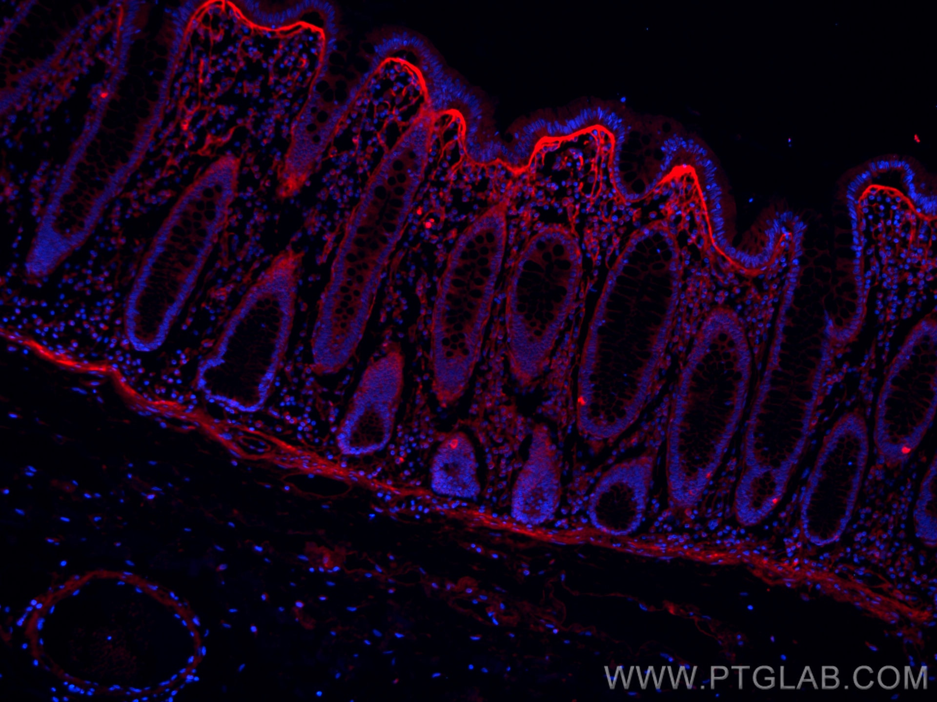
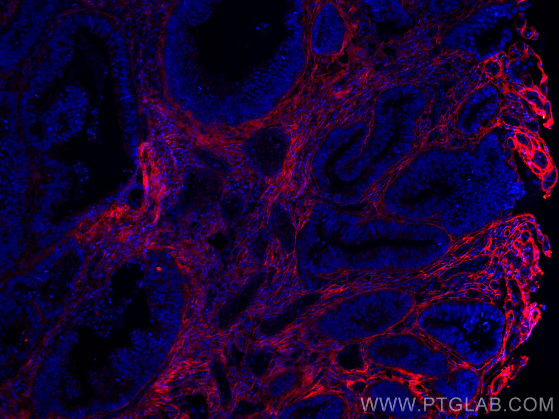
| Résultats positifs en IF | tissu de cancer du côlon humain, |
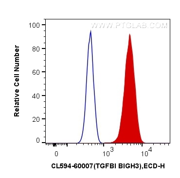
| Résultats positifs en cytométrie | cellules Y79 |
Dilution recommandée
| Application | Dilution |
|---|---|
| Immunofluorescence (IF) | IF : 1:50-1:500 |
| Flow Cytometry (FC) | FC : 0.40 ug per 10^6 cells in a 100 µl suspension |
| It is recommended that this reagent should be titrated in each testing system to obtain optimal results. | |
| Sample-dependent, check data in validation data gallery | |
Informations sur le produit
CL594-60007 cible TGFBI / BIGH3 dans les applications de IF, FC (Intra) et montre une réactivité avec des échantillons Humain
| Réactivité | Humain |
| Hôte / Isotype | Mouse / IgG2a |
| Clonalité | Monoclonal |
| Type | Anticorps |
| Immunogène | TGFBI / BIGH3 Protéine recombinante Ag0241 |
| Nom complet | transforming growth factor, beta-induced, 68kDa |
| Masse moléculaire calculée | 683 aa, 75 kDa |
| Numéro d’acquisition GenBank | BC000097 |
| Symbole du gène | TGFBI |
| Identification du gène (NCBI) | 7045 |
| Conjugaison | CoraLite®594 Fluorescent Dye |
| Excitation/Emission maxima wavelengths | 588 nm / 604 nm |
| Forme | Liquide |
| Méthode de purification | Purification par protéine A |
| Tampon de stockage | PBS avec glycérol à 50 %, Proclin300 à 0,05 % et BSA à 0,5 %, pH 7,3. |
| Conditions de stockage | Stocker à -20 °C. Éviter toute exposition à la lumière. L'aliquotage n'est pas nécessaire pour le stockage à -20oC Les 20ul contiennent 0,1% de BSA. |
Informations générales
TGFBI, also named as BIGH3, Kerato-epithelin and RGD-CAP, binds to type I, II, and IV collagens. TGFBI is an adhesion protein which may play an important role in cell-collagen interactions. In cartilage, it may be involved in endochondral bone formation. TGFBI is an extracellular matrix adaptor protein, it has been reported to be differentially expressed in transformed tissues. TGFBI is a predictive factor of the response to chemotherapy, and suggest the use of TGFBI-derived peptides as possible therapeutic adjuvants for the enhancement of responses to chemotherapy.(PMID:20509890) Defects in TGFBI are the cause of epithelial basement membrane corneal dystrophy (EBMD). Defects in TGFBI are the cause of corneal dystrophy Groenouw type 1 (CDGG1). Defects in TGFBI are the cause of corneal dystrophy lattice type 1 (CDL1). Defects in TGFBI are a cause of corneal dystrophy Thiel-Behnke type (CDTB). Defects in TGFBI are the cause of Reis-Buecklers corneal dystrophy (CDRB). Defects in TGFBI are the cause of lattice corneal dystrophy type 3A (CDL3A). Defects in TGFBI are the cause of Avellino corneal dystrophy (ACD).
Protocole
| Product Specific Protocols | |
|---|---|
| IF protocol for CL594 TGFBI / BIGH3 antibody CL594-60007 | Download protocol |
| Standard Protocols | |
|---|---|
| Click here to view our Standard Protocols |


