- Phare
- Validé par KD/KO
Anticorps Monoclonal anti-RHOA
RHOA Monoclonal Antibody for IF, IHC, WB, ELISA
Hôte / Isotype
Mouse / IgG1
Réactivité testée
Humain, porc, rat, souris
Applications
WB, IHC, IF, ELISA
Conjugaison
Non conjugué
CloneNo.
1B3D7
N° de cat : 66733-1-Ig
Synonymes
Galerie de données de validation
Applications testées
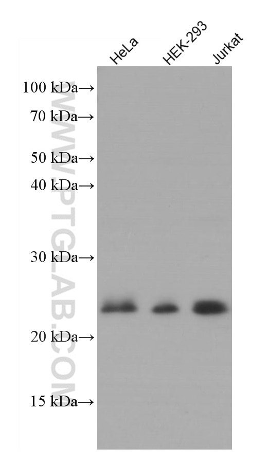
| Résultats positifs en WB | cellules HeLa, cellules HEK-293, cellules Jurkat |
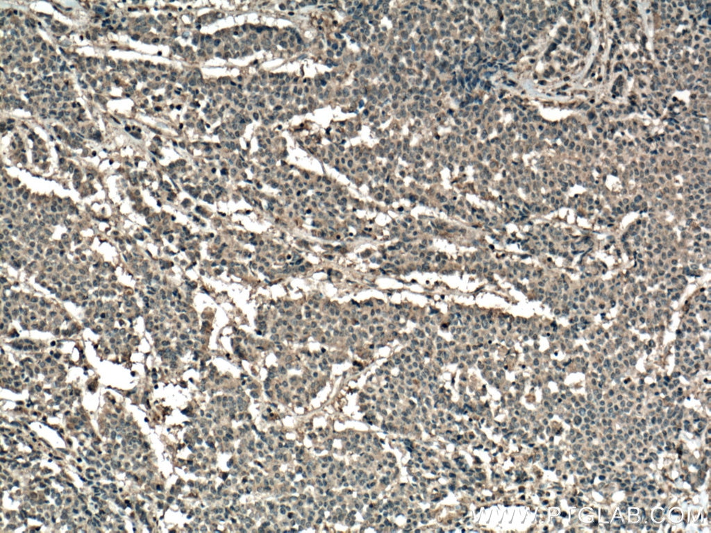
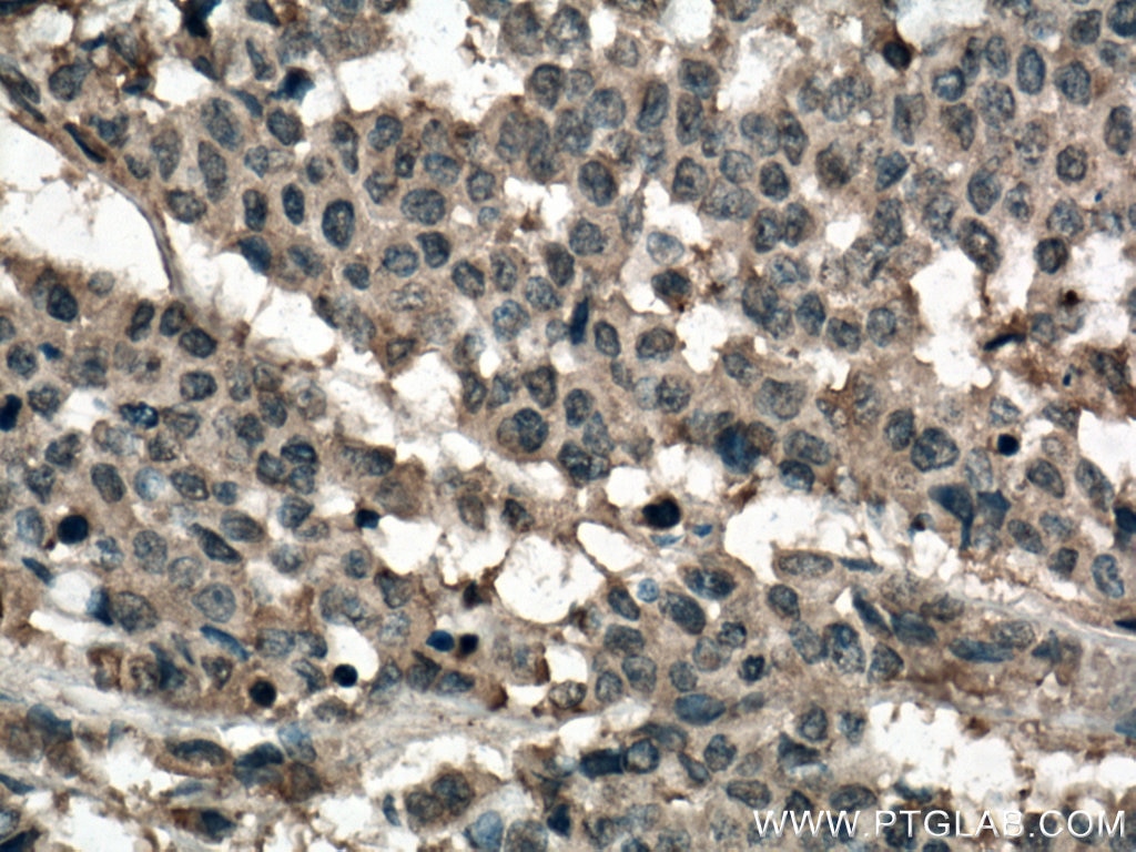
| Résultats positifs en IHC | tissu de cancer du côlon humain, il est suggéré de démasquer l'antigène avec un tampon de TE buffer pH 9.0; (*) À défaut, 'le démasquage de l'antigène peut être 'effectué avec un tampon citrate pH 6,0. |
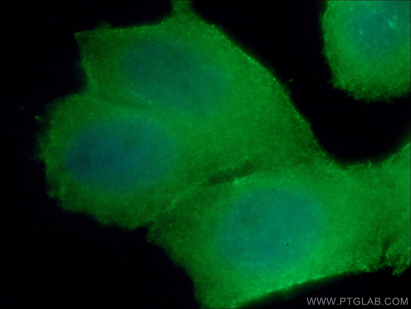
| Résultats positifs en IF | cellules HeLa, |
Dilution recommandée
| Application | Dilution |
|---|---|
| Western Blot (WB) | WB : 1:500-1:2000 |
| Immunohistochimie (IHC) | IHC : 1:50-1:500 |
| Immunofluorescence (IF) | IF : 1:50-1:500 |
| It is recommended that this reagent should be titrated in each testing system to obtain optimal results. | |
| Sample-dependent, check data in validation data gallery | |
Applications publiées
| KD/KO | See 1 publications below |
| WB | See 8 publications below |
| IHC | See 1 publications below |
| IF | See 1 publications below |
Informations sur le produit
66733-1-Ig cible RHOA dans les applications de WB, IHC, IF, ELISA et montre une réactivité avec des échantillons Humain, porc, rat, souris
| Réactivité | Humain, porc, rat, souris |
| Réactivité citée | Humain, porc |
| Hôte / Isotype | Mouse / IgG1 |
| Clonalité | Monoclonal |
| Type | Anticorps |
| Immunogène | RHOA Protéine recombinante Ag1141 |
| Nom complet | ras homolog gene family, member A |
| Masse moléculaire calculée | 22 kDa |
| Poids moléculaire observé | 22 kDa |
| Numéro d’acquisition GenBank | BC005976 |
| Symbole du gène | RHOA |
| Identification du gène (NCBI) | 387 |
| Conjugaison | Non conjugué |
| Forme | Liquide |
| Méthode de purification | Purification par protéine A |
| Tampon de stockage | PBS avec azoture de sodium à 0,02 % et glycérol à 50 % pH 7,3 |
| Conditions de stockage | Stocker à -20°C. Stable pendant un an après l'expédition. L'aliquotage n'est pas nécessaire pour le stockage à -20oC Les 20ul contiennent 0,1% de BSA. |
Informations générales
RhoA is a member of the Rho family of small GTPases, which cycle between inactive GDP-bound and active GTP-bound states and function as molecular switches in signal transduction cascades. Rho proteins promote reorganization of the actin cytoskeleton and regulate cell shape, attachment, and motility. Overexpression of RhoA is associated with tumor cell proliferation and metastasis. RhoA signalling is critical to many cellular processes including migration, mechanotransduction, and is often disrupted in carcinogenesis.
Protocole
| Product Specific Protocols | |
|---|---|
| WB protocol for RHOA antibody 66733-1-Ig | Download protocol |
| IHC protocol for RHOA antibody 66733-1-Ig | Download protocol |
| IF protocol for RHOA antibody 66733-1-Ig | Download protocol |
| Standard Protocols | |
|---|---|
| Click here to view our Standard Protocols |
Publications
| Species | Application | Title |
|---|---|---|
Cell Death Differ Mirtronic miR-4646-5p promotes gastric cancer metastasis by regulating ABHD16A and metabolite lysophosphatidylserines. | ||
Clin Sci (Lond) The long noncoding RNA KTN1-AS1 promotes bladder cancer tumorigenesis via KTN1 cis-activation and the consequent initiation of Rho GTPase-mediated signaling. | ||
J Biol Chem SARS-CoV-2 hijacks macropinocytosis to facilitate its entry and promote viral spike-mediated cell-to-cell fusion
| ||
Front Mol Biosci CUX1 Facilitates the Development of Oncogenic Properties Via Activating Wnt/β-Catenin Signaling Pathway in Glioma. | ||
J Cancer GTPBP2 positively regulates the invasion, migration and proliferation of non-small cell lung cancer. | ||
Exp Cell Res RALY may cause an aggressive biological behavior and a dismal prognosis in non-small-cell lung cancer. |
Avis
The reviews below have been submitted by verified Proteintech customers who received an incentive forproviding their feedback.
FH Udesh (Verified Customer) (12-22-2023) | Worked well for WB in Glioma cells at 1:1000 in BSA
|


