Anticorps Polyclonal de lapin anti-PARP1
PARP1 Polyclonal Antibody for FC (Intra)
Hôte / Isotype
Lapin / IgG
Réactivité testée
Humain, rat, souris
Applications
FC (Intra)
Conjugaison
CoraLite® Plus 647 Fluorescent Dye
N° de cat : CL647-13371
Synonymes
Galerie de données de validation
Applications testées
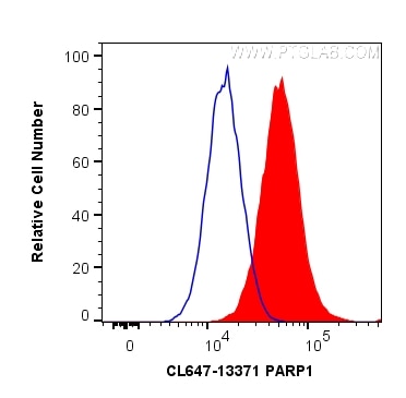
| Résultats positifs en cytométrie | cellules HeLa |
Dilution recommandée
| Application | Dilution |
|---|---|
| Flow Cytometry (FC) | FC : 0.20 ug per 10^6 cells in a 100 µl suspension |
| It is recommended that this reagent should be titrated in each testing system to obtain optimal results. | |
| Sample-dependent, check data in validation data gallery | |
Informations sur le produit
CL647-13371 cible PARP1 dans les applications de FC (Intra) et montre une réactivité avec des échantillons Humain, rat, souris
| Réactivité | Humain, rat, souris |
| Hôte / Isotype | Lapin / IgG |
| Clonalité | Polyclonal |
| Type | Anticorps |
| Immunogène | PARP1 Protéine recombinante Ag4193 |
| Nom complet | poly (ADP-ribose) polymerase 1 |
| Masse moléculaire calculée | 1014 aa, 113 kDa |
| Poids moléculaire observé | 113-116 kDa, 89 kDa |
| Numéro d’acquisition GenBank | BC037545 |
| Symbole du gène | PARP1 |
| Identification du gène (NCBI) | 142 |
| Conjugaison | CoraLite® Plus 647 Fluorescent Dye |
| Excitation/Emission maxima wavelengths | 654 nm / 674 nm |
| Forme | Liquide |
| Méthode de purification | Purification par affinité contre l'antigène |
| Tampon de stockage | PBS avec glycérol à 50 %, Proclin300 à 0,05 % et BSA à 0,5 %, pH 7,3. |
| Conditions de stockage | Stocker à -20 °C. Éviter toute exposition à la lumière. Stable pendant un an après l'expédition. L'aliquotage n'est pas nécessaire pour le stockage à -20oC Les 20ul contiennent 0,1% de BSA. |
Informations générales
PARP1 (poly(ADP-ribose) polymerase 1) is a nuclear enzyme catalyzing the poly(ADP-ribosyl)ation of many key proteins in vivo. The normal function of PARP1 is the routine repair of DNA damage. Activated by DNA strand breaks, the PARP1 is cleaved into an 85 to 89-kDa COOH-terminal fragment and a 24-kDa NH2-terminal peptide by caspases during the apoptotic process. The appearance of PARP fragments is commonly considered as an important biomarker of apoptosis. In addition to caspases, other proteases like calpains, cathepsins, granzymes and matrix metalloproteinases (MMPs) have also been reported to cleave PARP1 and gave rise to fragments ranging from 42-89-kDa. This antibody was generated against the C-terminal region of human PARP1 and it recognizes the full-length as well as the cleavage of the PARP1.
Protocole
| Product Specific Protocols | |
|---|---|
| FC protocol for CL Plus 647 PARP1 antibody CL647-13371 | Download protocol |
| Standard Protocols | |
|---|---|
| Click here to view our Standard Protocols |


