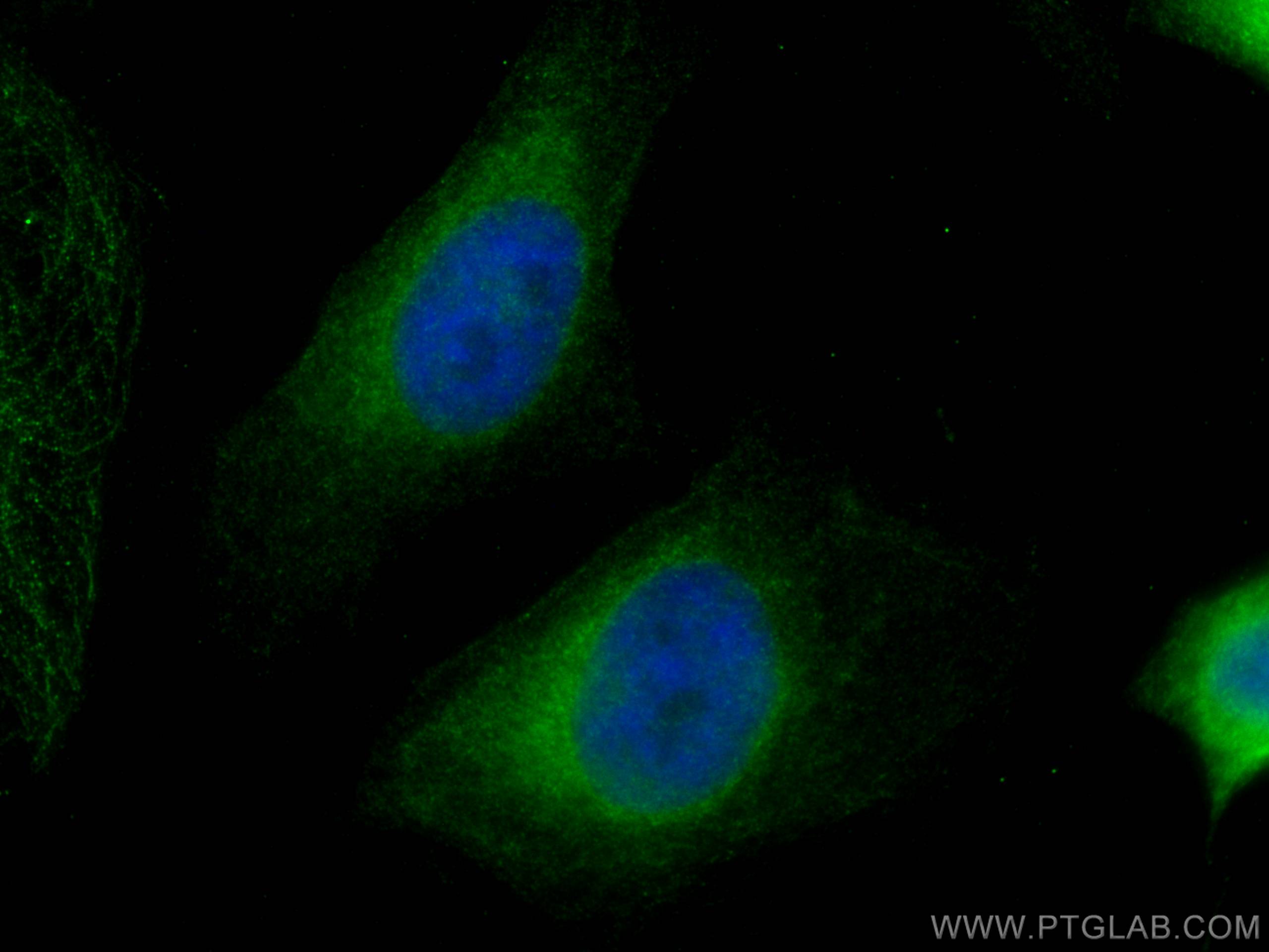
Anticorps Monoclonal anti-HRD1/SYVN1
HRD1/SYVN1 Monoclonal Antibody for IF, IHC, WB,ELISA
Hôte / Isotype
Mouse / IgG2b
Réactivité testée
Humain, rat, souris et plus (1)
Applications
WB, IHC, IF, ELISA
Conjugaison
Non conjugué
CloneNo.
2C8A7
N° de cat : 67488-1-Ig
Synonymes
Galerie de données de validation
Applications testées
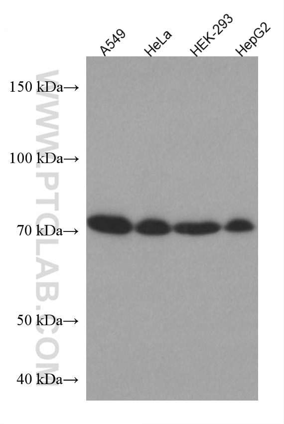
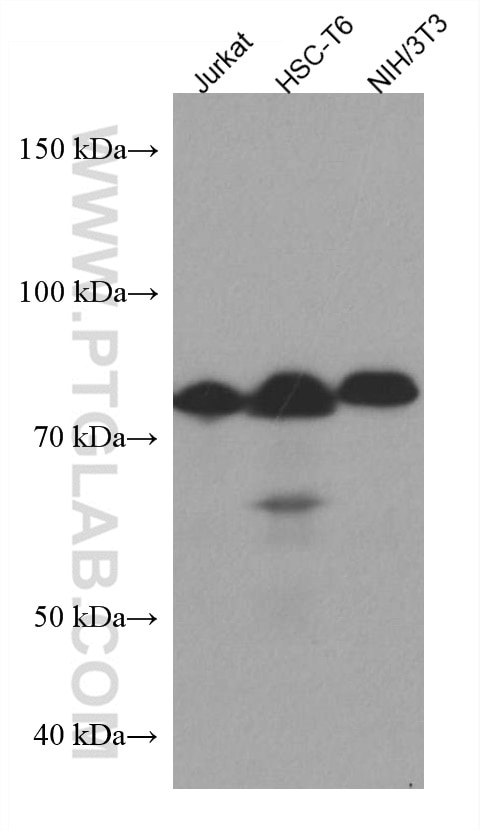
| Résultats positifs en WB | cellules A549, cellules HEK-293, cellules HeLa, cellules HepG2, cellules HSC-T6, cellules Jurkat, cellules NIH/3T3 |
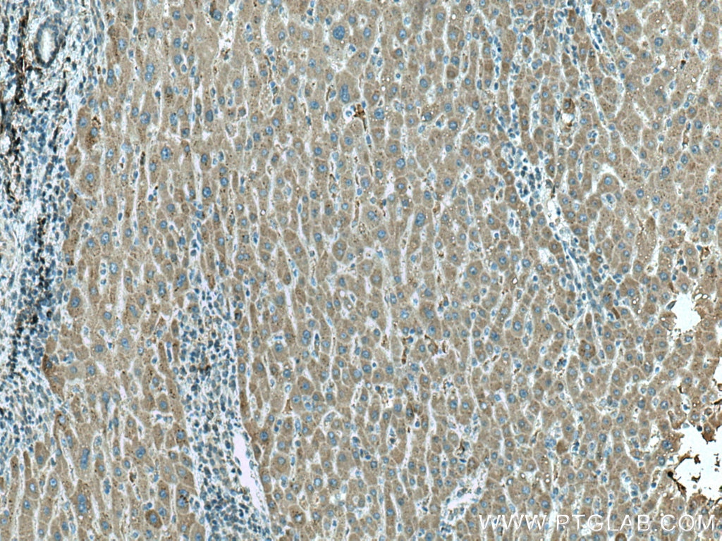
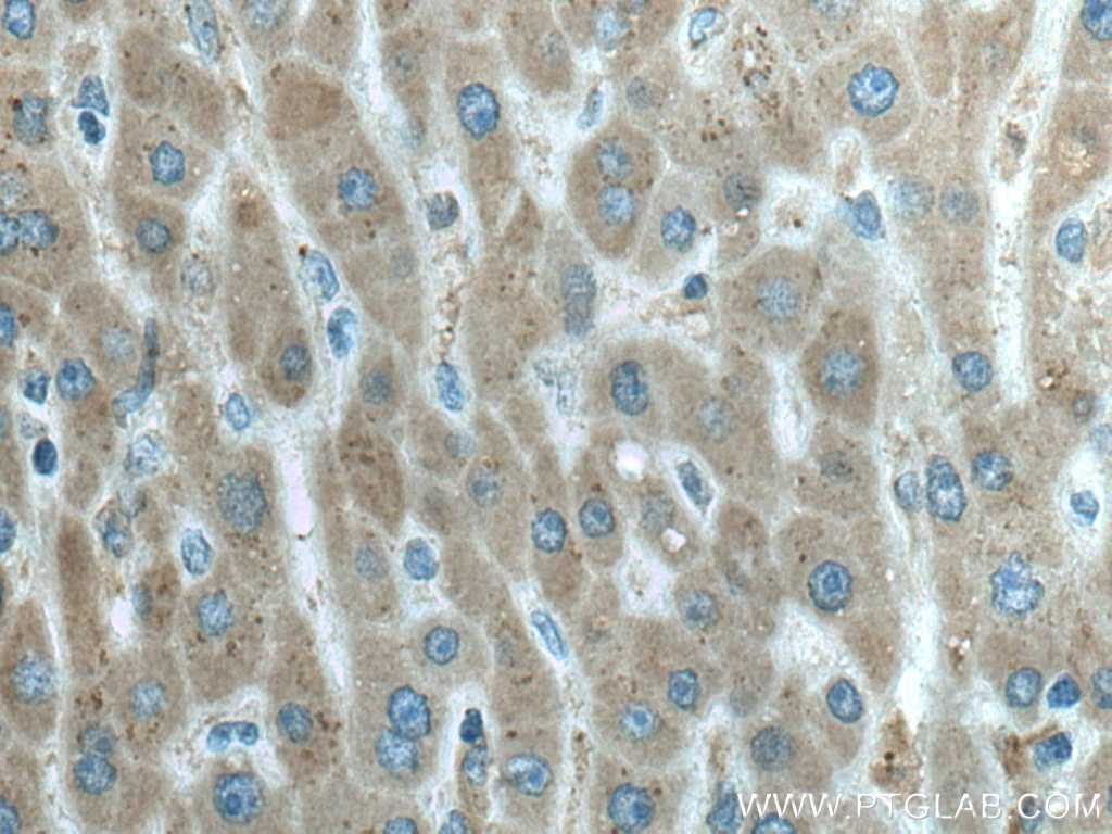
| Résultats positifs en IHC | tissu de cancer du foie humain, il est suggéré de démasquer l'antigène avec un tampon de TE buffer pH 9.0; (*) À défaut, 'le démasquage de l'antigène peut être 'effectué avec un tampon citrate pH 6,0. |
| Résultats positifs en IF | cellules HeLa, |
Dilution recommandée
| Application | Dilution |
|---|---|
| Western Blot (WB) | WB : 1:1000-1:6000 |
| Immunohistochimie (IHC) | IHC : 1:500-1:2000 |
| Immunofluorescence (IF) | IF : 1:200-1:800 |
| It is recommended that this reagent should be titrated in each testing system to obtain optimal results. | |
| Sample-dependent, check data in validation data gallery | |
Applications publiées
| KD/KO | See 1 publications below |
| WB | See 4 publications below |
| IF | See 3 publications below |
Informations sur le produit
67488-1-Ig cible HRD1/SYVN1 dans les applications de WB, IHC, IF, ELISA et montre une réactivité avec des échantillons Humain, rat, souris
| Réactivité | Humain, rat, souris |
| Réactivité citée | Chèvre, Humain, souris |
| Hôte / Isotype | Mouse / IgG2b |
| Clonalité | Monoclonal |
| Type | Anticorps |
| Immunogène | HRD1/SYVN1 Protéine recombinante Ag4456 |
| Nom complet | synovial apoptosis inhibitor 1, synoviolin |
| Masse moléculaire calculée | 617 aa, 68 kDa |
| Poids moléculaire observé | 68-76 kDa |
| Numéro d’acquisition GenBank | BC030530 |
| Symbole du gène | SYVN1 |
| Identification du gène (NCBI) | 84447 |
| Conjugaison | Non conjugué |
| Forme | Liquide |
| Méthode de purification | Purification par protéine A |
| Tampon de stockage | PBS avec azoture de sodium à 0,02 % et glycérol à 50 % pH 7,3 |
| Conditions de stockage | Stocker à -20 ℃. L'aliquotage n'est pas nécessaire pour le stockage à -20oC Les 20ul contiennent 0,1% de BSA. |
Informations générales
HRD1is also named as SYVN1(Synovial apoptosis inhibitor 1) , KIAA1810. It acts as an E3 ubiquitin-protein ligase which accepts ubiquitin specifically from endoplasmic reticulum-associated UBC7 E2 ligase and transfers it to substrates, promoting their degradation. Two distinct binding sites mediate Hrd1 dimerization or oligomerization, one located within the transmembrane region and another within the cytosolic domain. (PMID:19864457).Western blot analysis detected abundant HRD1 expression in liver and kidney. Mouse liver and spleen expressed Hrd1 as an 85-kD protein(PMID:12646171).
Protocole
| Product Specific Protocols | |
|---|---|
| WB protocol for HRD1/SYVN1 antibody 67488-1-Ig | Download protocol |
| IHC protocol for HRD1/SYVN1 antibody 67488-1-Ig | Download protocol |
| IF protocol for HRD1/SYVN1 antibody 67488-1-Ig | Download protocol |
| Standard Protocols | |
|---|---|
| Click here to view our Standard Protocols |
Publications
| Species | Application | Title |
|---|---|---|
Cell Mol Life Sci S100A16 promotes acute kidney injury by activating HRD1-induced ubiquitination and degradation of GSK3β and CK1α. | ||
Int J Mol Sci Integrative Proteomics and Transcriptomics Profiles of the Oviduct Reveal the Prolificacy-Related Candidate Biomarkers of Goats (Capra hircus) in Estrous Periods | ||
Eur J Pharmacol Down-regulation of Hrd1 protects against myocardial ischemia-reperfusion injury by regulating PPARα to prevent oxidative stress, endoplasmic reticulum stress, and cellular apoptosis
| ||
Clin Transl Med Circular RNA circARPC1B functions as a stabilisation enhancer of Vimentin to prevent high cholesterol-induced articular cartilage degeneration |
