Anticorps Monoclonal anti-HDAC4
HDAC4 Monoclonal Antibody for IHC, WB, ELISA
Hôte / Isotype
Mouse / IgG2a
Réactivité testée
Humain et plus (1)
Applications
WB, IP, IHC, IF, ChIP, ELISA
Conjugaison
Non conjugué
CloneNo.
1D2G12
N° de cat : 66838-1-Ig
Synonymes
Galerie de données de validation
Applications testées
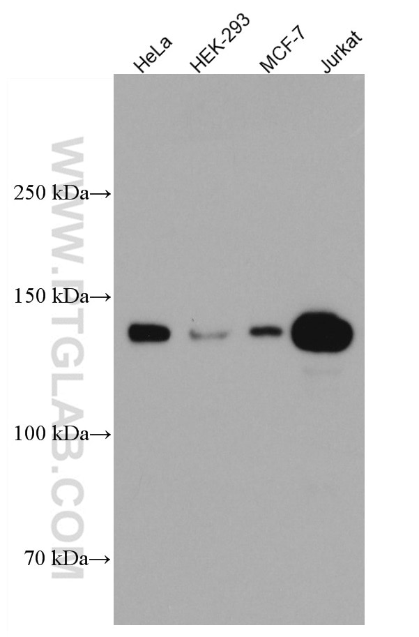
| Résultats positifs en WB | cellules HeLa, cellules HEK-293, cellules Jurkat, cellules MCF-7 |
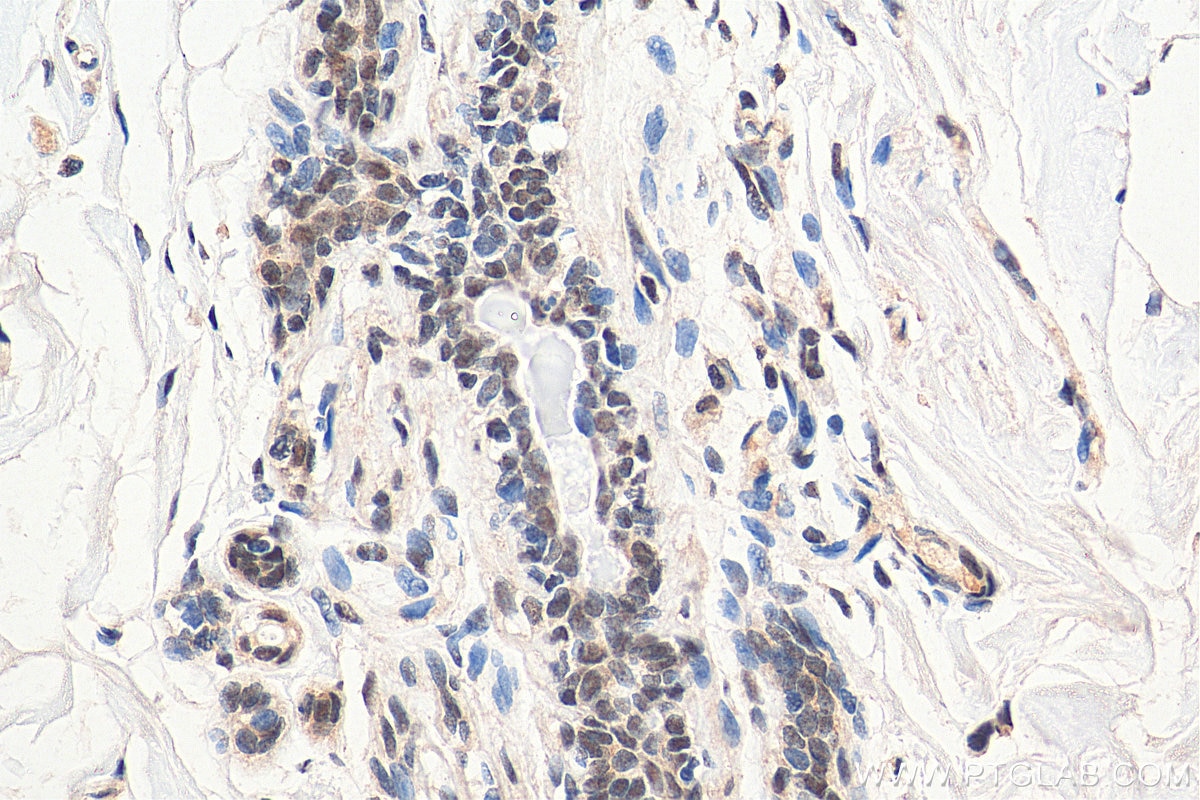
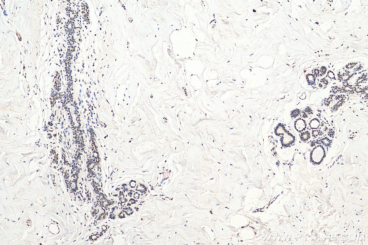
| Résultats positifs en IHC | tissu de cancer du sein humain, il est suggéré de démasquer l'antigène avec un tampon de TE buffer pH 9.0; (*) À défaut, 'le démasquage de l'antigène peut être 'effectué avec un tampon citrate pH 6,0. |
Dilution recommandée
| Application | Dilution |
|---|---|
| Western Blot (WB) | WB : 1:1000-1:3000 |
| Immunohistochimie (IHC) | IHC : 1:300-1:1200 |
| It is recommended that this reagent should be titrated in each testing system to obtain optimal results. | |
| Sample-dependent, check data in validation data gallery | |
Applications publiées
| KD/KO | See 2 publications below |
| WB | See 5 publications below |
| IF | See 2 publications below |
| IP | See 1 publications below |
| ChIP | See 1 publications below |
Informations sur le produit
66838-1-Ig cible HDAC4 dans les applications de WB, IP, IHC, IF, ChIP, ELISA et montre une réactivité avec des échantillons Humain
| Réactivité | Humain |
| Réactivité citée | Humain, souris |
| Hôte / Isotype | Mouse / IgG2a |
| Clonalité | Monoclonal |
| Type | Anticorps |
| Immunogène | HDAC4 Protéine recombinante Ag11345 |
| Nom complet | histone deacetylase 4 |
| Masse moléculaire calculée | 972 aa, 106 kDa |
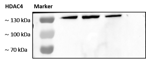
| Poids moléculaire observé | 140 kDa |
| Numéro d’acquisition GenBank | BC039904 |
| Symbole du gène | HDAC4 |
| Identification du gène (NCBI) | 9759 |
| Conjugaison | Non conjugué |
| Forme | Liquide |
| Méthode de purification | Purification par protéine A |
| Tampon de stockage | PBS avec azoture de sodium à 0,02 % et glycérol à 50 % pH 7,3 |
| Conditions de stockage | Stocker à -20°C. Stable pendant un an après l'expédition. L'aliquotage n'est pas nécessaire pour le stockage à -20oC Les 20ul contiennent 0,1% de BSA. |
Informations générales
HDAC4, also named as HDACA, belongs to the histone deacetylase family. HDAC4 is responsible for the deacetylation of lysine residues on the N-terminal part of the core histones (H2A, H2B, H3 and H4). Histone deacetylation gives a tag for epigenetic repression and plays an important role in transcriptional regulation, cell cycle progression and developmental events. Histone deacetylases act via the formation of large multiprotein complexes. HDAC4 is Involved in muscle maturation via its interaction with the myocyte enhancer factors such as MEF2A, MEF2C and MEF2D.
Protocole
| Product Specific Protocols | |
|---|---|
| WB protocol for HDAC4 antibody 66838-1-Ig | Download protocol |
| IHC protocol for HDAC4 antibody 66838-1-Ig | Download protocol |
| Standard Protocols | |
|---|---|
| Click here to view our Standard Protocols |
Publications
| Species | Application | Title |
|---|---|---|
Oncogenesis USP38 regulates the stemness and chemoresistance of human colorectal cancer via regulation of HDAC3. | ||
Virulence Inhibition of cell proliferation by Tas of foamy viruses through cell cycle arrest or apoptosis underlines the different mechanisms of virus-host interactions. | ||
J Mol Cell Cardiol Cellular heterogeneity and immune microenvironment revealed by single-cell transcriptome in venous malformation and cavernous venous malformation. | ||
Appl Physiol Nutr Metab Exercise improves high-fat diet-induced metabolic disorder by promoting HDAC5 degradation through the ubiquitin-proteasome system in skeletal muscle
| ||
J Toxicol Sci Propofol improves ischemia reperfusion-induced liver fibrosis by regulating lncRNA HOXA11-AS
| ||
Adv Clin Exp Med Tasquinimod enhances the sensitivity of ovarian cancer cells to cisplatin by regulating the Nur77-Bcl-2 apoptotic pathway |
Avis
The reviews below have been submitted by verified Proteintech customers who received an incentive forproviding their feedback.
FH Shinford (Verified Customer) (10-26-2023) | In our screening of basic expression in lung, breast, and gastric cancer cell lines, antibody concentrations ranging from 1:1000 to 2000 all displayed clear bands, indicating stable working conditions.
 |


