- Phare
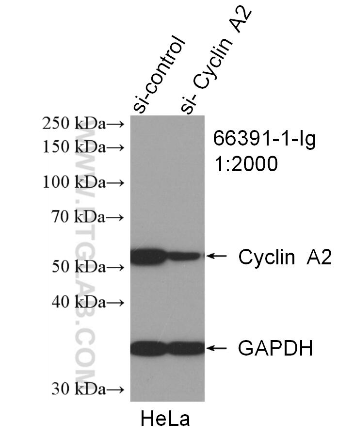
- Validé par KD/KO
Anticorps Monoclonal anti-Cyclin A2
Cyclin A2 Monoclonal Antibody for IF, IHC, WB, ELISA
Hôte / Isotype
Mouse / IgG2a
Réactivité testée
Humain et plus (2)
Applications
WB, IHC, IF, ELISA
Conjugaison
Non conjugué
CloneNo.
1C6C11
N° de cat : 66391-1-Ig
Synonymes
Galerie de données de validation
Applications testées
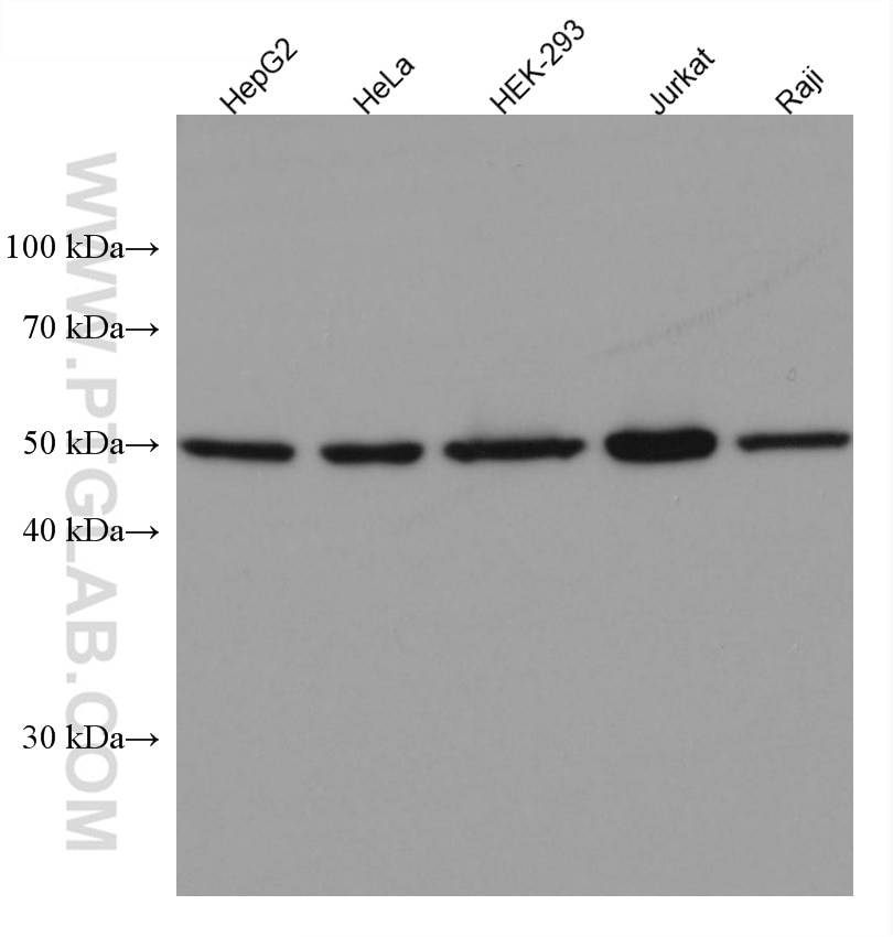
| Résultats positifs en WB | cellules HepG2, cellules HEK-293, cellules HeLa, cellules Jurkat, cellules Raji |
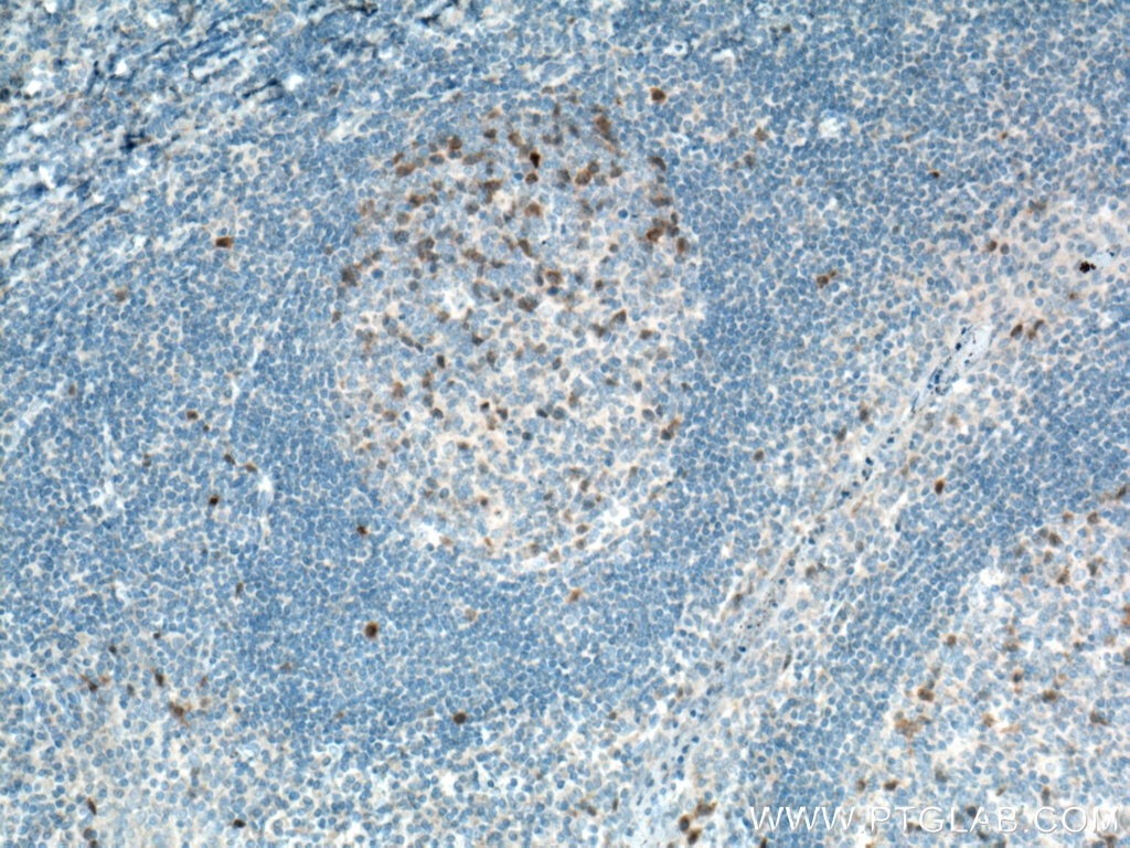
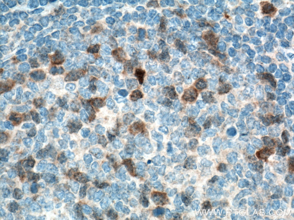
| Résultats positifs en IHC | tissu d'amygdalite humain, il est suggéré de démasquer l'antigène avec un tampon de TE buffer pH 9.0; (*) À défaut, 'le démasquage de l'antigène peut être 'effectué avec un tampon citrate pH 6,0. |
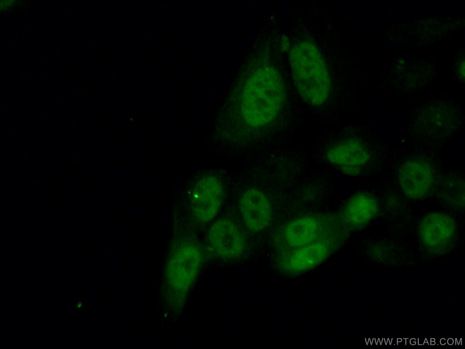
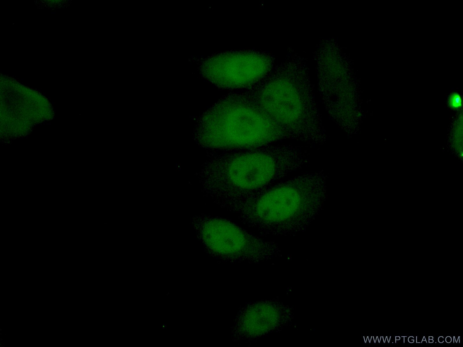
| Résultats positifs en IF | cellules HeLa, cellules MCF-7 |
Dilution recommandée
| Application | Dilution |
|---|---|
| Western Blot (WB) | WB : 1:2000-1:10000 |
| Immunohistochimie (IHC) | IHC : 1:1000-1:4000 |
| Immunofluorescence (IF) | IF : 1:50-1:500 |
| It is recommended that this reagent should be titrated in each testing system to obtain optimal results. | |
| Sample-dependent, check data in validation data gallery | |
Applications publiées
| WB | See 20 publications below |
| IF | See 2 publications below |
Informations sur le produit
66391-1-Ig cible Cyclin A2 dans les applications de WB, IHC, IF, ELISA et montre une réactivité avec des échantillons Humain
| Réactivité | Humain |
| Réactivité citée | rat, Humain, souris |
| Hôte / Isotype | Mouse / IgG2a |
| Clonalité | Monoclonal |
| Type | Anticorps |
| Immunogène | Cyclin A2 Protéine recombinante Ag12765 |
| Nom complet | cyclin A2 |
| Masse moléculaire calculée | 432 aa, 49 kDa |
| Poids moléculaire observé | 52 kDa |
| Numéro d’acquisition GenBank | BC104783 |
| Symbole du gène | CCNA2 |
| Identification du gène (NCBI) | 890 |
| Conjugaison | Non conjugué |
| Forme | Liquide |
| Méthode de purification | Purification par protéine A |
| Tampon de stockage | PBS avec azoture de sodium à 0,02 % et glycérol à 50 % pH 7,3 |
| Conditions de stockage | Stocker à -20°C. Stable pendant un an après l'expédition. L'aliquotage n'est pas nécessaire pour le stockage à -20oC Les 20ul contiennent 0,1% de BSA. |
Informations générales
CCNA2 belongs to the highly conserved cyclin family, whose members are characterized by a dramatic periodicity in protein abundance through the cell cycle. Cyclins function as regulators of CDK kinases. Different cyclins exhibit distinct expression and degradation patterns which contribute to the temporal coordination of each mitotic event. In contrast to cyclin A1, which is present only in germ cells, this cyclin is expressed in all tissues tested. CCNA2 binds and activates CDC2 or CDK2 kinases, and thus promotes both cell cycle G1/S and G2/M transitions.
Protocole
| Product Specific Protocols | |
|---|---|
| WB protocol for Cyclin A2 antibody 66391-1-Ig | Download protocol |
| IHC protocol for Cyclin A2 antibody 66391-1-Ig | Download protocol |
| IF protocol for Cyclin A2 antibody 66391-1-Ig | Download protocol |
| Standard Protocols | |
|---|---|
| Click here to view our Standard Protocols |
Publications
| Species | Application | Title |
|---|---|---|
Dev Cell Cell cycle regulation of ER membrane biogenesis protects against chromosome missegregation. | ||
Oncogene KCTD12 promotes tumorigenesis by facilitating CDC25B/CDK1/Aurora A-dependent G2/M transition. | ||
Elife METTL3 promotes homologous recombination repair and modulates chemotherapeutic response in breast cancer by regulating the EGF/RAD51 axis. | ||
J Cell Mol Med Cell division cycle associated 8: A novel diagnostic and prognostic biomarker for hepatocellular carcinoma | ||
Cell Cycle Enterovirus 71 mediates cell cycle arrest in S phase through non-structural protein 3D. | ||
Molecules A Potential Anti-Glioblastoma Compound LH20 Induces Apoptosis and Arrest of Human Glioblastoma Cells via CDK4/6 Inhibition |
Avis
The reviews below have been submitted by verified Proteintech customers who received an incentive forproviding their feedback.
FH Juri (Verified Customer) (01-20-2024) | Signal present after ICC, but expressing from all the cells.
|
