Anticorps Polyclonal de lapin anti-ATG14/Barkor
ATG14/Barkor Polyclonal Antibody for IHC, IP, WB, ELISA
Hôte / Isotype
Lapin / IgG
Réactivité testée
Humain, rat, souris
Applications
WB, IP, IHC, ELISA
Conjugaison
Non conjugué
N° de cat : 28021-1-AP
Synonymes
Galerie de données de validation
Applications testées
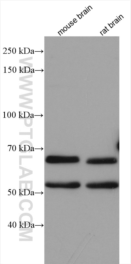
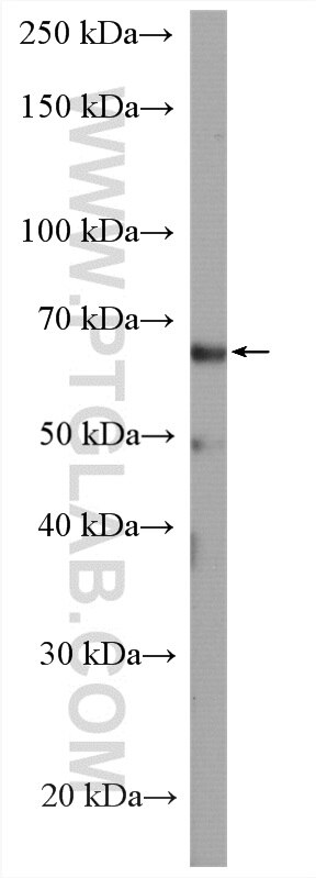
| Résultats positifs en WB | tissu cérébral de souris, cellules A549, tissu cérébral de rat |
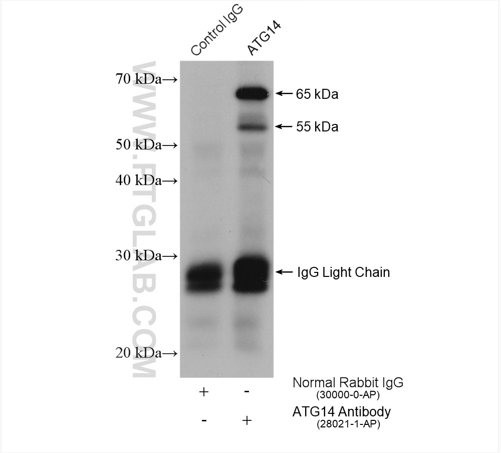
| Résultats positifs en IP | tissu cérébral de rat, |
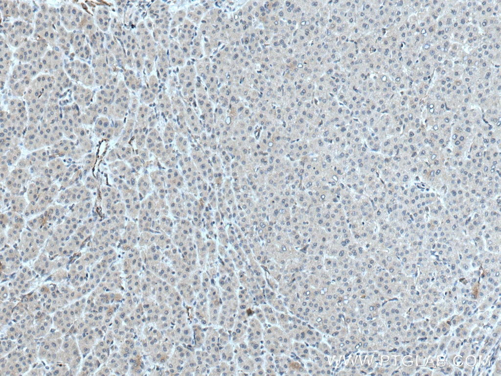
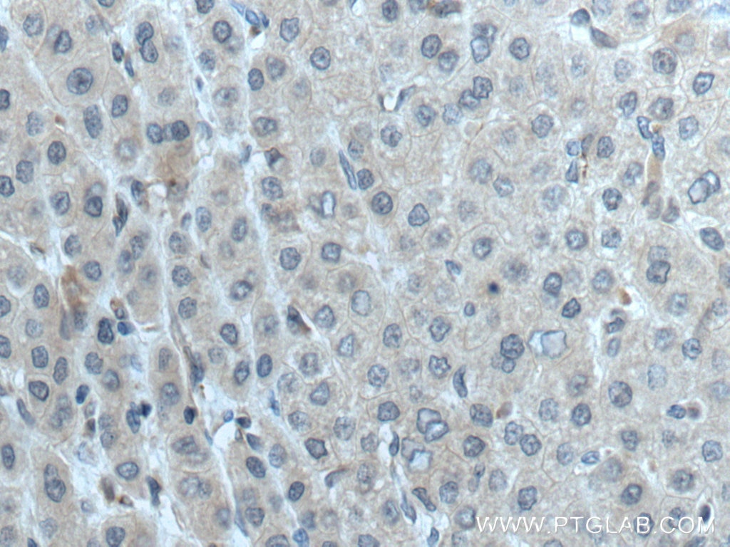
| Résultats positifs en IHC | tissu de cancer du foie humain, il est suggéré de démasquer l'antigène avec un tampon de TE buffer pH 9.0; (*) À défaut, 'le démasquage de l'antigène peut être 'effectué avec un tampon citrate pH 6,0. |
Dilution recommandée
| Application | Dilution |
|---|---|
| Western Blot (WB) | WB : 1:500-1:2000 |
| Immunoprécipitation (IP) | IP : 0.5-4.0 ug for 1.0-3.0 mg of total protein lysate |
| Immunohistochimie (IHC) | IHC : 1:200-1:800 |
| It is recommended that this reagent should be titrated in each testing system to obtain optimal results. | |
| Sample-dependent, check data in validation data gallery | |
Applications publiées
| WB | See 8 publications below |
| IHC | See 1 publications below |
Informations sur le produit
28021-1-AP cible ATG14/Barkor dans les applications de WB, IP, IHC, ELISA et montre une réactivité avec des échantillons Humain, rat, souris
| Réactivité | Humain, rat, souris |
| Réactivité citée | Humain, souris |
| Hôte / Isotype | Lapin / IgG |
| Clonalité | Polyclonal |
| Type | Anticorps |
| Immunogène | ATG14/Barkor Protéine recombinante Ag27671 |
| Nom complet | KIAA0831 |
| Masse moléculaire calculée | 55 kDa |
| Poids moléculaire observé | 55 and 65 kDa |
| Numéro d’acquisition GenBank | NM_014924 |
| Symbole du gène | KIAA0831 |
| Identification du gène (NCBI) | 22863 |
| Conjugaison | Non conjugué |
| Forme | Liquide |
| Méthode de purification | Purification par affinité contre l'antigène |
| Tampon de stockage | PBS avec azoture de sodium à 0,02 % et glycérol à 50 % pH 7,3 |
| Conditions de stockage | Stocker à -20°C. Stable pendant un an après l'expédition. L'aliquotage n'est pas nécessaire pour le stockage à -20oC Les 20ul contiennent 0,1% de BSA. |
Informations générales
ATG14 plays a critical role in autophagy which is required for both basal and inducible autophagy. It has been reported that the loss of ATG14 completely blocks autophagic fusion with lysosomes. ATG14 is associated with the location of PI3-kinase complex PI3KC3-C1 and the formation of autophagosome. Besides, BECN2 can form the BECN2:ATG14 heterodimer with ATG14. 28021-1-AP antibody recognizes the 55 kDa protein and the phosphorylated 65 kDa protein in SDS-PAGE. (PMID: 28218432, 24597603, 30894050)
Protocole
| Product Specific Protocols | |
|---|---|
| WB protocol for ATG14/Barkor antibody 28021-1-AP | Download protocol |
| IHC protocol for ATG14/Barkor antibody 28021-1-AP | Download protocol |
| IP protocol for ATG14/Barkor antibody 28021-1-AP | Download protocol |
| Standard Protocols | |
|---|---|
| Click here to view our Standard Protocols |
Publications
| Species | Application | Title |
|---|---|---|
Cancer Lett Targeting PP2A with lomitapide suppresses colorectal tumorigenesis through the activation of AMPK/Beclin1-mediated autophagy. | ||
Ecotoxicol Environ Saf Weakened interaction of ATG14 and the SNARE complex blocks autophagosome-lysosome fusion contributes to fluoride-induced developmental neurotoxicity. | ||
Cells Cytosolic HMGB1 Mediates LPS-Induced Autophagy in Microglia by Interacting with NOD2 and Suppresses Its Proinflammatory Function | ||
Life (Basel) MiR-371a-5p Positively Associates with Hepatocellular Carcinoma Malignancy but Sensitizes Cancer Cells to Oxaliplatin by Suppressing BECN1-Dependent Autophagy | ||
FASEB J Melatonin ameliorates cerebral ischemia-reperfusion injury in diabetic mice by enhancing autophagy via the SIRT1-BMAL1 pathway | ||
Heliyon Elevated GABRP expression is correlated to the excessive autophagy in intrahepatic cholestasis of pregnancy |
Avis
The reviews below have been submitted by verified Proteintech customers who received an incentive forproviding their feedback.
FH Balawant (Verified Customer) (12-25-2023) | I have used this antibody, and it is working great. this antibody has specific western blot band and no background.
|
