GFP tag Polyklonaler Antikörper
GFP tag Polyklonal Antikörper für IF, IP, WB, ELISA
Wirt / Isotyp
Kaninchen / IgG
Getestete Reaktivität
aequorea victoria, rekombinanten Protein und mehr (3)
Anwendung
WB, RIP, IP-MS, IP, IHC, IF, CoIP, ChIP, ELISA
Konjugation
Unkonjugiert
Kat-Nr. : 50430-2-AP
Synonyme
Galerie der Validierungsdaten
 at various dilutions.")
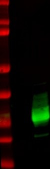
 at dilution of 1:1000 incubated at room temperature for 1.5 hours.")
 with Transfected HEK-293 cells lysate 300ug.")
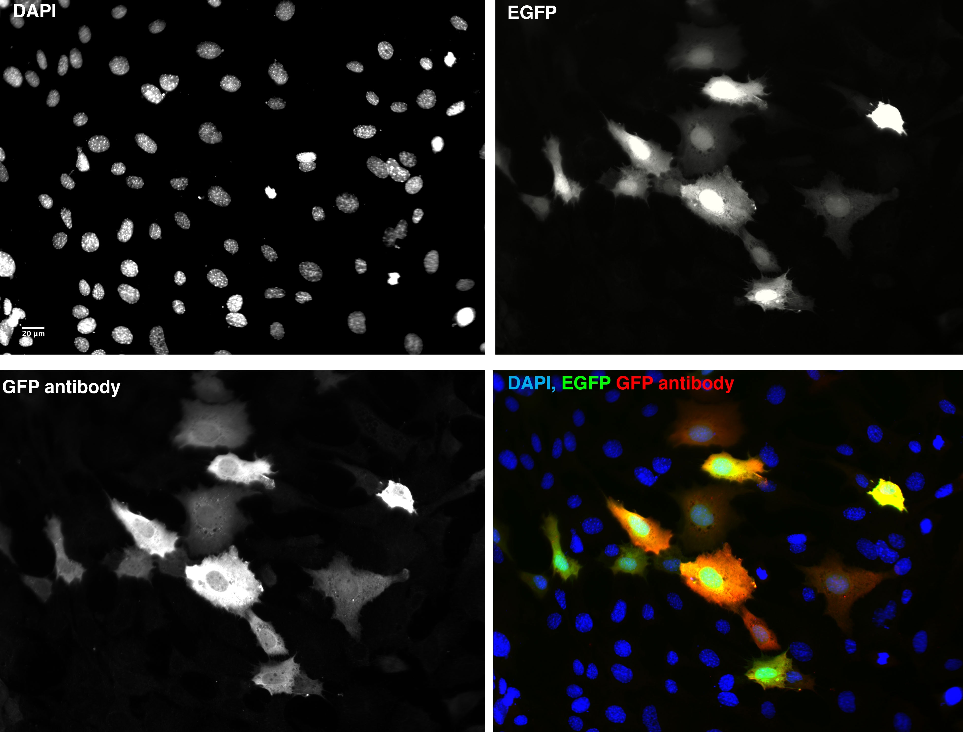
 fixed Transfected HEK-293 cells using 50430-2-AP (GFP tag antibody) at dilution of 1:100 and Alexa Fluor 594-conjugated AffiniPure Goat Anti-Rabbit IgG(H+L).")
 fixed Transfected HEK-293 cells using GFP tag antibody (50430-2-AP) at dilution of 1:1000 and CoraLite®594-Conjugated AffiniPure Goat Anti-Rabbit IgG(H+L).")
Geprüfte Anwendungen
| Erfolgreiche Detektion in WB | recombinant protein, Transfizierte HEK-293-Zellen |
| Erfolgreiche IP | Transfizierte HEK-293-Zellen |
| Erfolgreiche Detektion in IF | Transfizierte HEK-293-Zellen |
Empfohlene Verdünnung
| Anwendung | Verdünnung |
|---|---|
| Western Blot (WB) | WB : 1:1000-1:4000 |
| Immunpräzipitation (IP) | IP : 0.5-4.0 ug for 1.0-3.0 mg of total protein lysate |
| Immunfluoreszenz (IF) | IF : 1:20-1:200 |
| It is recommended that this reagent should be titrated in each testing system to obtain optimal results. | |
| Sample-dependent, check data in validation data gallery | |
Veröffentlichte Anwendungen
| WB | See 301 publications below |
| IHC | See 13 publications below |
| IF | See 119 publications below |
| IP | See 51 publications below |
| CoIP | See 27 publications below |
| ChIP | See 3 publications below |
| RIP | See 1 publications below |
Produktinformation
50430-2-AP bindet in WB, RIP, IP-MS, IP, IHC, IF, CoIP, ChIP, ELISA GFP tag und zeigt Reaktivität mit aequorea victoria, rekombinanten Protein
| Getestete Reaktivität | aequorea victoria, rekombinanten Protein |
| In Publikationen genannte Reaktivität | Affe, Maus, Ratte |
| Wirt / Isotyp | Kaninchen / IgG |
| Klonalität | Polyklonal |
| Typ | Antikörper |
| Immunogen | GFP tag fusion protein Ag2128 |
| Vollständiger Name | GFP tag |
| Berechnetes Molekulargewicht | 26 kDa |
| GenBank-Zugangsnummer | U73901 |
| Gene symbol | |
| Gene ID (NCBI) | |
| Konjugation | Unkonjugiert |
| Form | Liquid |
| Reinigungsmethode | Antigen-Affinitätsreinigung |
| Lagerungspuffer | PBS mit 0.02% Natriumazid und 50% Glycerin pH 7.3. |
| Lagerungsbedingungen | Bei -20°C lagern. Nach dem Versand ein Jahr lang stabil Aliquotieren ist bei -20oC Lagerung nicht notwendig. 20ul Größen enthalten 0,1% BSA. |
Hintergrundinformationen
Protein tags are protein or peptide sequences located either on the C- or N- terminal of the target protein, which facilitates one or several of the following characteristics: solubility, detection, purification, localization and expression. Green fluorescence protein(GFP) is a protein composed of 238 amino acid residues(26.9kDa) derived from the Jellyfish Aequorea victoria, which emits green light(emission peak at 509nm) when excited by blue light(excitation peak at 395nm). GFP has become an invaluable tool in cell biology research, since its intrinsic fluorescence can be visualized in living cells. EGFP contains the double-amino-acid substitutions Phe-64 to Leu and Ser-65 to Thr(previously published as GFPmut1; PMID: 8707053). In contrast to wtGFP, EGFP has a single, strong, red-shifted excitation peak at 488nm. GFPmut1 fluoresces 35-fold more intensely than wtGFP when excited at 488nm, due to an increase in its extinction coefficient(Em). This antibody is a rabbit polyclonal antibody raised against full-length eGFP and reactive against all variants of Aequorea victoria GFP such as S65T-GFP, RS-GFP, YFP, CFP and eGFP.
Protokolle
| Produktspezifische Protokolle | |
|---|---|
| WB protocol for GFP tag antibody 50430-2-AP | Protokoll herunterladen |
| IF protocol for GFP tag antibody 50430-2-AP | Protokoll herunterladen |
| IP protocol for GFP tag antibody 50430-2-AP | Protokoll herunterladen |
| Standard-Protokolle | |
|---|---|
| Klicken Sie hier, um unsere Standardprotokolle anzuzeigen |
Publikationen
| Species | Application | Title |
|---|---|---|
Gastroenterology PTEN deficiency facilitates exosome secretion and metastasis in cholangiocarcinoma by impairing TFEB-mediated lysosome biogenesis | ||
Nat Genet Pathogenic SPTBN1 variants cause an autosomal dominant neurodevelopmental syndrome. | ||
Cell Stem Cell DUX-miR-344-ZMYM2-Mediated Activation of MERVL LTRs Induces a Totipotent 2C-like State. | ||
Nat Methods TALEN-mediated precise genome modification by homologous recombination in zebrafish. | ||
Immunity Intestinal Tuft-2 cells exert antimicrobial immunity via sensing bacterial metabolite N-undecanoylglycine. |
Rezensionen
The reviews below have been submitted by verified Proteintech customers who received an incentive forproviding their feedback.
FH S (Verified Customer) (05-26-2023) | Excellent
 |
FH PK (Verified Customer) (03-20-2023) | Very Good
 |
FH Stephen (Verified Customer) (08-02-2022) | HeLa cells transfected with plasmid expressing EGFP fixed with 4% PFA for 15 mins and immunostained with rabbit GFP antibody at 4 degrees incubated for 20h washed then incubated secondary alexa 594 and DAPI for 1 hour room temp imaged on wide field fluorescence microscope.
 |
FH A (Verified Customer) (01-04-2022) | Works for IP
|
FH Rinalda (Verified Customer) (06-21-2021) | Great product
|
FH Lana (Verified Customer) (12-22-2020) | SDS-PAGE: 15 ug/ul RIPA protein lysate, 4-12% Bis-Tris gradient gel.Transfer: Immobilon-FL transfer membranes (Millipore) for 2h at 80V, 4C.Blocking: SEA Block Blocking Buffer 1h, room T.Primary Ab: O/N incubation at 4C, 1:2500.Secondary Ab: IRDye 800CW Goat anti-Rabbit, 1:15000.Lines of WB image: 1 – protein ladder, 2 – HEK293 whole cell lysate, negative transfection, 3 – whole cell lysate of cells transfected with eGFP.
 |
FH Paul (Verified Customer) (01-15-2020) | Works well for WB.
|
FH Laura (Verified Customer) (01-14-2020) | Good sensitivity for Western blot.
|
FH Aamir (Verified Customer) (01-08-2020) | Works well for WB and IF
|
FH Erica (Verified Customer) (09-26-2019) | This antibody works very well with both western blots and IP. We previously used GFP antibody from another company and it didn't work well. We then switched to Proteinntech's and the signal was very strong! Highly recommend!
|
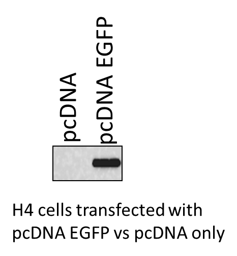
FH Mayur (Verified Customer) (04-30-2019) | The H4 cells were transfected with pcDNA only and pcDNA EGFP . The expression of the EGFP was measured using the GFP antibody from ProteinTech. Great antibody.
 |

