- Featured Product
- KD/KO Validated
SLC3A1 Polyklonaler Antikörper
SLC3A1 Polyklonal Antikörper für IHC, IP, WB, ELISA
Wirt / Isotyp
Kaninchen / IgG
Getestete Reaktivität
human, Maus, Ratte und mehr (1)
Anwendung
WB, IP, IHC, IF, ELISA
Konjugation
Unkonjugiert
Kat-Nr. : 16343-1-AP
Synonyme
Galerie der Validierungsdaten
Geprüfte Anwendungen
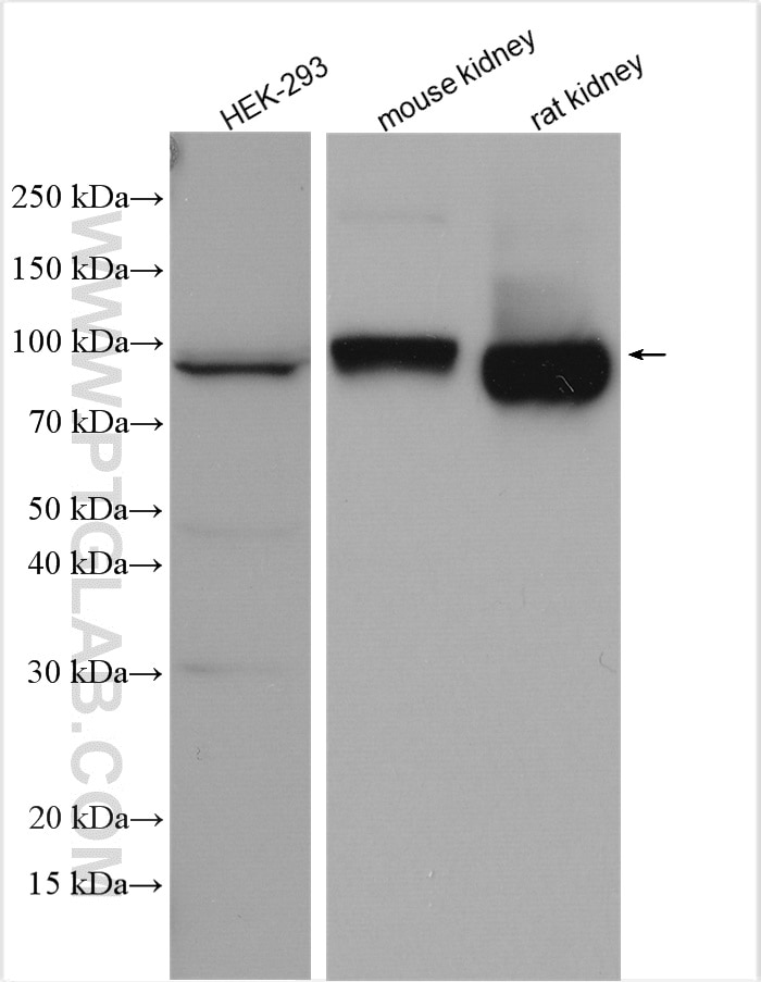
| Erfolgreiche Detektion in WB | HEK-293-Zellen, Mausnierengewebe, Rattennierengewebe |
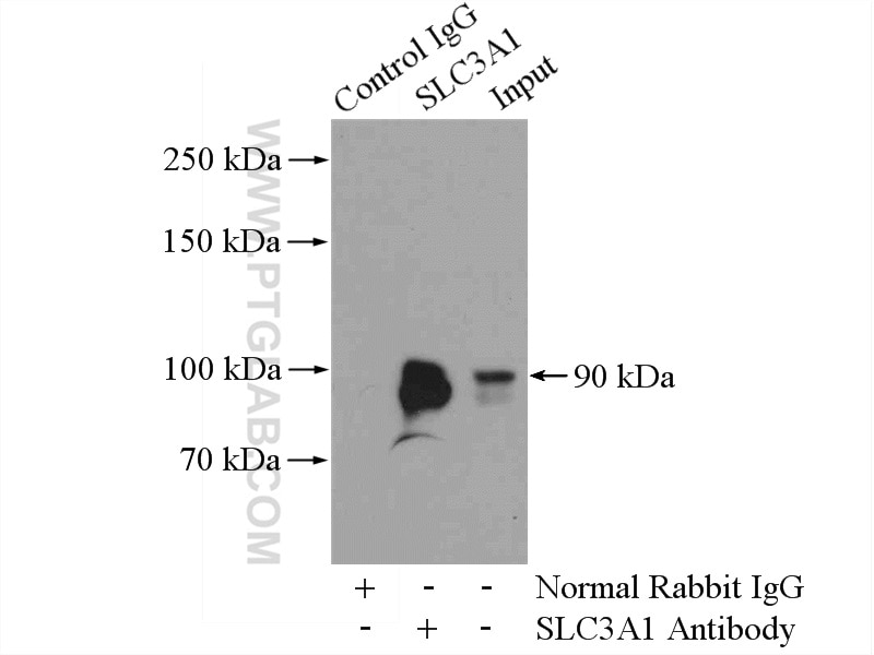
| Erfolgreiche IP | Mausnierengewebe |
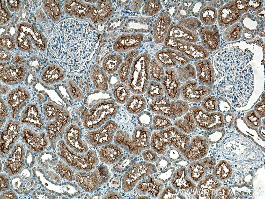
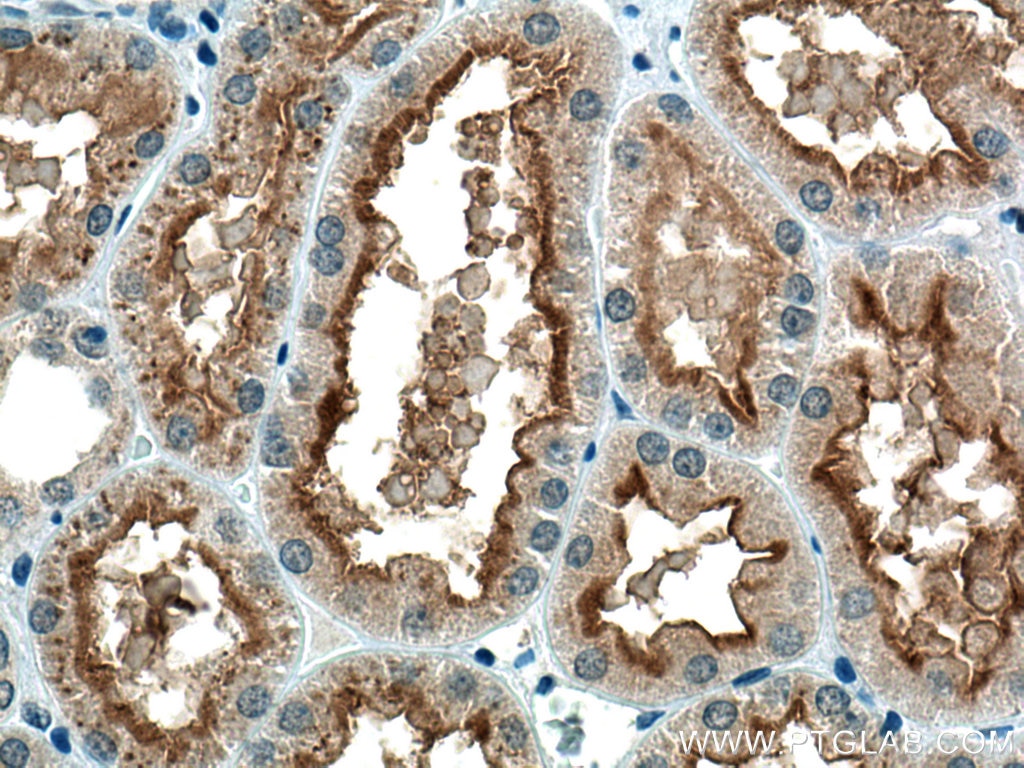
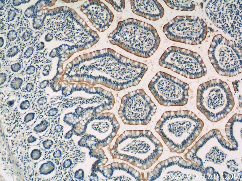
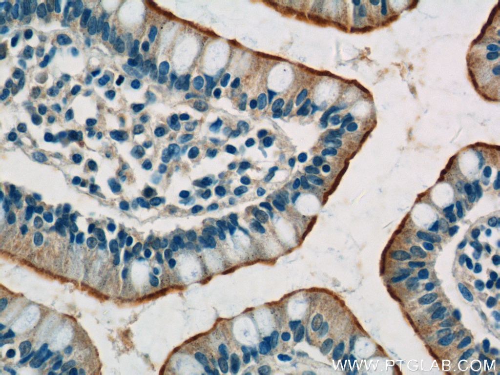
| Erfolgreiche Detektion in IHC | humanes Nierengewebe, humanes Dünndarmgewebe Hinweis: Antigendemaskierung mit TE-Puffer pH 9,0 empfohlen. (*) Wahlweise kann die Antigendemaskierung auch mit Citratpuffer pH 6,0 erfolgen. |
Empfohlene Verdünnung
| Anwendung | Verdünnung |
|---|---|
| Western Blot (WB) | WB : 1:500-1:2000 |
| Immunpräzipitation (IP) | IP : 0.5-4.0 ug for 1.0-3.0 mg of total protein lysate |
| Immunhistochemie (IHC) | IHC : 1:50-1:500 |
| It is recommended that this reagent should be titrated in each testing system to obtain optimal results. | |
| Sample-dependent, check data in validation data gallery | |
Veröffentlichte Anwendungen
| KD/KO | See 2 publications below |
| WB | See 4 publications below |
| IHC | See 1 publications below |
| IF | See 5 publications below |
Produktinformation
16343-1-AP bindet in WB, IP, IHC, IF, ELISA SLC3A1 und zeigt Reaktivität mit human, Maus, Ratten
| Getestete Reaktivität | human, Maus, Ratte |
| In Publikationen genannte Reaktivität | human, Maus, Ziege |
| Wirt / Isotyp | Kaninchen / IgG |
| Klonalität | Polyklonal |
| Typ | Antikörper |
| Immunogen | SLC3A1 fusion protein Ag9525 |
| Vollständiger Name | solute carrier family 3 (cystine, dibasic and neutral amino acid transporters, activator of cystine, dibasic and neutral amino acid transport), member 1 |
| Berechnetes Molekulargewicht | 685 aa, 79 kDa |
| Beobachtetes Molekulargewicht | 79 kDa |
| GenBank-Zugangsnummer | BC022386 |
| Gene symbol | SLC3A1 |
| Gene ID (NCBI) | 6519 |
| Konjugation | Unkonjugiert |
| Form | Liquid |
| Reinigungsmethode | Antigen-Affinitätsreinigung |
| Lagerungspuffer | PBS mit 0.02% Natriumazid und 50% Glycerin pH 7.3. |
| Lagerungsbedingungen | Bei -20°C lagern. Nach dem Versand ein Jahr lang stabil Aliquotieren ist bei -20oC Lagerung nicht notwendig. 20ul Größen enthalten 0,1% BSA. |
Hintergrundinformationen
SLC3A1(Neutral and basic amino acid transport protein rBAT) is also named as NBAT,D2h,RBAT. It is involved in the high-affinity, sodium-independent transport of cystine and neutral and dibasic amino acids (system B(0,+)-like activity).SLC3A1 can be degraded, at least in part, via the endoplasmic reticulum-associated degradation (ERAD) pathway(PMID:18332091). SLC3A1 has different isoforms vary from 79 kDa to 36kDa and the 85 kDa glycosylated protein can be detected in SDS-PAGE. (PMID:11546643)
Protokolle
| Produktspezifische Protokolle | |
|---|---|
| WB protocol for SLC3A1 antibody 16343-1-AP | Protokoll herunterladen |
| IHC protocol for SLC3A1 antibody 16343-1-AP | Protokoll herunterladen |
| IP protocol for SLC3A1 antibody 16343-1-AP | Protokoll herunterladen |
| Standard-Protokolle | |
|---|---|
| Klicken Sie hier, um unsere Standardprotokolle anzuzeigen |
Publikationen
| Species | Application | Title |
|---|---|---|
Nat Cell Biol Directing human embryonic stem cell differentiation towards a renal lineage generates a self-organizing kidney. | ||
Nat Commun Transcription factors AP-2α and AP-2β regulate distinct segments of the distal nephron in the mammalian kidney. | ||
Dev Cell AP-2β/KCTD1 Control Distal Nephron Differentiation and Protect against Renal Fibrosis. | ||
Theranostics Cysteine transporter SLC3A1 promotes breast cancer tumorigenesis.
| ||
Rezensionen
The reviews below have been submitted by verified Proteintech customers who received an incentive forproviding their feedback.
FH Felisha (Verified Customer) (01-03-2020) | The primary antibody worked great to localize the derived human stem cells in the mouse tissue samples that were sectioned and paraffin embedded. Wonderful for immunohistochemistry!
|
