Phospho-AKT (Ser473) Monoklonaler Antikörper
Phospho-AKT (Ser473) Monoklonal Antikörper für FC (Intra)
Wirt / Isotyp
Maus / IgG1
Getestete Reaktivität
human, Maus
Anwendung
FC (Intra)
Konjugation
CoraLite® Plus 488 Fluorescent Dye
CloneNo.
1C10B8
Kat-Nr. : CL488-66444
Synonyme
Galerie der Validierungsdaten
Geprüfte Anwendungen
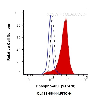
| Erfolgreiche Detektion in FC | Mit Calyculin A behandelte PC-3-Zellen |
Empfohlene Verdünnung
| Anwendung | Verdünnung |
|---|---|
| Durchflusszytometrie (FC) | FC : 0.25 ug per 10^6 cells in a 100 µl suspension |
| It is recommended that this reagent should be titrated in each testing system to obtain optimal results. | |
| Sample-dependent, check data in validation data gallery | |
Produktinformation
CL488-66444 bindet in FC (Intra) Phospho-AKT (Ser473) und zeigt Reaktivität mit human, Maus
| Getestete Reaktivität | human, Maus |
| Wirt / Isotyp | Maus / IgG1 |
| Klonalität | Monoklonal |
| Typ | Antikörper |
| Immunogen | Peptid |
| Vollständiger Name | v-akt murine thymoma viral oncogene homolog 1 |
| Beobachtetes Molekulargewicht | 60-62 kDa |
| GenBank-Zugangsnummer | NM_005163 |
| Gene symbol | AKT1 |
| Gene ID (NCBI) | 207 |
| Konjugation | CoraLite® Plus 488 Fluorescent Dye |
| Excitation/Emission maxima wavelengths | 493 nm / 522 nm |
| Form | Liquid |
| Reinigungsmethode | Protein-A-Reinigung |
| Lagerungspuffer | BS mit 50% Glyzerin, 0,05% Proclin300, 0,5% BSA, pH 7,3. |
| Lagerungsbedingungen | Bei -20°C lagern. Vor Licht schützen. Aliquotieren ist bei -20oC Lagerung nicht notwendig. 20ul Größen enthalten 0,1% BSA. |
Hintergrundinformationen
1) What is AKT?
The serine/threonine kinase B AKT pathway (also known as the PI3K-Akt pathway) plays a vital role in the regulation of cellular processes, including cell proliferation, survival, and growth - processes that are essential for oncogenesis. Mutation of the regulator proteins PI3K and PTEN causes uncontrolled disruption within the PI3-kinase pathway, leading to the development of human cancers (1,2; see also AKT pathway poster for more details).
2) phospho-AKT and FAQs
A) What is the best way to normalize phosphorylated proteins analyzed by western blot?
Normalize phospho-AKT and total AKT with your loading control (e.g. Actin, tubulin), then calculate the phospho/total ratio using these normalized values.
Put more simply:
1. Calculate the ratio of band intensities of a phospho-AKT band: the loading control.
2. Calculate the ratio of band intensities of total AKT: loading control.
3. Divide ratio obtained #1 by #2 to obtain a normalized value for comparison among different conditions. This procedure allows one to distinguish between a change in AKT expression and a change in the ratio of phospho-AKT.
* If you are looking at the differences in a phospho-AKT expression resulting from an experimental condition (e.g., knockdown), you should also show the expression of total AKT to distinguish between a change in AKT expression (transcription/translation level) and a change in the AKT phosphorylation status.
B) What is the observed molecular weight for AKT and phospho-AKT?
Molecular Weight AKT - 56 kDa
Molecular Weight phospho-AKT - 60 kDa (Figure 1)

Figure 1. WB: HEK-293 cell lysate was subjected to SDS PAGE followed by western blot with 60203-2-Ig (AKT antibody) and 66444-1-Ig (AKT-phospho-S473 antibody) at a dilution of 1:4000 incubated at room temperature for 1.5 hours.
C) Are there any special WB conditions to optimize staining of a phospho-AKT?
Since this is a phosphorylated protein, 5% BSA is recommended over non-fat milk as a blocking agent.
D) What are good positive and negative controls for a phospho-AKT?
- Positive Control: HEK293 cells
- Negative Control: Treatment with PI3K inhibitors (e.g. wortmannin)
E) What species does this antibody react with?
Our internal testing has confirmed that it reacts with the human and mouse forms of phospho-AKT.Reactivity with the human form is also supported by the literature's citations of this antibody.
References:
1. Perturbations of the AKT signaling pathway in human cancer.
2. Targeting the PI3K-Akt pathway in human cancer: rationale and promise.


